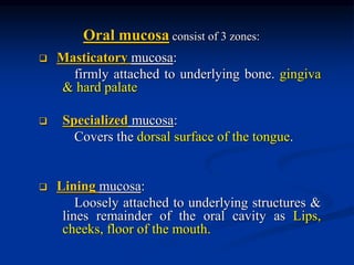
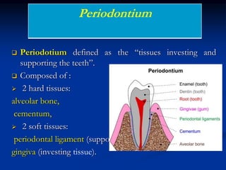
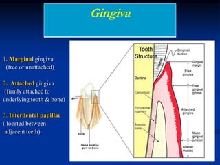
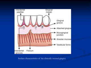
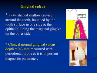
SlideShare a Scribd company logo
1 of 82
An Introduction to Periodontology Course&
normal anatomy of periodontium
Prepared by
Dr. Hisham Abozaid
Lecturer of oral Medicine & periodontology
 Normal periodontium : (Clinical and Histological Characteristics):
Gingiva, Periodontal ligament, Cementum, alveolar bone.
 Pathogenesis of periodontal disease
 Etiology of Periodontal disease: local & systemic
 Classification of Periodontal diseases
 Gingival lesions
 Periodontal lesions
 Diagnosis, Prognosis and Treatment plan
 Treatment :
 Non-surgical Periodontal Therapy
 Surgical Periodontal Therapy
Anatomy of the Periodontium
Clinical and Histological Characteristics
Objectives
 What is meant by periodontium? What are its various
components?
 Describe criteria & functions of each component of
periodontium.
The term periodontium arises from the Greek word peri
meaning around & odont meaning tooth.
 The various diseases of the periodontium are
collectively termed as periodontal diseases.
 Their treatment is referred to as periodontal therapy.
 The clinical science that deals with the periodontium in
health & disease is called periodontology.
 The branch of dentistry concerned with prevention &
treatment of periodontal disease is termed
periodontics .
 Masticatory mucosa:
firmly attached to underlying bone. gingiva
& hard palate
 Specialized mucosa:
Covers the dorsal surface of the tongue.
 Lining mucosa:
Loosely attached to underlying structures &
lines remainder of the oral cavity as Lips,
cheeks, floor of the mouth.
Oral mucosa consist of 3 zones:
 Periodotium defined as the “tissues investing and
supporting the teeth”.
 Composed of :
 2 hard tissues:
alveolar bone,
cementum,
 2 soft tissues:
periodontal ligament (supporting tissues)
gingiva (investing tissue).
Periodontium
 Normal gingiva is that part of oral mucosa, which
covers the alveolar bone & tooth root to a level just
coronal to the cementoenamel junction.
 The main function of the gingiva is to protect the
surrounding tissues from the oral environment.
Gingiva
The gingiva divided into three parts:
1. Marginal gingiva
(free or unattached)
2. Attached gingiva
(firmly attached to
underlying tooth & bone)
3. Interdental papillae
( located between
adjacent teeth).
Gingiva
Surface characteristics of the clinically-normal gingiva
Clinically
1)Mucogingival junction.
2)Interdental gingiva.
3)Free gingival groove
4)Attached gingiva.
5)Alveolar mucosa
6) Marginal gingiva.
Marginal(free) gingiva
• The most coronal portion of gingiva.
• well-adapted to tooth surface but not
attached to it.
• creates soft tissue wall of the gingival
sulcus.
• In health, knife-edged in contour &
smooth in texture.
Marginal(free) gingiva
• extends apically to free gingival
groove, approximately 1 mm wide.
• It may be separated from
the tooth surface with
a periodontal probe.
• The most apical point of the marginal
gingival scallop is called the gingival
zenith.
Attached Gingiva
• firmly bound to the underlying tooth & alveolar bone.
• stippled in texture, and firm & resilient in consistency and
covered by keratinized epith.
• Bound coronally by free gingival
groove & apically by
mucogingival junction.
• can withstand
mechanical forces
of brushing & tension
applied on mucosa.
Width of Attached Gingiva
• important clinical parameter.
• the distance between
mucogingival junction &
the projection on the external surface
of the bottom of the gingival sulcus.
• should not be confused with the width of the keratinized gingiva
because the latter also includes marginal gingiva
• Greatest in incisor region (3.5-4.5 in maxilla, 3.3-3.9 in
mandible). & Least in premolar area (1.9 mm in maxilla, 1.8 in
mandible).
Interdental gingiva (papilla)
 portion of free gingiva located in inter-proximal space created by
contact between two adjacent teeth.
 pyramidal in anterior region & tent shape in posterior region.
 The shape of interdental papilla depends on :
 the presence or absence of a contact point between the adjacent
teeth,
 the distance between the contact point
& the osseous crest
 Presence or absence of some degree of
recession.
 Facial & lingual papillae are connected together by a valley-like
depression called “Col” made up from non keratinized epithelium
& represents the most frequent site for initiation of disease process.
• If a diastema is present,
the gingiva is firmly bound
over the interdental bone
& forms a smooth,
rounded surface without
interdental papillae
Gingival sulcus
* a -V- shaped shallow crevice
around the tooth, bounded by the
tooth surface in one side & the
epithelial lining the marginal gingiva
on the other side.
* Clinical normal gingival sulcus
depth = 0-3 mm measured with
periodontal probe & it is important
diagnostic parameter.
 There is a fluid secreted from gingival C.T into the sulcus
through the thin sulcular wall termed as gingival fluid.
 Its secretion increases with tissue inflammation and during
chewing.
 contains a vast array of biochemical factors, thereby offering its
potential use as a diagnostic or prognostic biomarker of the
biologic state of the periodontium in health & disease.
Gingival (Crevicular) Fluid
Components of sulcular fluid
1. Cellular elements : bacteria, desquamated epithelial cells, PMNs,
lymphocytes and monocytes.
2. Electrolytes: potassium, sodium and calcium,
3. Organic compounds: as carbohydrates, proteins and
immunoglobulin (IgA, IgM), complement C3 and C4, plasma proteins,
4. Metabolic & bacterial products: lactic acid, urea, hydrogen
sulfide, endotoxinns.
5.Enzymes: alkaline phosphatase, lysozomes, proteases, lactic
dehydrogenase.
Functions of Gingival (Crevicular) Fluid
 Its flow washes out irritants & prevent bacterial penetration.
 Its plasma proteins improve epithelial adhesion to the teeth.
 Has antibacterial effect upon many oral microorganisms.
 Has defense mechanism by its immunoglobulin contents.
 Essential growth factor for many microorganisms.
The understanding of clinical features
of periodontium is enhanced by a
knowledge of histological component of
tissue.
Microanatomy of the gingiva
Histological Features
 The gingiva consist of central core of connective tissue
covered by stratified squamous
epithelium.
 the epith. is predominantly
cellular in nature.
 C.T is less cellular & composed
primarily of collagen fibers &
ground substance
Gingival Epithelium
 Functions of gingival epith.
 provide a physical barrier to infection & the
underlying gingival attachment
 play an active role in innate & acquired host
defense.
 Composed of
1)Oral (outer) Epithelium: facing oral cavity
2) Sulcular Epithelium: facing tooth surface
3) Junctional Epithelium: forms
the junction between gingiva & tooth surface
Cells of the gingival epithelium
􀂆Keratinocytes(keratin producer cells): 90% of gingival
epithelial cells.
􀂆Non keratinocytes :
• Melanocytes: produce melanin, which is a pigment found
in the skin, eyes, hair, and gingiva
• Langerhans cells : important role in the immune
reaction as antigen-presenting cells for lymphocytes
• Merkel cells: harbor nerve endings& identified as tactile
perceptors
Oral Epithelium
covers the crest, outer surfaces of marginal & attached gingivae;
consists of keratinized stratified squamous epithelium arranged in 4
layers:
 Stratum basale, Basal cell layer (The deepest): possessing ability to
undergo mitotic division.
 Stratum Spinosum , prickle cell layer: cells are polyhedral and
attached by desmosomes.
 Stratum granulosum, granular cell layer: contains keratohyaline
bodies and glycogen granules.
 Stratum corneum, Keratinized cell layer (Keratinized, para, or
nonkeratinized) : cytoplasm is filled with keratin
Layers of Squamous epithelium
 lines the gingival sulcus facing the tooth extending from
coronal limit of junctional epith. to the crest of gingival
margin.
 acts as a semipermeable membrane through which bacterial
products can passes & tissue fluid can seeps.
 It is thin non-keratinized stratified squamous
epith. without rete pegs & lacks stratum
granuolosum and corneum
Sulcular Epithelium
Junctional Epithelium (Epithelial attachment)
 the tissue that joins the tooth on one side and to sulcular epith. & C.T
on the other. It forms the base of the sulcus.
 consists of collar-like band of thin non-keratinized stratified squamous
epith.
 The junctional epithelium is attached to the tooth
by internal basal lamina & to gingival C.T by
external basal lamina.
Junctional Epithelium (Epithelial attachment)
Exhibits several unique structural & functional features that contribute to
preventing pathogenic bacteria from colonizing subgingival tooth
surface:
 firmly attached to the tooth surface, forming an epithelial barrier
against plaque bacteria.
 allows access of gingival fluid, inflammatory cells, and components of
the immunologic host defense to the gingival margin.
 junctional epithelial cells exhibit rapid turnover, which contributes to
the host-parasite equilibrium & rapid repair of damaged tissue.
Junctional Epithelium (Epithelial attachment)
 Attachment of the junctional epith. to the tooth is reinforced by the
gingival fibers, which brace the marginal gingiva against the tooth
surface. For this, the junctional epith. and the gingival fibers are
considered a functional unit, referred to as the dentogingival unit.
 Three zones in junctional epith. have been described, apical, coronal
and middle. Apical is for germination, middle is for adhesion and
coronal is permeable.
Connective Tissue(lamina propria)
Divided into two layers:
(1) superficial papillary layer between epithelial retepegs,
(2) deep reticular layer, over the periosteium of the
alveolar bone.
 Lamina propria consist of:
 mainly Collagen fibers (60%)
 Cells mainly fibroblasts(5%)
 Intercellular ground substance
 Blood vessels, lymphatic,
Nerves
Gingival collagen fibers
 Functions:
 brace the free gingiva firmly against the tooth
 Provide rigidity to resist the force of mastication
 unite the free marginal gingiva with the cementum of the root &
adjacent attached gingiva.
 Composed mainly of collagen type I.
 Gingival collagen fibers may be:
Primary Fibers
Secondary Fibers
Primary gingival fibers
 Circular fibers: surround tooth in
ring like fashion & course through C.T of
marginal & attached gingiva
 Dentogingival fibers: from
cementum in a fan-like towards the crest
& outer surface of marginal gingiva.
 Alveologingival fibers: from
periosteum of alveolar crest coronally
into lamina propria.
 Dentoperiosteal fibers: from the
cementum near CEJ & insert into
periosteum of alveolar bone
 Transseptal fibers: interproximally
from cementum of one tooth to
cementum of the neighboring tooth
Secondary gingival Fibers
Semicircular fibers (SCF): from mesial
surface of a tooth to distal surface of
same tooth in a half circle
Transgingival fibers (TGF): these fibers
traversing from CEJ to free gingiva of
adjacent tooth
Intergingival fibers (IGF): extend along
facial and lingual marginal gingiva from
tooth to tooth.
Fibroblast: They are the predominant cells, they are
functioning to
1.Synthesize and secrete collagen fibers, elastin, non-collagenous
proteins, glycoprotein
2. Renewal of collagen fibers and other constituents.
3. Regulation of gingival wound healing.
Plasma cells
Mast cells
Lymphocytes
PMNs
Cellular Elements of Gingival Connective Tissue
• The extracellular matrix (ground substance) fill
the space between fibers and cells, and has high
content of water.
• It is composed of proteoglycans (mainly hyaluronic
acid) and glycoproteins (mainly fibronectin).
Vascular Supply
It’s derived from the branches of the superior and inferior
alveolar arteries:
 Supraperiosteal vessels: along facial and lingual surface of the
alveolar bone sending branches to the sulcular epithelium and
C.T. papillae between retepegs.
 Periodontal ligament vessels: extend into gingiva and
anastomose with the capillaries in the sulcular area.
 Terminal branches of intraseptal artery: extend parallel to the
crest of the bone and anastomose with supraperiosteal and
periodontal ligament vessels.
Blood supply of the gingiva: (1) inferior alveolar artery, (2) dental artery, (3) intraaseptal artery,
(4) periodontal ligament vessels, (5) terminal branches of intraseptal artery, (6) supraperiosteal
vessels, (7) subepithelial plexus, and (8) dentogingival plexus.
The lymphatic drainage usually
follows the blood supply, the major
portion of the lymph drainage from the
gingiva going to the submandibular
lymph nodes.
Nerve Supply
Innervations of the gingiva is derived from maxillary and
mandibular branches of trigeminal nerve. Thus, these
innervations are derived from fibers arising from nerves in
periodontal ligament and from the labial, buccal and palatal
nerves as following:
Surface Maxillary Gingiva Mandibular Gingiva
Anterior
Teeth
Posterior
Teeth
Anterior
Teeth
Posterior
Teeth
Buccal (facial) Iinfra-orbital
nerve (Labial
branch)
Superior
Alveolar
Nerve
Mental Nerve Long Buccal
Nerve
Lingual
(palatal)
Nasopalatine
Nerve
Anterior
Palatal
Nerve
Lingual Nerve Lingual Nerve
Clinical Descriptive Criteria of healthy gingiva
Gingival color
 It is pale pink depends on:
- amount of melanin
- thickness of epithelium
- vascularity
- degree of keratinization
 Melanin pigmentation is prominent in black
individuals occurs as a diffuse, deep-purplish
discoloration or as irregularly shaped, brown &
light-brown patches. (racial pigmentaation)
Gingival contour
-Free gingiva overlaps the teeth in a collar-
like fashion, ending as knife edge and follows
scalloped outline.
-Pointed interdental papillae
-Festooned appearance
Gingival Consistency
Usually resilient and firm because of the
dense collagenous nature of the gingival
connective tissue.
Gingival size
absence of swelling & edema. Changes
in gingival size indicate periodontal disease.
Gingival Surface Texture
• Orange peel or stippling, mainly in the attached gingiva
and the center of interdental papillae.
• It results from alternate elevations and depressions
produced by papillary connective tissue between epithelial
rete pegs .
Shape
the interdental papillae are pyramidal in anterior teeth & tent in
posterior teeth.
Position (refers to the level at which the gingival margin is attached to
the tooth):
Clinical crown (which not covered with gingiva) is approximately 2/3
of anatomical crown (tooth portion covered by enamel).
Gingival sulcus : normal (0-3mm)
Bleeding: No bleeding on probing
Periodontal ligament(PDL)
 a complex vascular & highly cellular C.T that surrounds the tooth root
& connects it to the inner wall of the alveolar
bone.
 In the coronal direction, PDL is continuous
with C.T of the gingiva & communicates with
the marrow spaces of the alveolar bone.
 Average width of PDL space is about 0.25mm.
 The periodontal space is diminished around teeth
that are not in function & in unerupted teeth, but is
increased in teeth subjected to hyperfunction.
Periodontal ligament components
 Periodontal fibers
-the principal fibers
-Secondary fibers
 Cellular elements
 Ground substance
-Proteoglycans
-Glycoproteins
Periodontal fibers
The most important elements of PDL are the principal fibers, which are collagenous
(mainly type I) & arranged in bundles in six groups that develop sequentially in the
developing root & follow a wavy course.
1)Transseptal group
2)Alveolar crest group
-to retain tooth in socket
-resist lateral tooth movement
3)Horizontal group
4)Oblique group(the largest group)
resist axially directed forces
5)Apical group
prevents tooth tipping;
resists luxation
6) Interradicular group
resists luxation &
tipping & torquing.
Periodontal fibers
 The ends of the principal fibers are embedded in cementum on the tooth side &
in the alveolar bone proper on the opposite side. The embedded calcified
portions of the principal fibers are the Sharpey’s fibers
Secondary fibers of periodontal ligament
 In addition to the principal fiber groups, PDL contains other
well-formed fiber bundles, that inter-digitate at right angles or
splay around & between the regular fiber bundles.
 These fibers are associated with blood vessels & nerves of the
PDL.
Cellular elements of PDL
Four types of cells have been identified in PDL:
 Connective tissue cells,
 Epithelial rest cells,
 Immune system cells,
 Cells associated with neurovascular elements.
Cellular elements of PDL
1) Connective tissue cells
 Fibroblasts
 (65% of total cell population) the most prominent cells in PDL.
 synthesize collagen & possess the capacity to phagocytose old collagen fibers &
degrade them by enzyme hydrolysis.
 The main function of fibroblasts is the production of various types of fibers(
Collagen , Reticulin ,Oxytalan ,Elastin fibers) & in the synthesis of connective
tissue matrix.
 Other cells:
Osteoblasts & Osteoclasts,Cementoblasts & odontoclasts: seen in
the cemental & osseous surfaces of PDL.
Cellular elements of PDL
2) The epithelial rests of Malassez
 considered remnants of the Hertwig root sheath, which disintegrates during root
development.
 Although their functional properties are still unclear, the epithelial rests are
reported to contain keratinocyte growth factors & In addition, epithelial rests
proliferate when stimulated & they participate in the formation of periapical cysts
& lateral root cysts.
3) immune system cells: neutrophils,lymphocytes, macrophages, mast cells &
eosinophils.
4) cells associated with neurovascular elements.
Functions of PDL
 Physical Functions
1. Provision of a soft-tissue “casing” to protect the vessels & nerves
from injury by mechanical forces
2. Transmission of occlusal forces to the bone
3. Attachment of the teeth to the bone
4. Maintenance of the gingival tissues in their proper relationship to the
teeth
5. Resistance to the impact of occlusal forces (shock absorption)
Functions of PDL
 Formative& remodelling : provide cells that has the ability to form
as well as resorb all the tissues that make up the attachment apparatus.
 Sensory: PDL is supplied by nerve fibers that can transmit sensation
of touch, pressure & pain to higher centers.
 Nutritive: supplies nutrients to cementum, bone& gingiva by way of
the blood vessels.
PDL clinical considerations
 PDL cells are capable of remodeling the ligament & adjacent bone
when functional forces are altered or the ligament is damaged as
in Orthodontic Tooth Movement
-Depends on resorption & formation of both bone & PDL. These
activities can be stimulated by properly regulated pressure & tension.
-If the movement of tooth is within physiological limits, the
compression of PDL on pressure side results in bone resorption,
where as on the tension side, bone apposition is seen.
-Application of large forces results in necrosis of PDL & alveolar
bone.
 PDL plays a key role in protecting the tooth from being resorbed
by the normal remodeling process that affects the adjacent
alveolar bone.
 Accidentally exfoliated teeth can be replanted.
 PDL is unique among the periodontal tissues, in that it contains
precursor cells for the production of the entire attachment
apparatus of the tooth, i.e. cementum, PDL & bone
PDL clinical considerations
Cementum
 Calcified avascular tissue that forms the outer covering of the
root of the teeth & provide attachment to the periodontal
ligament.
 Consist of collagen fibers in a ground substance consist
of 45-50% inorganic materials. 50-55% organic
materials.
 Has no vascular or nerve connection.
 Characterized by continous deposition throughout life.
Two types of cementum
Acellular (primary) & Cellular (secondary)
 Both consist of a calcified interfibrillar matrix & collagen fibrils.
Functions of cementum
a. Primary function of cementum is to provide anchorage to the
tooth in its alveolus. This is achieved through the collagen fiber
bundles of PDL, whose ends are embedded in cementum.
b. Cementum also plays an important role in maintaining occlusal
relationships, whenever the incisal & occlusal surfaces are abraded
due to attrition, the tooth supra erupts in order to compensate for the
loss & deposition of new cementum occurs at the apical root area.
Cemento Enamel Junction
 The area where cementum and enamel meet (cervical area).
 Three different relationships
• 60-65 %  cementum
overlaps enamel
• 30% edge to edge
• 5%-10% cementum
fail to meet enamel resulting
in exposed dentin &hypersensitivity.
Hypercementosis or cemental hyperplasia:
 a prominent thickening of cementum, localized or generalized. Exact etiology is
unknown.
 Hypercementosis of entire dentition may be hereditary & occurs in Paget’s disease.
 does not require treatment. It could pose a problem if an affected tooth requires
extraction. In a multirooted tooth, sectioning of the tooth may be required before
extraction
Cementicles:
 are globular calcified masses adherent to or detached from the root surface.
 They may be developed from calcified epithelial rests, calcified Sharpey’s fibers, &
calcified thrombosed vessels within PDL.
Cementomas:
 They are masses of cementum, situated apical to tooth & may or may not attach to
the apex.
Development & acquired abnormalities of cementum
 Cementum resorption may be due to local or systemic causes:
 Local: trauma from occlusion; orthodontic movement; cysts,
tumors, replanted and transplanted teeth, teeth without functional
antagonist, periapical disease & periodontal disease.
Systemic: calcium deficiency, hypothyroidism, Paget's disease
Idiopathic
 Cementum resorption is not continous, may alternate with periods
of repair.
Cementum Resorption & Repair
 Fusion of the cementum & alveolar bone with
obliteration of PDL.
 occurs in teeth with resorbed cementum which may represent a
form of abnormal repair , it may develop following chronic
periapical inflammation & occlusal trauma.
 Clinically, ankylosed teeth lack the physiologic
mobility of normal teeth, which is one diagnostic sign
for ankylotic resorption.
Ankylosis
 Ankylosis results in the resorption of the root &
its gradual replacement by bone tissue. For this reason,
reimplanted teeth that ankylose will lose their roots
after 4 to 5 years and will be exfoliated.
 Treatment modalities range from a conservative approach, such as
restorative intervention, to surgical, such as the extraction of the
affected tooth.
Ankylosis
Clinical considerations
 It provides a protective function to the tooth itself, as it is less susceptible to
resorption than bone. This allows movement of the tooth through bone, as in
orthodontics, while minimizing resorptive damage to the tooth.
 Cementum is similar to bone but has no nerves. Therefore it is non-sensitive to
pain. Scaling produces no pain, but if cementum is removed, dentin is exposed
causes sensitivity.
 New cementum formation is a key process during therapeutic procedures aimed
at gaining new fibers of PDL.
 Cemental deposition in apical portion of the root compensates to some degree
for the slow tooth eruption that takes place throughout life to compensate for
occlusal attrition.
Alveolar bone
the portion of the maxilla & mandible that forms & supports the tooth socket (alveoli).
Alveolar bone
􀂆Consists of
• Alveolar bone proper (inner socket wall). cribriform plate or lamina
dura (x-ray term). thin compact bone arranged in layers parallel to
the root surface & contains Sharpey’s fibers of PDL.
• Outer layer of compact bone
• Supporting alveolar bone
cancellous (trabecular) bone, contains
marrow spaces between the two
compact layers.
Alveolar processes are divided anatomically into various
parts
depending on relationship to teeth they surround:
1. Interproximal bone (interdental septum): between
the adjacent teeth.
2. Radicular bone: located on
facial or lingual tooth surface.
3. Interadicular bone: located
between roots.
Bone components
1. Extracellular matrix
• 2/3 inorganic matter mainly calcium + phosphate in the form of
hydroxyapatite crystals.
• 1/3 organic matrix mainly collagen type I.
2. Bone cells:
 Osteoblast: deposits organic matrix that will be mineralized later
& form bone.
 Osteoclast: bone resorbing cell.
 Osteocytes
Periosteum & Endosteum
 The tissue covering the outer surface of bone is termed as
periosteum, where as the tissue lining the internal bone cavities is
called endosteum.
 The periosteum consists of two layers,
 the inner layer: next to the bone surface, consists of bone cells
that have the potential to differentiate into osteoblasts.
 an outer layer which is more fibrous containing blood vessels
& nerves.
 The endosteum is composed of a single layer of
osteoprogenitor cells & a small amount of C.T.
Alveolar bone processes & cementoenamel junction
The coronal margin of the alveolar bone shows a wavy
configuration that corresponds to the course of
cementoenamel junctions & it is situated 1 to 2 mm from
these junctions.
• Isolated areas in which the root is denuded of bone & the root surface is covered
only by periosteum & overlying gingiva are termed fenestrations. marginal bone
is intact.
• When the denuded areas extend through the marginal bone, the defect is called
dehiscences.
Fenestrations & Dehiscences
Variations in Normal Structure of Alveolar Bone
• Site : Such defects occur on approximately 20% of the teeth
more often on the facial bone than on the lingual & more
common in anterior teeth than on the posterior.
• The etiologies of these defects are not clear;
• predisposing factors : root prominence, malposition, and teeth in
labial version with thin bony plates.
• Importance: Fenestration & dehiscence are important, because
they may complicate the outcome of periodontal surgery.
Fenestrations & Dehiscences
Clinical Considerations
 Bone although one of the hardest tissue of human body, is
biologically a highly plastic tissue. Bone is resorbed on the side of
pressure & apposed on the side of tension.
 The most frequent & harmful change in alveolar process is that
which is associated with periodontal disease. The bone
resorption caused by this is usually symmetrical, occurs in episodic
manner, & is both of the horizontal and vertical type. Once lost, this
bone is very difficult to regenerate. Regeneration of just a few
millimeters of bone is the greatest challenge to the periodontists
across the world.
Defense mechanisms of the gingiva
1.The intact status of epithelium coverage.
2. Gingival crevicular fluid (GCF).
3. The saliva.
4. Initial stage of inflammatory response.
Gingival Epithelium
Gingiva has an intact surface, constitutes a continuous lining
of stratified squamous epithelium. Its main function is to
protect the deep structures & allowing selective interchanges
with the oral environment.
Gingival crevicular fluid
 contains a vast array of biochemical factors, offering potential use
as a diagnostic or prognostic biomarkers of the biologic state of the
periodontium in health and disease.
 In the healthy sulcus, the amount of gingival fluid is very small,
while during inflammation, the gingival fluid flow increases & its
composition start to resemble that of an inflammatory exudate.
 It seeps from the gingival C.T through the thin sulcular epitehlium
into the gingival sulcus.
The Saliva
Thank you

More Related Content

What's hot

Periodontal ligament in disease
Periodontal ligament in disease Periodontal ligament in disease
Periodontal ligament in disease Manu Bhaskaran
 
Chlorhexidine : Gold standard for chemical plaque control
Chlorhexidine : Gold standard for chemical plaque controlChlorhexidine : Gold standard for chemical plaque control
Chlorhexidine : Gold standard for chemical plaque controlNavneet Randhawa
 
Gingiva in health and disease
Gingiva in health and diseaseGingiva in health and disease
Gingiva in health and diseaseGanesh Nair
 
Cementum : An integral part of the Periodontium
Cementum : An integral part of the PeriodontiumCementum : An integral part of the Periodontium
Cementum : An integral part of the PeriodontiumNavneet Randhawa
 
Development of periodontium
Development of periodontiumDevelopment of periodontium
Development of periodontiumJignesh Patel
 
TMJ and Its Applied Aspects in Prosthodontics
TMJ and Its Applied Aspects in ProsthodonticsTMJ and Its Applied Aspects in Prosthodontics
TMJ and Its Applied Aspects in ProsthodonticsTanmay Popat
 
3.periodontal ligament Dr.Manisha
3.periodontal ligament Dr.Manisha3.periodontal ligament Dr.Manisha
3.periodontal ligament Dr.ManishaManishaDeol1
 
Bonding to Enamel and Dentin (ramkumaradhikari)
Bonding to Enamel and Dentin (ramkumaradhikari)Bonding to Enamel and Dentin (ramkumaradhikari)
Bonding to Enamel and Dentin (ramkumaradhikari)Ramkumar Adhikari
 
Periodontal flap surgeries by Dr. Jerry
Periodontal flap surgeries by Dr. JerryPeriodontal flap surgeries by Dr. Jerry
Periodontal flap surgeries by Dr. JerryDeepesh Mehta
 
Probiotics in periodontal health and disease
Probiotics in periodontal health and diseaseProbiotics in periodontal health and disease
Probiotics in periodontal health and diseaseShrutiPatil123
 
Development of periodontium. periodontics
Development of periodontium. periodonticsDevelopment of periodontium. periodontics
Development of periodontium. periodonticsGururam MDS
 
Ultrastructure of gingiva
Ultrastructure of gingiva Ultrastructure of gingiva
Ultrastructure of gingiva naseemashraf2
 
Cytokines in Periodontal Diseaase
Cytokines in Periodontal DiseaaseCytokines in Periodontal Diseaase
Cytokines in Periodontal DiseaaseDr. Kritika Jangid
 
Development of periodontium 1
Development of periodontium 1Development of periodontium 1
Development of periodontium 1Mehul Shinde
 

What's hot (20)

Periodontal ligament in disease
Periodontal ligament in disease Periodontal ligament in disease
Periodontal ligament in disease
 
Periodontium
Periodontium Periodontium
Periodontium
 
Chlorhexidine : Gold standard for chemical plaque control
Chlorhexidine : Gold standard for chemical plaque controlChlorhexidine : Gold standard for chemical plaque control
Chlorhexidine : Gold standard for chemical plaque control
 
Gingiva in health and disease
Gingiva in health and diseaseGingiva in health and disease
Gingiva in health and disease
 
Cementum : An integral part of the Periodontium
Cementum : An integral part of the PeriodontiumCementum : An integral part of the Periodontium
Cementum : An integral part of the Periodontium
 
Development of periodontium
Development of periodontiumDevelopment of periodontium
Development of periodontium
 
Periodontal pocket
Periodontal pocketPeriodontal pocket
Periodontal pocket
 
Biologic width 2
Biologic width 2Biologic width 2
Biologic width 2
 
Gingiva
GingivaGingiva
Gingiva
 
TMJ and Its Applied Aspects in Prosthodontics
TMJ and Its Applied Aspects in ProsthodonticsTMJ and Its Applied Aspects in Prosthodontics
TMJ and Its Applied Aspects in Prosthodontics
 
alveolar bone
alveolar bonealveolar bone
alveolar bone
 
3.periodontal ligament Dr.Manisha
3.periodontal ligament Dr.Manisha3.periodontal ligament Dr.Manisha
3.periodontal ligament Dr.Manisha
 
Bonding to Enamel and Dentin (ramkumaradhikari)
Bonding to Enamel and Dentin (ramkumaradhikari)Bonding to Enamel and Dentin (ramkumaradhikari)
Bonding to Enamel and Dentin (ramkumaradhikari)
 
Periodontal flap surgeries by Dr. Jerry
Periodontal flap surgeries by Dr. JerryPeriodontal flap surgeries by Dr. Jerry
Periodontal flap surgeries by Dr. Jerry
 
The periodontal flap
The periodontal flapThe periodontal flap
The periodontal flap
 
Probiotics in periodontal health and disease
Probiotics in periodontal health and diseaseProbiotics in periodontal health and disease
Probiotics in periodontal health and disease
 
Development of periodontium. periodontics
Development of periodontium. periodonticsDevelopment of periodontium. periodontics
Development of periodontium. periodontics
 
Ultrastructure of gingiva
Ultrastructure of gingiva Ultrastructure of gingiva
Ultrastructure of gingiva
 
Cytokines in Periodontal Diseaase
Cytokines in Periodontal DiseaaseCytokines in Periodontal Diseaase
Cytokines in Periodontal Diseaase
 
Development of periodontium 1
Development of periodontium 1Development of periodontium 1
Development of periodontium 1
 

Similar to 1 normal periodontium.ppt

Similar to 1 normal periodontium.ppt (20)

Gingiva
Gingiva Gingiva
Gingiva
 
Introduction to Periodontics
Introduction to PeriodonticsIntroduction to Periodontics
Introduction to Periodontics
 
Gingiva
GingivaGingiva
Gingiva
 
Gingiva
GingivaGingiva
Gingiva
 
Gingiva
GingivaGingiva
Gingiva
 
Gingiva (2)
Gingiva (2)Gingiva (2)
Gingiva (2)
 
Gingiva (Macroscopic features)
Gingiva (Macroscopic features)Gingiva (Macroscopic features)
Gingiva (Macroscopic features)
 
Gingiva / rotary endodontic courses by indian dental academy
Gingiva / rotary endodontic courses by indian dental academyGingiva / rotary endodontic courses by indian dental academy
Gingiva / rotary endodontic courses by indian dental academy
 
Anatomy of the Periodontium
Anatomy of the PeriodontiumAnatomy of the Periodontium
Anatomy of the Periodontium
 
Gingiva
GingivaGingiva
Gingiva
 
Surgical procedures in fixed partial denture prostheses/ General orthodontics
Surgical procedures in fixed partial denture prostheses/ General orthodonticsSurgical procedures in fixed partial denture prostheses/ General orthodontics
Surgical procedures in fixed partial denture prostheses/ General orthodontics
 
Normal periodontium
Normal periodontiumNormal periodontium
Normal periodontium
 
Gingiva Macroscopic Features
Gingiva Macroscopic FeaturesGingiva Macroscopic Features
Gingiva Macroscopic Features
 
Gingiva
Gingiva Gingiva
Gingiva
 
Gingiva
GingivaGingiva
Gingiva
 
GINGIVA SEMINAR .pptx
GINGIVA SEMINAR .pptxGINGIVA SEMINAR .pptx
GINGIVA SEMINAR .pptx
 
1 тема Стоматология Лечебное дело на англ.pptx
1 тема Стоматология Лечебное дело на англ.pptx1 тема Стоматология Лечебное дело на англ.pptx
1 тема Стоматология Лечебное дело на англ.pptx
 
Gingiva
GingivaGingiva
Gingiva
 
Anatomy of periodontium
Anatomy of periodontiumAnatomy of periodontium
Anatomy of periodontium
 
GINGIVA IN HEALTH.ppt
GINGIVA IN HEALTH.pptGINGIVA IN HEALTH.ppt
GINGIVA IN HEALTH.ppt
 

Recently uploaded

government_intervention_in_business_ownership[1].pdf
government_intervention_in_business_ownership[1].pdfgovernment_intervention_in_business_ownership[1].pdf
government_intervention_in_business_ownership[1].pdfshaunmashale756
 
Stock Market Brief Deck for 4/24/24 .pdf
Stock Market Brief Deck for 4/24/24 .pdfStock Market Brief Deck for 4/24/24 .pdf
Stock Market Brief Deck for 4/24/24 .pdfMichael Silva
 
20240417-Calibre-April-2024-Investor-Presentation.pdf
20240417-Calibre-April-2024-Investor-Presentation.pdf20240417-Calibre-April-2024-Investor-Presentation.pdf
20240417-Calibre-April-2024-Investor-Presentation.pdfAdnet Communications
 
VIP High Class Call Girls Saharanpur Anushka 8250192130 Independent Escort Se...
VIP High Class Call Girls Saharanpur Anushka 8250192130 Independent Escort Se...VIP High Class Call Girls Saharanpur Anushka 8250192130 Independent Escort Se...
VIP High Class Call Girls Saharanpur Anushka 8250192130 Independent Escort Se...Suhani Kapoor
 
(办理学位证)加拿大萨省大学毕业证成绩单原版一比一
(办理学位证)加拿大萨省大学毕业证成绩单原版一比一(办理学位证)加拿大萨省大学毕业证成绩单原版一比一
(办理学位证)加拿大萨省大学毕业证成绩单原版一比一S SDS
 
Attachment Of Assets......................
Attachment Of Assets......................Attachment Of Assets......................
Attachment Of Assets......................AmanBajaj36
 
Andheri Call Girls In 9825968104 Mumbai Hot Models
Andheri Call Girls In 9825968104 Mumbai Hot ModelsAndheri Call Girls In 9825968104 Mumbai Hot Models
Andheri Call Girls In 9825968104 Mumbai Hot Modelshematsharma006
 
House of Commons ; CDC schemes overview document
House of Commons ; CDC schemes overview documentHouse of Commons ; CDC schemes overview document
House of Commons ; CDC schemes overview documentHenry Tapper
 
Log your LOA pain with Pension Lab's brilliant campaign
Log your LOA pain with Pension Lab's brilliant campaignLog your LOA pain with Pension Lab's brilliant campaign
Log your LOA pain with Pension Lab's brilliant campaignHenry Tapper
 
Vip B Aizawl Call Girls #9907093804 Contact Number Escorts Service Aizawl
Vip B Aizawl Call Girls #9907093804 Contact Number Escorts Service AizawlVip B Aizawl Call Girls #9907093804 Contact Number Escorts Service Aizawl
Vip B Aizawl Call Girls #9907093804 Contact Number Escorts Service Aizawlmakika9823
 
letter-from-the-chair-to-the-fca-relating-to-british-steel-pensions-scheme-15...
letter-from-the-chair-to-the-fca-relating-to-british-steel-pensions-scheme-15...letter-from-the-chair-to-the-fca-relating-to-british-steel-pensions-scheme-15...
letter-from-the-chair-to-the-fca-relating-to-british-steel-pensions-scheme-15...Henry Tapper
 
VIP Kolkata Call Girl Serampore 👉 8250192130 Available With Room
VIP Kolkata Call Girl Serampore 👉 8250192130  Available With RoomVIP Kolkata Call Girl Serampore 👉 8250192130  Available With Room
VIP Kolkata Call Girl Serampore 👉 8250192130 Available With Roomdivyansh0kumar0
 
(办理原版一样)QUT毕业证昆士兰科技大学毕业证学位证留信学历认证成绩单补办
(办理原版一样)QUT毕业证昆士兰科技大学毕业证学位证留信学历认证成绩单补办(办理原版一样)QUT毕业证昆士兰科技大学毕业证学位证留信学历认证成绩单补办
(办理原版一样)QUT毕业证昆士兰科技大学毕业证学位证留信学历认证成绩单补办fqiuho152
 
Q3 2024 Earnings Conference Call and Webcast Slides
Q3 2024 Earnings Conference Call and Webcast SlidesQ3 2024 Earnings Conference Call and Webcast Slides
Q3 2024 Earnings Conference Call and Webcast SlidesMarketing847413
 
原版1:1复刻堪萨斯大学毕业证KU毕业证留信学历认证
原版1:1复刻堪萨斯大学毕业证KU毕业证留信学历认证原版1:1复刻堪萨斯大学毕业证KU毕业证留信学历认证
原版1:1复刻堪萨斯大学毕业证KU毕业证留信学历认证jdkhjh
 
BPPG response - Options for Defined Benefit schemes - 19Apr24.pdf
BPPG response - Options for Defined Benefit schemes - 19Apr24.pdfBPPG response - Options for Defined Benefit schemes - 19Apr24.pdf
BPPG response - Options for Defined Benefit schemes - 19Apr24.pdfHenry Tapper
 
Mulki Call Girls 7001305949 WhatsApp Number 24x7 Best Services
Mulki Call Girls 7001305949 WhatsApp Number 24x7 Best ServicesMulki Call Girls 7001305949 WhatsApp Number 24x7 Best Services
Mulki Call Girls 7001305949 WhatsApp Number 24x7 Best Servicesnajka9823
 
fca-bsps-decision-letter-redacted (1).pdf
fca-bsps-decision-letter-redacted (1).pdffca-bsps-decision-letter-redacted (1).pdf
fca-bsps-decision-letter-redacted (1).pdfHenry Tapper
 
call girls in Nand Nagri (DELHI) 🔝 >༒9953330565🔝 genuine Escort Service 🔝✔️✔️
call girls in  Nand Nagri (DELHI) 🔝 >༒9953330565🔝 genuine Escort Service 🔝✔️✔️call girls in  Nand Nagri (DELHI) 🔝 >༒9953330565🔝 genuine Escort Service 🔝✔️✔️
call girls in Nand Nagri (DELHI) 🔝 >༒9953330565🔝 genuine Escort Service 🔝✔️✔️9953056974 Low Rate Call Girls In Saket, Delhi NCR
 

Recently uploaded (20)

government_intervention_in_business_ownership[1].pdf
government_intervention_in_business_ownership[1].pdfgovernment_intervention_in_business_ownership[1].pdf
government_intervention_in_business_ownership[1].pdf
 
Stock Market Brief Deck for 4/24/24 .pdf
Stock Market Brief Deck for 4/24/24 .pdfStock Market Brief Deck for 4/24/24 .pdf
Stock Market Brief Deck for 4/24/24 .pdf
 
20240417-Calibre-April-2024-Investor-Presentation.pdf
20240417-Calibre-April-2024-Investor-Presentation.pdf20240417-Calibre-April-2024-Investor-Presentation.pdf
20240417-Calibre-April-2024-Investor-Presentation.pdf
 
VIP High Class Call Girls Saharanpur Anushka 8250192130 Independent Escort Se...
VIP High Class Call Girls Saharanpur Anushka 8250192130 Independent Escort Se...VIP High Class Call Girls Saharanpur Anushka 8250192130 Independent Escort Se...
VIP High Class Call Girls Saharanpur Anushka 8250192130 Independent Escort Se...
 
(办理学位证)加拿大萨省大学毕业证成绩单原版一比一
(办理学位证)加拿大萨省大学毕业证成绩单原版一比一(办理学位证)加拿大萨省大学毕业证成绩单原版一比一
(办理学位证)加拿大萨省大学毕业证成绩单原版一比一
 
Attachment Of Assets......................
Attachment Of Assets......................Attachment Of Assets......................
Attachment Of Assets......................
 
Andheri Call Girls In 9825968104 Mumbai Hot Models
Andheri Call Girls In 9825968104 Mumbai Hot ModelsAndheri Call Girls In 9825968104 Mumbai Hot Models
Andheri Call Girls In 9825968104 Mumbai Hot Models
 
🔝+919953056974 🔝young Delhi Escort service Pusa Road
🔝+919953056974 🔝young Delhi Escort service Pusa Road🔝+919953056974 🔝young Delhi Escort service Pusa Road
🔝+919953056974 🔝young Delhi Escort service Pusa Road
 
House of Commons ; CDC schemes overview document
House of Commons ; CDC schemes overview documentHouse of Commons ; CDC schemes overview document
House of Commons ; CDC schemes overview document
 
Log your LOA pain with Pension Lab's brilliant campaign
Log your LOA pain with Pension Lab's brilliant campaignLog your LOA pain with Pension Lab's brilliant campaign
Log your LOA pain with Pension Lab's brilliant campaign
 
Vip B Aizawl Call Girls #9907093804 Contact Number Escorts Service Aizawl
Vip B Aizawl Call Girls #9907093804 Contact Number Escorts Service AizawlVip B Aizawl Call Girls #9907093804 Contact Number Escorts Service Aizawl
Vip B Aizawl Call Girls #9907093804 Contact Number Escorts Service Aizawl
 
letter-from-the-chair-to-the-fca-relating-to-british-steel-pensions-scheme-15...
letter-from-the-chair-to-the-fca-relating-to-british-steel-pensions-scheme-15...letter-from-the-chair-to-the-fca-relating-to-british-steel-pensions-scheme-15...
letter-from-the-chair-to-the-fca-relating-to-british-steel-pensions-scheme-15...
 
VIP Kolkata Call Girl Serampore 👉 8250192130 Available With Room
VIP Kolkata Call Girl Serampore 👉 8250192130  Available With RoomVIP Kolkata Call Girl Serampore 👉 8250192130  Available With Room
VIP Kolkata Call Girl Serampore 👉 8250192130 Available With Room
 
(办理原版一样)QUT毕业证昆士兰科技大学毕业证学位证留信学历认证成绩单补办
(办理原版一样)QUT毕业证昆士兰科技大学毕业证学位证留信学历认证成绩单补办(办理原版一样)QUT毕业证昆士兰科技大学毕业证学位证留信学历认证成绩单补办
(办理原版一样)QUT毕业证昆士兰科技大学毕业证学位证留信学历认证成绩单补办
 
Q3 2024 Earnings Conference Call and Webcast Slides
Q3 2024 Earnings Conference Call and Webcast SlidesQ3 2024 Earnings Conference Call and Webcast Slides
Q3 2024 Earnings Conference Call and Webcast Slides
 
原版1:1复刻堪萨斯大学毕业证KU毕业证留信学历认证
原版1:1复刻堪萨斯大学毕业证KU毕业证留信学历认证原版1:1复刻堪萨斯大学毕业证KU毕业证留信学历认证
原版1:1复刻堪萨斯大学毕业证KU毕业证留信学历认证
 
BPPG response - Options for Defined Benefit schemes - 19Apr24.pdf
BPPG response - Options for Defined Benefit schemes - 19Apr24.pdfBPPG response - Options for Defined Benefit schemes - 19Apr24.pdf
BPPG response - Options for Defined Benefit schemes - 19Apr24.pdf
 
Mulki Call Girls 7001305949 WhatsApp Number 24x7 Best Services
Mulki Call Girls 7001305949 WhatsApp Number 24x7 Best ServicesMulki Call Girls 7001305949 WhatsApp Number 24x7 Best Services
Mulki Call Girls 7001305949 WhatsApp Number 24x7 Best Services
 
fca-bsps-decision-letter-redacted (1).pdf
fca-bsps-decision-letter-redacted (1).pdffca-bsps-decision-letter-redacted (1).pdf
fca-bsps-decision-letter-redacted (1).pdf
 
call girls in Nand Nagri (DELHI) 🔝 >༒9953330565🔝 genuine Escort Service 🔝✔️✔️
call girls in  Nand Nagri (DELHI) 🔝 >༒9953330565🔝 genuine Escort Service 🔝✔️✔️call girls in  Nand Nagri (DELHI) 🔝 >༒9953330565🔝 genuine Escort Service 🔝✔️✔️
call girls in Nand Nagri (DELHI) 🔝 >༒9953330565🔝 genuine Escort Service 🔝✔️✔️
 

1 normal periodontium.ppt

  • 1. An Introduction to Periodontology Course& normal anatomy of periodontium Prepared by Dr. Hisham Abozaid Lecturer of oral Medicine & periodontology
  • 2.  Normal periodontium : (Clinical and Histological Characteristics): Gingiva, Periodontal ligament, Cementum, alveolar bone.  Pathogenesis of periodontal disease  Etiology of Periodontal disease: local & systemic  Classification of Periodontal diseases  Gingival lesions  Periodontal lesions  Diagnosis, Prognosis and Treatment plan  Treatment :  Non-surgical Periodontal Therapy  Surgical Periodontal Therapy
  • 3. Anatomy of the Periodontium Clinical and Histological Characteristics
  • 4. Objectives  What is meant by periodontium? What are its various components?  Describe criteria & functions of each component of periodontium. The term periodontium arises from the Greek word peri meaning around & odont meaning tooth.
  • 5.  The various diseases of the periodontium are collectively termed as periodontal diseases.  Their treatment is referred to as periodontal therapy.  The clinical science that deals with the periodontium in health & disease is called periodontology.  The branch of dentistry concerned with prevention & treatment of periodontal disease is termed periodontics .
  • 6.  Masticatory mucosa: firmly attached to underlying bone. gingiva & hard palate  Specialized mucosa: Covers the dorsal surface of the tongue.  Lining mucosa: Loosely attached to underlying structures & lines remainder of the oral cavity as Lips, cheeks, floor of the mouth. Oral mucosa consist of 3 zones:
  • 7.  Periodotium defined as the “tissues investing and supporting the teeth”.  Composed of :  2 hard tissues: alveolar bone, cementum,  2 soft tissues: periodontal ligament (supporting tissues) gingiva (investing tissue). Periodontium
  • 8.  Normal gingiva is that part of oral mucosa, which covers the alveolar bone & tooth root to a level just coronal to the cementoenamel junction.  The main function of the gingiva is to protect the surrounding tissues from the oral environment. Gingiva
  • 9. The gingiva divided into three parts: 1. Marginal gingiva (free or unattached) 2. Attached gingiva (firmly attached to underlying tooth & bone) 3. Interdental papillae ( located between adjacent teeth). Gingiva
  • 10. Surface characteristics of the clinically-normal gingiva
  • 11. Clinically 1)Mucogingival junction. 2)Interdental gingiva. 3)Free gingival groove 4)Attached gingiva. 5)Alveolar mucosa 6) Marginal gingiva.
  • 12. Marginal(free) gingiva • The most coronal portion of gingiva. • well-adapted to tooth surface but not attached to it. • creates soft tissue wall of the gingival sulcus. • In health, knife-edged in contour & smooth in texture.
  • 13. Marginal(free) gingiva • extends apically to free gingival groove, approximately 1 mm wide. • It may be separated from the tooth surface with a periodontal probe. • The most apical point of the marginal gingival scallop is called the gingival zenith.
  • 14. Attached Gingiva • firmly bound to the underlying tooth & alveolar bone. • stippled in texture, and firm & resilient in consistency and covered by keratinized epith. • Bound coronally by free gingival groove & apically by mucogingival junction. • can withstand mechanical forces of brushing & tension applied on mucosa.
  • 15. Width of Attached Gingiva • important clinical parameter. • the distance between mucogingival junction & the projection on the external surface of the bottom of the gingival sulcus. • should not be confused with the width of the keratinized gingiva because the latter also includes marginal gingiva • Greatest in incisor region (3.5-4.5 in maxilla, 3.3-3.9 in mandible). & Least in premolar area (1.9 mm in maxilla, 1.8 in mandible).
  • 16. Interdental gingiva (papilla)  portion of free gingiva located in inter-proximal space created by contact between two adjacent teeth.  pyramidal in anterior region & tent shape in posterior region.  The shape of interdental papilla depends on :  the presence or absence of a contact point between the adjacent teeth,  the distance between the contact point & the osseous crest  Presence or absence of some degree of recession.
  • 17.  Facial & lingual papillae are connected together by a valley-like depression called “Col” made up from non keratinized epithelium & represents the most frequent site for initiation of disease process.
  • 18. • If a diastema is present, the gingiva is firmly bound over the interdental bone & forms a smooth, rounded surface without interdental papillae
  • 19. Gingival sulcus * a -V- shaped shallow crevice around the tooth, bounded by the tooth surface in one side & the epithelial lining the marginal gingiva on the other side. * Clinical normal gingival sulcus depth = 0-3 mm measured with periodontal probe & it is important diagnostic parameter.
  • 20.  There is a fluid secreted from gingival C.T into the sulcus through the thin sulcular wall termed as gingival fluid.  Its secretion increases with tissue inflammation and during chewing.  contains a vast array of biochemical factors, thereby offering its potential use as a diagnostic or prognostic biomarker of the biologic state of the periodontium in health & disease. Gingival (Crevicular) Fluid
  • 21. Components of sulcular fluid 1. Cellular elements : bacteria, desquamated epithelial cells, PMNs, lymphocytes and monocytes. 2. Electrolytes: potassium, sodium and calcium, 3. Organic compounds: as carbohydrates, proteins and immunoglobulin (IgA, IgM), complement C3 and C4, plasma proteins, 4. Metabolic & bacterial products: lactic acid, urea, hydrogen sulfide, endotoxinns. 5.Enzymes: alkaline phosphatase, lysozomes, proteases, lactic dehydrogenase.
  • 22. Functions of Gingival (Crevicular) Fluid  Its flow washes out irritants & prevent bacterial penetration.  Its plasma proteins improve epithelial adhesion to the teeth.  Has antibacterial effect upon many oral microorganisms.  Has defense mechanism by its immunoglobulin contents.  Essential growth factor for many microorganisms.
  • 23. The understanding of clinical features of periodontium is enhanced by a knowledge of histological component of tissue.
  • 24. Microanatomy of the gingiva Histological Features  The gingiva consist of central core of connective tissue covered by stratified squamous epithelium.  the epith. is predominantly cellular in nature.  C.T is less cellular & composed primarily of collagen fibers & ground substance
  • 25. Gingival Epithelium  Functions of gingival epith.  provide a physical barrier to infection & the underlying gingival attachment  play an active role in innate & acquired host defense.  Composed of 1)Oral (outer) Epithelium: facing oral cavity 2) Sulcular Epithelium: facing tooth surface 3) Junctional Epithelium: forms the junction between gingiva & tooth surface
  • 26. Cells of the gingival epithelium 􀂆Keratinocytes(keratin producer cells): 90% of gingival epithelial cells. 􀂆Non keratinocytes : • Melanocytes: produce melanin, which is a pigment found in the skin, eyes, hair, and gingiva • Langerhans cells : important role in the immune reaction as antigen-presenting cells for lymphocytes • Merkel cells: harbor nerve endings& identified as tactile perceptors
  • 27. Oral Epithelium covers the crest, outer surfaces of marginal & attached gingivae; consists of keratinized stratified squamous epithelium arranged in 4 layers:  Stratum basale, Basal cell layer (The deepest): possessing ability to undergo mitotic division.  Stratum Spinosum , prickle cell layer: cells are polyhedral and attached by desmosomes.  Stratum granulosum, granular cell layer: contains keratohyaline bodies and glycogen granules.  Stratum corneum, Keratinized cell layer (Keratinized, para, or nonkeratinized) : cytoplasm is filled with keratin
  • 28. Layers of Squamous epithelium
  • 29.  lines the gingival sulcus facing the tooth extending from coronal limit of junctional epith. to the crest of gingival margin.  acts as a semipermeable membrane through which bacterial products can passes & tissue fluid can seeps.  It is thin non-keratinized stratified squamous epith. without rete pegs & lacks stratum granuolosum and corneum Sulcular Epithelium
  • 30. Junctional Epithelium (Epithelial attachment)  the tissue that joins the tooth on one side and to sulcular epith. & C.T on the other. It forms the base of the sulcus.  consists of collar-like band of thin non-keratinized stratified squamous epith.  The junctional epithelium is attached to the tooth by internal basal lamina & to gingival C.T by external basal lamina.
  • 31. Junctional Epithelium (Epithelial attachment) Exhibits several unique structural & functional features that contribute to preventing pathogenic bacteria from colonizing subgingival tooth surface:  firmly attached to the tooth surface, forming an epithelial barrier against plaque bacteria.  allows access of gingival fluid, inflammatory cells, and components of the immunologic host defense to the gingival margin.  junctional epithelial cells exhibit rapid turnover, which contributes to the host-parasite equilibrium & rapid repair of damaged tissue.
  • 32. Junctional Epithelium (Epithelial attachment)  Attachment of the junctional epith. to the tooth is reinforced by the gingival fibers, which brace the marginal gingiva against the tooth surface. For this, the junctional epith. and the gingival fibers are considered a functional unit, referred to as the dentogingival unit.  Three zones in junctional epith. have been described, apical, coronal and middle. Apical is for germination, middle is for adhesion and coronal is permeable.
  • 33. Connective Tissue(lamina propria) Divided into two layers: (1) superficial papillary layer between epithelial retepegs, (2) deep reticular layer, over the periosteium of the alveolar bone.  Lamina propria consist of:  mainly Collagen fibers (60%)  Cells mainly fibroblasts(5%)  Intercellular ground substance  Blood vessels, lymphatic, Nerves
  • 34. Gingival collagen fibers  Functions:  brace the free gingiva firmly against the tooth  Provide rigidity to resist the force of mastication  unite the free marginal gingiva with the cementum of the root & adjacent attached gingiva.  Composed mainly of collagen type I.  Gingival collagen fibers may be: Primary Fibers Secondary Fibers
  • 35. Primary gingival fibers  Circular fibers: surround tooth in ring like fashion & course through C.T of marginal & attached gingiva  Dentogingival fibers: from cementum in a fan-like towards the crest & outer surface of marginal gingiva.  Alveologingival fibers: from periosteum of alveolar crest coronally into lamina propria.  Dentoperiosteal fibers: from the cementum near CEJ & insert into periosteum of alveolar bone  Transseptal fibers: interproximally from cementum of one tooth to cementum of the neighboring tooth
  • 36. Secondary gingival Fibers Semicircular fibers (SCF): from mesial surface of a tooth to distal surface of same tooth in a half circle Transgingival fibers (TGF): these fibers traversing from CEJ to free gingiva of adjacent tooth Intergingival fibers (IGF): extend along facial and lingual marginal gingiva from tooth to tooth.
  • 37. Fibroblast: They are the predominant cells, they are functioning to 1.Synthesize and secrete collagen fibers, elastin, non-collagenous proteins, glycoprotein 2. Renewal of collagen fibers and other constituents. 3. Regulation of gingival wound healing. Plasma cells Mast cells Lymphocytes PMNs Cellular Elements of Gingival Connective Tissue
  • 38. • The extracellular matrix (ground substance) fill the space between fibers and cells, and has high content of water. • It is composed of proteoglycans (mainly hyaluronic acid) and glycoproteins (mainly fibronectin).
  • 39. Vascular Supply It’s derived from the branches of the superior and inferior alveolar arteries:  Supraperiosteal vessels: along facial and lingual surface of the alveolar bone sending branches to the sulcular epithelium and C.T. papillae between retepegs.  Periodontal ligament vessels: extend into gingiva and anastomose with the capillaries in the sulcular area.  Terminal branches of intraseptal artery: extend parallel to the crest of the bone and anastomose with supraperiosteal and periodontal ligament vessels.
  • 40. Blood supply of the gingiva: (1) inferior alveolar artery, (2) dental artery, (3) intraaseptal artery, (4) periodontal ligament vessels, (5) terminal branches of intraseptal artery, (6) supraperiosteal vessels, (7) subepithelial plexus, and (8) dentogingival plexus.
  • 41. The lymphatic drainage usually follows the blood supply, the major portion of the lymph drainage from the gingiva going to the submandibular lymph nodes.
  • 42. Nerve Supply Innervations of the gingiva is derived from maxillary and mandibular branches of trigeminal nerve. Thus, these innervations are derived from fibers arising from nerves in periodontal ligament and from the labial, buccal and palatal nerves as following: Surface Maxillary Gingiva Mandibular Gingiva Anterior Teeth Posterior Teeth Anterior Teeth Posterior Teeth Buccal (facial) Iinfra-orbital nerve (Labial branch) Superior Alveolar Nerve Mental Nerve Long Buccal Nerve Lingual (palatal) Nasopalatine Nerve Anterior Palatal Nerve Lingual Nerve Lingual Nerve
  • 43. Clinical Descriptive Criteria of healthy gingiva Gingival color  It is pale pink depends on: - amount of melanin - thickness of epithelium - vascularity - degree of keratinization  Melanin pigmentation is prominent in black individuals occurs as a diffuse, deep-purplish discoloration or as irregularly shaped, brown & light-brown patches. (racial pigmentaation)
  • 44. Gingival contour -Free gingiva overlaps the teeth in a collar- like fashion, ending as knife edge and follows scalloped outline. -Pointed interdental papillae -Festooned appearance
  • 45. Gingival Consistency Usually resilient and firm because of the dense collagenous nature of the gingival connective tissue. Gingival size absence of swelling & edema. Changes in gingival size indicate periodontal disease.
  • 46. Gingival Surface Texture • Orange peel or stippling, mainly in the attached gingiva and the center of interdental papillae. • It results from alternate elevations and depressions produced by papillary connective tissue between epithelial rete pegs .
  • 47. Shape the interdental papillae are pyramidal in anterior teeth & tent in posterior teeth. Position (refers to the level at which the gingival margin is attached to the tooth): Clinical crown (which not covered with gingiva) is approximately 2/3 of anatomical crown (tooth portion covered by enamel). Gingival sulcus : normal (0-3mm) Bleeding: No bleeding on probing
  • 48. Periodontal ligament(PDL)  a complex vascular & highly cellular C.T that surrounds the tooth root & connects it to the inner wall of the alveolar bone.  In the coronal direction, PDL is continuous with C.T of the gingiva & communicates with the marrow spaces of the alveolar bone.  Average width of PDL space is about 0.25mm.  The periodontal space is diminished around teeth that are not in function & in unerupted teeth, but is increased in teeth subjected to hyperfunction.
  • 49. Periodontal ligament components  Periodontal fibers -the principal fibers -Secondary fibers  Cellular elements  Ground substance -Proteoglycans -Glycoproteins
  • 50. Periodontal fibers The most important elements of PDL are the principal fibers, which are collagenous (mainly type I) & arranged in bundles in six groups that develop sequentially in the developing root & follow a wavy course. 1)Transseptal group 2)Alveolar crest group -to retain tooth in socket -resist lateral tooth movement 3)Horizontal group 4)Oblique group(the largest group) resist axially directed forces 5)Apical group prevents tooth tipping; resists luxation 6) Interradicular group resists luxation & tipping & torquing.
  • 51. Periodontal fibers  The ends of the principal fibers are embedded in cementum on the tooth side & in the alveolar bone proper on the opposite side. The embedded calcified portions of the principal fibers are the Sharpey’s fibers
  • 52. Secondary fibers of periodontal ligament  In addition to the principal fiber groups, PDL contains other well-formed fiber bundles, that inter-digitate at right angles or splay around & between the regular fiber bundles.  These fibers are associated with blood vessels & nerves of the PDL.
  • 53. Cellular elements of PDL Four types of cells have been identified in PDL:  Connective tissue cells,  Epithelial rest cells,  Immune system cells,  Cells associated with neurovascular elements.
  • 54. Cellular elements of PDL 1) Connective tissue cells  Fibroblasts  (65% of total cell population) the most prominent cells in PDL.  synthesize collagen & possess the capacity to phagocytose old collagen fibers & degrade them by enzyme hydrolysis.  The main function of fibroblasts is the production of various types of fibers( Collagen , Reticulin ,Oxytalan ,Elastin fibers) & in the synthesis of connective tissue matrix.  Other cells: Osteoblasts & Osteoclasts,Cementoblasts & odontoclasts: seen in the cemental & osseous surfaces of PDL.
  • 55. Cellular elements of PDL 2) The epithelial rests of Malassez  considered remnants of the Hertwig root sheath, which disintegrates during root development.  Although their functional properties are still unclear, the epithelial rests are reported to contain keratinocyte growth factors & In addition, epithelial rests proliferate when stimulated & they participate in the formation of periapical cysts & lateral root cysts. 3) immune system cells: neutrophils,lymphocytes, macrophages, mast cells & eosinophils. 4) cells associated with neurovascular elements.
  • 56. Functions of PDL  Physical Functions 1. Provision of a soft-tissue “casing” to protect the vessels & nerves from injury by mechanical forces 2. Transmission of occlusal forces to the bone 3. Attachment of the teeth to the bone 4. Maintenance of the gingival tissues in their proper relationship to the teeth 5. Resistance to the impact of occlusal forces (shock absorption)
  • 57. Functions of PDL  Formative& remodelling : provide cells that has the ability to form as well as resorb all the tissues that make up the attachment apparatus.  Sensory: PDL is supplied by nerve fibers that can transmit sensation of touch, pressure & pain to higher centers.  Nutritive: supplies nutrients to cementum, bone& gingiva by way of the blood vessels.
  • 58. PDL clinical considerations  PDL cells are capable of remodeling the ligament & adjacent bone when functional forces are altered or the ligament is damaged as in Orthodontic Tooth Movement -Depends on resorption & formation of both bone & PDL. These activities can be stimulated by properly regulated pressure & tension. -If the movement of tooth is within physiological limits, the compression of PDL on pressure side results in bone resorption, where as on the tension side, bone apposition is seen. -Application of large forces results in necrosis of PDL & alveolar bone.
  • 59.  PDL plays a key role in protecting the tooth from being resorbed by the normal remodeling process that affects the adjacent alveolar bone.  Accidentally exfoliated teeth can be replanted.  PDL is unique among the periodontal tissues, in that it contains precursor cells for the production of the entire attachment apparatus of the tooth, i.e. cementum, PDL & bone PDL clinical considerations
  • 60. Cementum  Calcified avascular tissue that forms the outer covering of the root of the teeth & provide attachment to the periodontal ligament.  Consist of collagen fibers in a ground substance consist of 45-50% inorganic materials. 50-55% organic materials.  Has no vascular or nerve connection.  Characterized by continous deposition throughout life.
  • 61. Two types of cementum Acellular (primary) & Cellular (secondary)  Both consist of a calcified interfibrillar matrix & collagen fibrils.
  • 62. Functions of cementum a. Primary function of cementum is to provide anchorage to the tooth in its alveolus. This is achieved through the collagen fiber bundles of PDL, whose ends are embedded in cementum. b. Cementum also plays an important role in maintaining occlusal relationships, whenever the incisal & occlusal surfaces are abraded due to attrition, the tooth supra erupts in order to compensate for the loss & deposition of new cementum occurs at the apical root area.
  • 63. Cemento Enamel Junction  The area where cementum and enamel meet (cervical area).  Three different relationships • 60-65 %  cementum overlaps enamel • 30% edge to edge • 5%-10% cementum fail to meet enamel resulting in exposed dentin &hypersensitivity.
  • 64. Hypercementosis or cemental hyperplasia:  a prominent thickening of cementum, localized or generalized. Exact etiology is unknown.  Hypercementosis of entire dentition may be hereditary & occurs in Paget’s disease.  does not require treatment. It could pose a problem if an affected tooth requires extraction. In a multirooted tooth, sectioning of the tooth may be required before extraction Cementicles:  are globular calcified masses adherent to or detached from the root surface.  They may be developed from calcified epithelial rests, calcified Sharpey’s fibers, & calcified thrombosed vessels within PDL. Cementomas:  They are masses of cementum, situated apical to tooth & may or may not attach to the apex. Development & acquired abnormalities of cementum
  • 65.  Cementum resorption may be due to local or systemic causes:  Local: trauma from occlusion; orthodontic movement; cysts, tumors, replanted and transplanted teeth, teeth without functional antagonist, periapical disease & periodontal disease. Systemic: calcium deficiency, hypothyroidism, Paget's disease Idiopathic  Cementum resorption is not continous, may alternate with periods of repair. Cementum Resorption & Repair
  • 66.  Fusion of the cementum & alveolar bone with obliteration of PDL.  occurs in teeth with resorbed cementum which may represent a form of abnormal repair , it may develop following chronic periapical inflammation & occlusal trauma.  Clinically, ankylosed teeth lack the physiologic mobility of normal teeth, which is one diagnostic sign for ankylotic resorption. Ankylosis
  • 67.  Ankylosis results in the resorption of the root & its gradual replacement by bone tissue. For this reason, reimplanted teeth that ankylose will lose their roots after 4 to 5 years and will be exfoliated.  Treatment modalities range from a conservative approach, such as restorative intervention, to surgical, such as the extraction of the affected tooth. Ankylosis
  • 68. Clinical considerations  It provides a protective function to the tooth itself, as it is less susceptible to resorption than bone. This allows movement of the tooth through bone, as in orthodontics, while minimizing resorptive damage to the tooth.  Cementum is similar to bone but has no nerves. Therefore it is non-sensitive to pain. Scaling produces no pain, but if cementum is removed, dentin is exposed causes sensitivity.  New cementum formation is a key process during therapeutic procedures aimed at gaining new fibers of PDL.  Cemental deposition in apical portion of the root compensates to some degree for the slow tooth eruption that takes place throughout life to compensate for occlusal attrition.
  • 69. Alveolar bone the portion of the maxilla & mandible that forms & supports the tooth socket (alveoli).
  • 70. Alveolar bone 􀂆Consists of • Alveolar bone proper (inner socket wall). cribriform plate or lamina dura (x-ray term). thin compact bone arranged in layers parallel to the root surface & contains Sharpey’s fibers of PDL. • Outer layer of compact bone • Supporting alveolar bone cancellous (trabecular) bone, contains marrow spaces between the two compact layers.
  • 71. Alveolar processes are divided anatomically into various parts depending on relationship to teeth they surround: 1. Interproximal bone (interdental septum): between the adjacent teeth. 2. Radicular bone: located on facial or lingual tooth surface. 3. Interadicular bone: located between roots.
  • 72. Bone components 1. Extracellular matrix • 2/3 inorganic matter mainly calcium + phosphate in the form of hydroxyapatite crystals. • 1/3 organic matrix mainly collagen type I. 2. Bone cells:  Osteoblast: deposits organic matrix that will be mineralized later & form bone.  Osteoclast: bone resorbing cell.  Osteocytes
  • 73. Periosteum & Endosteum  The tissue covering the outer surface of bone is termed as periosteum, where as the tissue lining the internal bone cavities is called endosteum.  The periosteum consists of two layers,  the inner layer: next to the bone surface, consists of bone cells that have the potential to differentiate into osteoblasts.  an outer layer which is more fibrous containing blood vessels & nerves.  The endosteum is composed of a single layer of osteoprogenitor cells & a small amount of C.T.
  • 74. Alveolar bone processes & cementoenamel junction The coronal margin of the alveolar bone shows a wavy configuration that corresponds to the course of cementoenamel junctions & it is situated 1 to 2 mm from these junctions.
  • 75. • Isolated areas in which the root is denuded of bone & the root surface is covered only by periosteum & overlying gingiva are termed fenestrations. marginal bone is intact. • When the denuded areas extend through the marginal bone, the defect is called dehiscences. Fenestrations & Dehiscences Variations in Normal Structure of Alveolar Bone
  • 76. • Site : Such defects occur on approximately 20% of the teeth more often on the facial bone than on the lingual & more common in anterior teeth than on the posterior. • The etiologies of these defects are not clear; • predisposing factors : root prominence, malposition, and teeth in labial version with thin bony plates. • Importance: Fenestration & dehiscence are important, because they may complicate the outcome of periodontal surgery. Fenestrations & Dehiscences
  • 77. Clinical Considerations  Bone although one of the hardest tissue of human body, is biologically a highly plastic tissue. Bone is resorbed on the side of pressure & apposed on the side of tension.  The most frequent & harmful change in alveolar process is that which is associated with periodontal disease. The bone resorption caused by this is usually symmetrical, occurs in episodic manner, & is both of the horizontal and vertical type. Once lost, this bone is very difficult to regenerate. Regeneration of just a few millimeters of bone is the greatest challenge to the periodontists across the world.
  • 78. Defense mechanisms of the gingiva 1.The intact status of epithelium coverage. 2. Gingival crevicular fluid (GCF). 3. The saliva. 4. Initial stage of inflammatory response.
  • 79. Gingival Epithelium Gingiva has an intact surface, constitutes a continuous lining of stratified squamous epithelium. Its main function is to protect the deep structures & allowing selective interchanges with the oral environment.
  • 80. Gingival crevicular fluid  contains a vast array of biochemical factors, offering potential use as a diagnostic or prognostic biomarkers of the biologic state of the periodontium in health and disease.  In the healthy sulcus, the amount of gingival fluid is very small, while during inflammation, the gingival fluid flow increases & its composition start to resemble that of an inflammatory exudate.  It seeps from the gingival C.T through the thin sulcular epitehlium into the gingival sulcus.