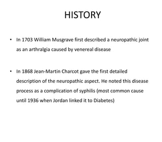
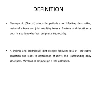
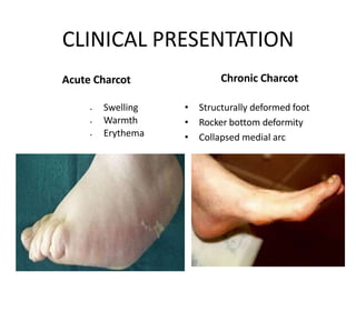
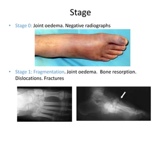
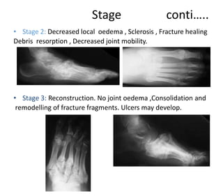
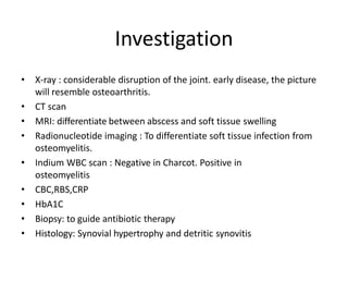
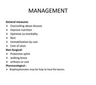
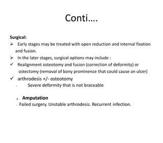
Welcome to short note
presentation
Charcot disease
Dr. Ram kishor goit
Phase-B, Resident
ORTHOPAEDIC SURGERY
HISTORY
• In 1703 William Musgrave first described a neuropathic joint
as an arthralgia caused by venereal disease
• In 1868 Jean-Martin Charcot gave the first detailed
description of the neuropathic aspect. He noted this disease
process as a complication of syphilis (most common cause
until 1936 when Jordan linked it to Diabetes)
DEFINITION
• Neuropathic (Charcot) osteoarthropathy is a non infective, destructive,
lesion of a bone and joint resulting from a fracture or dislocation or
both in a patient who has peripheral neuropathy.
• A chronic and progressive joint disease following loss of protective
sensation and leads to destruction of joints and surrounding bony
structures. May lead to amputation if left untreated.
Also known as,
 Charcot arthropathy
 Charcot foot
 Neuropathic arthropathy
 Neuropathic joints
 Neuropathic osteoarthropathy
RISK FACTORS
 Diabetic neuropathy
 Alcoholism
 Leprosy
 Meningomyelocele
 Tabes dorsalis/syphilis
 Syringomyelia
 Any condition that causes sensory or autonomic neuropathy
EPIDEMIOLOGY
 In diabetic patients: 0.1-1.4%
 In diabetics with neuropathy 7.5%
 Bilateral disease occurs in <10%
 Type 1 DM: 20-25 years post diagnosis
 Type 2 DM: 5-10 years post diagnosis
 Gender : Male predominance
PATHOPHYSIOLOGY
Neurotraumatic theory: German theory 1946.
Peripheral neuropathy  loss of protective sensation 
increase susceptibility to injuries (repeated minor or acute)
 progressive destruction and damage to bone and joints.
Neurovascular theory: French theory 1868.
Spinal cord lesion  autonomic neuropathy  AV shunting
 increased blood flow (warm foot and dilated veins)  Increased
osteoclast activity  bone resorption and mechanical weakening 
fractures and deformity
Symptoms
• Swelling foot and ankle
• Pain 50%
• Loss of function
Acute Charcot
• Swelling
• Warmth (3.3° warmer)
• Erythema (will decrease with Charcot but not with infection on elevation)
Chronic Charcot
• Structurally deformed foot
• Rocker bottom deformity
• Collapsed medial arch
CLINICAL PRESENTATION
Acute Charcot
 Swelling
 Warmth
 Erythema
Chronic Charcot
• Structurally deformed foot
• Rocker bottom deformity
• Collapsed medial arc
• Stage 0: Joint oedema. Negative radiographs
• Stage 1: Fragmentation. Joint oedema. Bone resorption.
Dislocations. Fractures
Stage
Stage conti…..
• Stage 2: Decreased local oedema , Sclerosis , Fracture healing
Debris resorption , Decreased joint mobility.
• Stage 3: Reconstruction. No joint oedema ,Consolidation and
remodelling of fracture fragments. Ulcers may develop.
Investigation
• X-ray : considerable disruption of the joint. early disease, the picture
will resemble osteoarthritis.
• CT scan
• MRI: differentiate between abscess and soft tissue swelling
• Radionucleotide imaging : To differentiate soft tissue infection from
osteomyelitis.
• Indium WBC scan : Negative in Charcot. Positive in
osteomyelitis
• CBC,RBS,CRP
• HbA1C
• Biopsy: to guide antibiotic therapy
• Histology: Synovial hypertrophy and detritic synovitis
MANAGEMENT
General measures
 Counselling about disease
 Improve nutrition
 Optimize co-morbidity
 Rest
 Immobilization by cast
 Care of ulcer.
Non-Surgical:
 Protective splint
 walking brace
 orthosis or cast
Pharmacological :
 Bisphosphonates may be help to heal the bones.
Conti….
Surgical:
 Early stages may be treated with open reduction and internal fixation
and fusion.
 In the later stages, surgical options may include :
 Realignment osteotomy and fusion (correction of deformity) or
ostectomy (removal of bony prominence that could cause an ulcer)
 arthrodesis +/- osteotomy
• Severe deformity that is not braceable
 Amputation
• Failed surgery. Unstable arthrodesis. Recurrent infection.
Complications
 Fractures
 Collapse of the midfoot arch (called rocker bottom
foot)
 Deformities of the foot and ankle
 Ulcers
 Non-union
 “floppy foot”( gross instability )
 Infection
 Soft tissue infection or osteomyelitis may occur.
THANK YOU

charcot disease.

  • 1.
    Welcome to shortnote presentation Charcot disease Dr. Ram kishor goit Phase-B, Resident ORTHOPAEDIC SURGERY
  • 2.
    HISTORY • In 1703William Musgrave first described a neuropathic joint as an arthralgia caused by venereal disease • In 1868 Jean-Martin Charcot gave the first detailed description of the neuropathic aspect. He noted this disease process as a complication of syphilis (most common cause until 1936 when Jordan linked it to Diabetes)
  • 3.
    DEFINITION • Neuropathic (Charcot)osteoarthropathy is a non infective, destructive, lesion of a bone and joint resulting from a fracture or dislocation or both in a patient who has peripheral neuropathy. • A chronic and progressive joint disease following loss of protective sensation and leads to destruction of joints and surrounding bony structures. May lead to amputation if left untreated.
  • 4.
    Also known as, Charcot arthropathy  Charcot foot  Neuropathic arthropathy  Neuropathic joints  Neuropathic osteoarthropathy
  • 5.
    RISK FACTORS  Diabeticneuropathy  Alcoholism  Leprosy  Meningomyelocele  Tabes dorsalis/syphilis  Syringomyelia  Any condition that causes sensory or autonomic neuropathy
  • 6.
    EPIDEMIOLOGY  In diabeticpatients: 0.1-1.4%  In diabetics with neuropathy 7.5%  Bilateral disease occurs in <10%  Type 1 DM: 20-25 years post diagnosis  Type 2 DM: 5-10 years post diagnosis  Gender : Male predominance
  • 7.
    PATHOPHYSIOLOGY Neurotraumatic theory: Germantheory 1946. Peripheral neuropathy  loss of protective sensation  increase susceptibility to injuries (repeated minor or acute)  progressive destruction and damage to bone and joints. Neurovascular theory: French theory 1868. Spinal cord lesion  autonomic neuropathy  AV shunting  increased blood flow (warm foot and dilated veins)  Increased osteoclast activity  bone resorption and mechanical weakening  fractures and deformity
  • 8.
    Symptoms • Swelling footand ankle • Pain 50% • Loss of function Acute Charcot • Swelling • Warmth (3.3° warmer) • Erythema (will decrease with Charcot but not with infection on elevation) Chronic Charcot • Structurally deformed foot • Rocker bottom deformity • Collapsed medial arch CLINICAL PRESENTATION
  • 9.
    CLINICAL PRESENTATION Acute Charcot Swelling  Warmth  Erythema Chronic Charcot • Structurally deformed foot • Rocker bottom deformity • Collapsed medial arc
  • 10.
    • Stage 0:Joint oedema. Negative radiographs • Stage 1: Fragmentation. Joint oedema. Bone resorption. Dislocations. Fractures Stage
  • 11.
    Stage conti….. • Stage2: Decreased local oedema , Sclerosis , Fracture healing Debris resorption , Decreased joint mobility. • Stage 3: Reconstruction. No joint oedema ,Consolidation and remodelling of fracture fragments. Ulcers may develop.
  • 12.
    Investigation • X-ray :considerable disruption of the joint. early disease, the picture will resemble osteoarthritis. • CT scan • MRI: differentiate between abscess and soft tissue swelling • Radionucleotide imaging : To differentiate soft tissue infection from osteomyelitis. • Indium WBC scan : Negative in Charcot. Positive in osteomyelitis • CBC,RBS,CRP • HbA1C • Biopsy: to guide antibiotic therapy • Histology: Synovial hypertrophy and detritic synovitis
  • 13.
    MANAGEMENT General measures  Counsellingabout disease  Improve nutrition  Optimize co-morbidity  Rest  Immobilization by cast  Care of ulcer. Non-Surgical:  Protective splint  walking brace  orthosis or cast Pharmacological :  Bisphosphonates may be help to heal the bones.
  • 14.
    Conti…. Surgical:  Early stagesmay be treated with open reduction and internal fixation and fusion.  In the later stages, surgical options may include :  Realignment osteotomy and fusion (correction of deformity) or ostectomy (removal of bony prominence that could cause an ulcer)  arthrodesis +/- osteotomy • Severe deformity that is not braceable  Amputation • Failed surgery. Unstable arthrodesis. Recurrent infection.
  • 15.
    Complications  Fractures  Collapseof the midfoot arch (called rocker bottom foot)  Deformities of the foot and ankle  Ulcers  Non-union  “floppy foot”( gross instability )  Infection  Soft tissue infection or osteomyelitis may occur.
  • 16.