
Recommended
Recommended
More Related Content
What's hot
What's hot (20)
Similar to General principles of arthroscopy kle, belgaum, dr utkarsh dwivedi
Similar to General principles of arthroscopy kle, belgaum, dr utkarsh dwivedi (20)
Recently uploaded
Recently uploaded (20)
General principles of arthroscopy kle, belgaum, dr utkarsh dwivedi
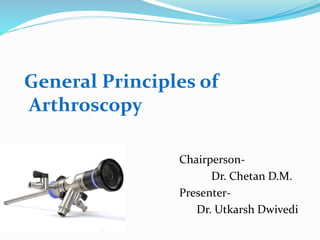
- 1. General Principles of Arthroscopy Chairperson- Dr. Chetan D.M. Presenter- Dr. Utkarsh Dwivedi
- 2. WHAT IS ARTHROSCOPY? This word arthroscopy came from GREEK , "arthro" (joint) and "skopein" (to look). The term literally means to look within the joint simply as if you see a room through a key – hole instead of opening doors. …. It offers a high degree of accuracy combined with low morbidity for making diagnosis and offering treatment.
- 4. INDEX Instruments and equipment Anesthesia Documentation Advantages & Disadvantages Indications & contraindications Basic arthroscopic techniques Complications
- 6. ARTHROSCOPE- It is a rigid optical instrument. Optical characteristics of an arthroscope are determined by diameter, angle of inclination and field of view. Diameter : 1.7-7 mm 4mm is the most commonly used, especially for knee joint. 1.9 & 2.7 mm useful for tighter joints like wrist & ankles.
- 7. Angle of inclination-is angle between axis of arthroscope and a line perpendicular to surface of the lens, varies from 0-120*. 25-30* is the most commonly used 70-90* is for seeing around corners or postero-lateral corners of the knee joint
- 9. Field of view-refers to viewing angle encompassed by lens and varies according to type of arthroscope. 1.9 mm scope has a 65* field of view. 2.7mm scope has 90* filed of view Wider viewing angles make orientation by observer much easier. Two designs- -Viewing -Operating, developed by O'Conner allows direct viewing , with a channel for the placement of operative instruments in line with the arthroscope.
- 13. FIRBREOPTIC LIGHT SOURCES LIGHT SOURCES : 300 – 350 watts required. Tungsten, halogen & xenon sources. Can produce low & high intensity output. FIBREOPTIC CABLE : -Fragile ,should be handled carefully. -One end connected to light source & another to Arthroscope. Works on the principle of “Total internal refraction”. -Length of cable also important as some amount of transmitted light is lost for each foot of cable. Now-a-days breakage of Fibreoptic cable has been eliminated with introduction of liquid (glycerin) light guides.
- 16. TELEVISION CAMERAS First introduced by McGinty and Johnson More comfortable Avoidance of contamination by the surgeon’s face Improvement offered by latest three chip technology- Decrease size of camera Increase resolution of image Cableless arthroscopic systems in which video signal is transmitted from an arthroscope with its own light source
- 18. BASIC INSTRUMENT KIT Arthroscopes 30 and 70 degrees Scissors Probes Basket forceps Grasping forceps Arthroscopic knives Motorized meniscus cutter and shaver Laser/radiofrequency instruments Miscellaneous epuipment
- 19. PROBE The extension of the arthroscopist’s finger. Used- To feel the consistency of a structure To determine the depth To identify and palpate loose structures To maneuver loose bodies into more accessible grasping position To probe fossae & recess To maneuver intraarticular structure To elevate meniscus
- 20. Most are right-angled 2 mm fixed tip size. This is used to measure length of structures inside joint cavity. Use the elbow of the probes for palpation Magnification occurs with the arthroscope; the closer it is the higher the magnification. So it can be placed close or far depending on the observer’s desire.
- 22. SCISSORS 3-4 mm in diameter JAWS : straight / hooked -hooked scissors preferred as jaws hook tissue & pull it between cutting edges of scissors rather than pushing materials as in straight scissors. CURVES : right / left ANGLES : right / left, usually with a rotating of jaw mechanism, actually cut at an angle to shaft of the scissors. -useful in detaching difficult-to-reach meniscal fragments.
- 26. BASKET FORCEPS One of the most commonly used arthroscopic instruments. Open base that permits the tissue to drop free within the joint & don’t require instrument to be removed from the joint & cleaned with each bite. The debris is subsequently removed from the joint by suction. 3-5mm sizes with straight or curved shaft Usually used for trimming the peripheral rim of the meniscus
- 27. Basket forceps specialized for meniscus are wide, low- profile baskets with hooked configuration. Shaft : straight / curved Jaws : straight / hooked Basket in assortment of 30 , 45 , 90 degree. Also as 15 degree up & down – biting.
- 29. GRASPING FORCEPS Retrieve material from the joint generally loose bodies from knee joint. Grasping tissue to cutting used to retrieve material from the joint, or to hold other tissue under tension to facilitate cutting. Rachet closure system for better hold. Jaws : single / double action with regular serrated interdigitating teeth / 1 – 2 sharp teeth Usually double side serrated forcep is used for securing loose bodies as it doesn’t slip from it.
- 32. ELECTROSURGICAL LASERS ELECTROCAUTERY :For cutting & hemostasis previously. Now a days only to obtain hemostasis after A’scopic synovectomy & subacromial decompression. Works in a non-electrolyte medium like distilled water, Carbon dioxide or glycine. Newer coated tip function in both NS / RL. LASER :role under investigation. CO2 laser ,YAG laser, excimer laser
- 35. RADIOSURGICAL SYSTEM Radiofrequency systems are used for tissue ablation, electrocautery, & capsular shrinkage. Monopolar uses a grounding pad & draw energy through the body. Bipolar in it energy is transferred b/w electrodes at the site of treatment. They are used for cutting and haemostasis for arthroscopic synovectomies and subacromial decompression. Complications include- articular cartilage damage, osteonecrosis, tissue damage.
- 36. KNIFE BLADES These should be inserted through cannula sheaths and cutting portion be exposed only when it enters the arthroscopic field. Available varities are- hooked or retrograde blades, regular down-cutting blades-straight and curved. Magnetic properties of blades are helpful in retrieving them when broken.
- 38. MOTORISED SHAVING SYSTEMS Consisting of Outer hollow sheath Inner hollow rotating cannula with corresponding windows & dia. of cutting tip usually 3 – 5.5 mm. principle : the window of inner sheath function as a two edged cylindrical blade ,that spins within the outer hollow tube. Suction through the cylinder brings the fragment of soft tissue in the window and as the blade rotates ,the fragments are amputated ,sucked to the outside ,and collected in the suction trap.
- 41. Special blade, for meniscal cutting or trimming, Synovial resection, and for shaving of articular cartilage. Special abraders & burrs for arthroscopic acromioplasty & cruciate Ligament reconstructions. Both clockwise & anticlockwise rotation. Reversing the rotation improves cutting efficiency & minimises Clogging with debris.
- 42. IMPLANTS Suture anchors Meniscal repair devices Devices for tendon and ligament fixation Articular cartilage repair
- 43. SUTURE ANCHORS Used to attach ligaments and tendons to bone without bony tunnel passage of sutures Desirable characteristics Must fix the suture to the bone Permit an easy surgical technique Not cause long-term problems
- 46. MENISCAL REPAIR DEVICES Allow an all-inside meniscal repair without the need for arthroscopic knot-tying 3 categories Arrows Darts Meniscal screws
- 49. IRRIGATION SYSTEMS Irrigation and distension Essential to all arthroscopic procedures Joint distension is maintained better by RL than NS. Inflow is via arthroscopic sheath: 6.2mm diameter with the cannula in separate portal with 68mm of pressure of water. Usually two 5 Lit plastic bags of RL , interconnevted with a Y-connector are suspended for use with the arthroscopy pump. Continuous irrigation is needed to- Keep clear viewing Maintain hydrostatic pressure and distension
- 51. DISTENSION PRESSURE It is optimal pressure required to distend the joint. Ingress = egress to maintain hydrostatic pressure & distention within joint. For each foot of elevation of solution bag above joint = 22 mm of hg pressure Varied according to joint as follows : Knee 60 -80 mm of hg Shoulder 30 mm of hg below systolic pressure Elbow 40 – 60 mm of hg Ankle 40 – 60 mm of hg
- 53. type of pump (arthrex AR 6450 , stryker 1.5L high flow pump , arthro FMS4 ,& acutex inteliject )all maintained a pressure of 60 mm of hg accurately. Sensor mechanism to check over distention. Distention is essential for arthroscopic viewing as it pushes synovial folds & other soft tissues out of the way in viewing area, expands internal capacity of joint, allowing greater maneuverability of arthroscope, defining proper portal entry points like posteromedial & posterolateral portals in knee.
- 54. TORNIQUET Contraindications History of thrombophlebitis Significant peripheral vascular disease Advantages Increased visibility Disadvantages Blanching of the synovium Difficult to diagnosis synovial disorders Ischemic damage if prolonged touniquet time (90- 120min)
- 55. LEG HOLDERS The biggest advantage of leg holders is that they permit application of stress primarily to open the posteromedial compartment for viewing or manipulation of the meniscus and posterior horn meniscuc surgery. The post does not confine knee and offers unlimited number of positions for the knee to be placed. Disadvantages Obstruct the operations in lateral compartment Use in case of medial compartment disease
- 58. METHOD OF STERILIZATION Ethylene oxide(best method) Low temperature sterilization process CIDEX is used for cold disinfection of equipments between successive procedures during whole day. Knives, forceps etc.: by steam autoclaving. Fibreoptic materials, camera, motorised instruments: by soaking in CIDEX sol. For 10 min. or in STERIS for 30 min.
- 59. ANESTHESIA Arthroscopy can be performed under Local Anesthesia Regional Anesthesia General Anesthesia
- 60. REGIONAL ANESTHESIA Usually used in lower extremities- Epidural or spinal anesthesia Femoral and sciatic blocks Features of peripheral blocks- Immediate ambulation Require experience anesthesiologist Longer time to prepare Generally use a 1:1 mixture of 1% lignocaine and 0.25% bupivacaine. Upper extremities Brachial Block
- 61. GENERAL ANESTHESIA Used in cases of- Not cooperative patients Allergy to local anesthetics Less experienced surgeon Increased pain (acutely injured knee)
- 62. POST-OP PAIN Oral NSAIDs or IM,IV administration Reduce swelling Increase ROM in early postoperative period 30mL of 0.25% bupivacaine +/-Morphine 3 mg intraarticular or subacromial flow Excellent postoperative pain relief Catheters should be removed in 48 hours
- 63. DOCUMENTATION Drawings and documentation are very essential 35-mm reflex camera photos Digital video recordings
- 64. INDICATIONS OF ARTHROSCOPY DIAGNOSTIC -For preoperative evaluation & confirmation of clinical diagnosis -For documentation in medicolegal cases THERAPEUTIC •Smoothening of Torn cartilage •Damaged ligaments reconstruction •Loose bodies removal •Joint effusions •Biopsy procedures •Fracture fixation •Sports Related Injuries
- 65. ADVANTAGES OF ARTHROSCOPY Reduced postoperative morbidity Smaller incision Less intense inflammatory response Improved thoroughness of diagnosis Absence of secondary effects Neuromas, scars
- 66. Reduced hospital cost Reduced complication rate Improved follow-up evaluation : second-look Possibility of performing surgical procedures that are difficult to perform through open arthrotomy
- 68. DISADVANTAGES OF ARTHROSCOPY Skill and temperament to perform arthroscopic surgery Need to maneuver within the tight confines of the intraarticular space Time-consuming procedures in cases of inexperienced surgeons and follows a steep learning curve Expensive equipment
- 69. INDICATIONS AND CONTRAINDICATIONS No absolute indications Diagnostic arthroscopy Preoperative evaluation and confirmation of the clinical diagnosis Documentation of specific lesions Contraindications Risk of joint sepsis, remote infection Ankylosis around the joint Capsular disruption
- 70. HOW IS ARTHROSCOPY PERFORMED? Under anesthesia make small incision in the skin around joint. Eg. Anteromedial and anterolateral entry points in the knee jnt. A sterile fluid is pumped into joint and then the arthroscope is inserted. Examine joint by images from arthroscope If necessary, other instruments inserted for procedure i.e. repair any damage or remove material that causes symptoms. Afterwards, the fluid is drained out, cuts are closed & dressed.
- 71. BASIC ARTHROSCOPIC TECHNIQUES Patience and persistence Techniques are mostly self-taught Artificial models or amputated specimens for initial practice Perform arthroscopic procedures in the company of an experienced arthroscopist. It has a steep Learning curve Keep in mind that open arthrotomy is to be preferred over poorly performed arthroscopic procedures
- 72. TRIANGULATION TECHNIQUE Involves use of one or more instruments inserted through separate portals and brought into the optical field of the arthroscope, the tip of the instruments and arthroscope forming apex of a triangle When the instrument is located, the scope and instrument are advanced together towards the intended area, reducing the field of vision and increasing the magnification.
- 73. If disoriented and difficulty in triangulation the instrument may be brought into the joint to contact the sheath and sliding to the tip Stereoscopic sense and two-handed ability are developed gradually
- 75. MOST COMMON CONDITIONS FOUND DURING ARTHROSCOPY Acute or Chronic Injury Shoulder: Rotator cuff tendon tears, impingement syndrome, and recurrent dislocations Knee: Meniscal (cartilage) tears, chondromalacia (wearing or injury of cartilage cushion), and anterior cruciate ligament tears with instability Wrist: Carpal tunnel syndrome Loose bodies of bone and/or cartilage: for example, knee, shoulder, elbow, ankle, or wrist Some problems associated with arthritis also can be treated.
- 76. COMMONLY DONE ARTHROSCOPIC SURGERIES Rotator cuff surgery Repair or resection of torn cartilage (meniscus) from knee or shoulder Reconstruction of anterior cruciate ligament in knee Removal of inflamed lining (synovium) in knee, shoulder, elbow, wrist, ankle Release of carpal tunnel Repair of torn ligaments Removal of loose bone or cartilage in knee, shoulder, elbow, ankle, wrist.
- 77. COMPLICATIONS Damage to intraarticular structures: most common Damage to Menisci and Fat Pad Damage to Cruciate Ligaments Damege to Extraarticular structures Hemathrosis Thrombophlebitis Infection Tourniquet Paresis Synovial Herniation and Fistulas Instrument Breakage
- 78. FOLLOW-UP AFTER ARTHROSCOPIC SURGERIES RECOVERY TIME DEPENDS UPON MANY FACTORS: severity of disease Type of surgery. Supports for 3 to 7 days, weight bearing on the operated leg as tolerated. Analgesics Rest, ice packs, and elevating the limb also recommended.
- 79. Physiotherapy not required in all patients, should be individualised. sitting job can be resumed one week after surgery. 3 weeks to recover fully for routine daily activities. 3 months before one can comfortably return to sports..
- 80. THANK YOU FOR LISTENING