Shoulder arthroscopy
•Download as PPTX, PDF•
12 likes•1,415 views

Arthroscopic approaches, patient positioning, portals

Recommended
More Related Content
What's hot
What's hot (20)
Similar to Shoulder arthroscopy
Similar to Shoulder arthroscopy (20)
Recently uploaded
Recently uploaded (20)
Shoulder arthroscopy
- 1. SHOULDER ARTHROSCOPY DR. MUHAMMAD JAWAD PGT ORTHOPEDICS SHL
- 2. INTRODUCTION • Modern era of management of shoulder pathology began in the 1930s with the work of Codman • Significant contributions to understanding of rotator cuff pathology originated with Neer • Bankart and Rowe - the major proponents of concepts underlying the current understanding of shoulder instability. • Arthroscopy of the shoulder did not become routine practice until the 1980s • Developments in technology - routine use of arthroscopy
- 3. PREOPERATIVE CONSIDERATIONS • Review all pertinent medical records • Athorough history and physical examination • Appropriate imaging to confirm the diagnosis to reduce the risk of surprises • Anesthesia - general, general with regional nerve blocks, or regional anesthesia alone. • Examination under anesthesia (EUA) • Systematic examination should be performed including evaluation of ROM and stability of the shoulder
- 4. PATIENT POSITIONING • THE LATERAL DECUBITUS. • In the supine position - general anesthesia and examination. • The patient is placed in the lateral decubitus position using a bean bag, often rotated backward 20 degrees. An axillary roll - to protect the brachial plexus and padding is placed between the down leg and table, as well as between the knees, as all bony prominences must be well padded • Arm is placed in sterile traction device with the arm in 45 to 70 degrees of abduction and 20 to 30 degrees of forward flexion. A 10 to 15 lbs of traction is usually adequate.
- 5. LATERAL DECUBITUS POSITION Dual traction for distraction of glenohumeral joint with minimal inferior subluxation. Wide 4-inch sling should be used; amount of traction and length of procedure should be monitored.
- 6. • All arthroscopic shoulder procedures can be performed from this position, with posterior shoulder stabilization a little easier in the lateral decubitus position • Higher risk of neuropraxia • Conversion to an open procedure may require repositioning and re- draping the patient • Regional anesthesia by itself is often not well tolerated in this position.
- 7. BEACH CHAIR • Patient is placed supine and an examination under anesthesia is performed . • Then placed in a beach chair position • Be careful to monitor the blood pressure • Be careful of the neck position and head support • Protect the elbow/ulnar nerve of both arm • Easier for anesthesia control during surgery • Position makes conversion to open procedure easier. • Risk of cerebral hypoperfusion with hypotensive anesthesia as well as the small risk of air embolism • Difficulty performing posterior stabilization • Easier to perform dynamic assessment (no traction to remove from the arm)
- 9. CONTROL OF BLEEDING • 3 techniques can help 1. Arthroscopy pump for inflow, maintaining a constant fluid flow and pressure of 60 to 70 mm Hg 2. second measure is to add 1 mL of 1:1000 epinephrine to each 3000-mL bag of irrigant, 3. The final technique, and perhaps the most effective, is to use hypotensive anesthesia, with a systolic BP of 90 to 100 mmHg. • Elevation of the fluid bags 3 feet above the level produces a similar pressure gradient of 66 mm fluid flow pressure
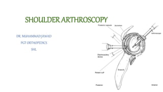
- 10. PORTAL PLACEMENT
- 11. POSTERIOR PORTAL • 1 to 2 cm distal and 1 to 2 cm medial to the posterolateral corner of the acromion • Verify position with spinal needle prior to incision • Primary viewing portal • Pierces posterior deltoid muscle and travels in the interval between the infraspinatus and the teres minor • The axillary nerve and suprascapular nerve are at risk with this portal, but are relatively safe
- 12. POSTEROINFERIOR 7-O’CLOCK PORTAL • 2cm above the posterior axillary fold • Used as inflow portal • Dangerous portal due to proximity of the axillary nerve, posterior humeral circumflex artery, and suprascapular nerve
- 13. ANTERIOR SUPERIOR PORTAL • Superior and lateral to the coracoid—just anterior to the AC joint • Localized with spinal needle in an outside-in technique just anterior to the biceps tendon next to the superior glenoid • Primarily an instrument portal but can be used to visualize the anterior glenoid rim and labrum, the posterior joint, as well as to visualize internal impingement dynamically • Pierces the anterior deltoid and travels through the rotator interval, avoiding injury to the rotator cuff • Neurovascular structures at risk include the musculocutaneous nerve, axillary nerve, as well as the cephalic vein, brachial plexus, and axillary artery
- 14. ANTERIOR INFERIOR PORTAL • Lateral to the coracoid • Pierces anterior deltoid muscle and enters joint just above subscapularis • If too low, risk of injury to the axillary nerve • If too medial, risk of injury to musculocutaneous nerve • Minimal risk of injury to cephalic vein, brachial plexus, and axillary artery
- 15. ANTEROINFERIOR PORTAL • Inferior and lateral to the coracoid • Localized with spinal needle using outside-in technique superior to the subscapularis • Used primarily for instrumentation for anterior Bankart repair suture anchor placement
- 16. SUPERIOR PORTAL (NEVIAZER) • Corner of the supraspinatus fossa • Enters joint just medial to the supra-glenoid tubercle • penetrates the trapezius muscle and passes through the supraspinatus muscle belly • Used for SLAP and rotator cuff repairs
- 17. LATERAL PORTAL • 1 to 2 cm distal to the acromion • Either mid acromion (anterior to posterior) or junction of the (anterior 1/3 , posterior 2/3) • Localized with spinal needle while viewing subacromial space • Used for subacromial decompression and rotator cuff repairs • Can be extended for mini-open deltoid splitting rotator cuff repairs • Pierces middle deltoid muscle • Axillary nerve at risk, because this averages 5 to 6 cm from the lateral acromial border, but may be as close as 3 cm from the acromion
- 18. POSTEROLATERAL PORTAL (WILMINGTON) • 1 cm anterior and 1 cm distal to the posterolateral corner of the acromion • Used for instrumentation during SLAP repair
- 21. COMMON ARTHROSCOPIC PROCEDURES • Rotator cuff repair • SLAP (superior labral anterior to posterior) repairs • Biceps tendon pathology • Repair of labral tears • Acromioplasty and distal clavicle resection • Adhesive capsulitis • Release of the suprascapular nerve
- 22. COMPLICATIONS • Neurovascular injury • most commonly when lateral decubitus position and traction are used • Infection • Deep vein thrombosis (DVT) • Anesthesia complications • Arthrofibrosis • More likely to happen with concomitant rotator cuff and SLAP repair • Iatrogenic chondromalacia