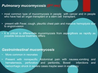
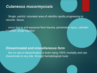
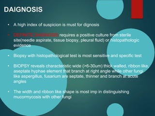
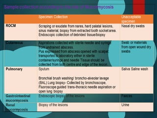
Mucormycosis is caused by fungi of the order Mucorales. It is an opportunistic infection seen in immunocompromised patients. The rhino-orbito-cerebral form presents as sinusitis that can invade the orbit and brain. Pulmonary mucormycosis is the second most common type seen in cancer and transplant patients. Diagnosis requires tissue biopsy demonstrating wide, ribbon-like hyphae. Treatment involves antifungal therapy with amphotericin B and surgical debridement of infected tissues. Prognosis depends on early diagnosis and treatment.
Mucormycosis is a life-threatening fungal infection of the nose and paranasal sinuses caused by fungi such as Rhizopus and Mucor. It has a high mortality rate of 50-80%. Risk factors include inhalation of fungal spores from soil and angioinvasion of blood vessels by the fungi. Symptoms include fever, nasal obstruction, vision loss, and facial swelling/pain. Diagnosis involves KOH mount of samples to view broad, aseptate hyphae and biopsy for histopathology and culture. Imaging like CT and MRI show soft tissue invasion and bone erosion. Aggressive surgical debridement and antifungal therapy are used for treatment.
Mucormycosis is an invasive fungal infection caused by fungi of the Mucoraceae family. It is an opportunistic infection seen predominantly in patients with diabetes, neutropenia, or other immunocompromised states. The rhinocerebral form involves the facial, orbital, paranasal sinus and cerebral regions. Diagnosis involves biopsy and culture. Treatment requires control of risk factors, aggressive surgical debridement of infected tissues, and antifungal therapy typically with amphotericin B. Despite treatment, mucormycosis has a high mortality rate of 50-85%.
Mucormycosis is a fungal infection caused by species in the orders Mucorales and Rhizopus. It most commonly affects immunocompromised patients via inhalation, ingestion, or traumatic inoculation. The fungi invade blood vessels, causing thrombosis, tissue necrosis, and different clinical forms including rhinocerebral, pulmonary, gastrointestinal, cutaneous, and disseminated mucormycosis. Rhinocerebral mucormycosis has the highest frequency and mortality, often occurring in diabetic patients experiencing ketoacidosis. Diagnosis involves biopsy and imaging of affected tissues showing broad, branching fungal hyphae. Treatment requires control of predisposing conditions and antif
Mucormycosis is a life-threatening fungal infection caused by fungi of the order Mucorales. It predominantly affects immunocompromised individuals, especially those with diabetes, hematological malignancies, or who have undergone transplants or immunosuppressive therapy. The infection spreads through inhalation or skin/mucous membrane contact with fungal spores and has a high mortality rate even with aggressive treatment. Management involves controlling underlying risk factors, surgical debridement of infected tissues when possible, and antifungal therapy primarily with polyene antifungals like amphotericin B.
Mucormycosis ppt by Dr. Bomkar bam ENT M.S.Bomkar Bam
mucormycosis in the covid era in India. it is mostly seen in the post-recovery patient of covid - 19. most of the data are derived from the 2nd wave of covid in India.
Mucormycosis and how it is related to Covid 19 disease - department seminar ...RubinaSubhani
Mucormycosis, also known as black fungus, is a serious fungal infection caused by a group of molds called mucormycetes. It affects people with health problems or those taking medications that lower the body's ability to fight germs and sickness. The infection is seen in some COVID-19 patients as well, likely due to them having diabetes, being on steroids, or having low immunity from the virus. Symptoms depend on the infected area but can include sinus congestion, black lesions, fever and breathing issues. Treatment requires antifungal medicines and sometimes surgery. People with diabetes should keep their blood sugar under control to reduce mucormycosis risk.
Mucormycosis is a rare fungal infection caused by organisms in the order Mucorales. It is life-threatening and most commonly affects immunocompromised individuals. There are several forms including rhinocerebral, pulmonary, cutaneous, and gastrointestinal depending on the route of exposure. Risk factors include uncontrolled diabetes, immunosuppressive medications, and neutropenia. Diagnosis involves imaging and biopsy of infected tissues. Treatment requires aggressive antifungal therapy typically with amphotericin B and surgical debridement of infected areas. Prognosis is poor with mortality rates over 50% even with treatment.
The document summarizes ENT manifestations of HIV infection. It describes how HIV attacks CD4 cells leading to opportunistic infections and malignancies. Common ENT issues seen include oral thrush, recurrent sinusitis, sensorineural hearing loss, and Kaposi sarcoma of the oral cavity, nose and larynx. Diagnosis involves CD4 counts and virus detection tests. Universal precautions are essential to prevent transmission among health workers.
This document discusses Rhinosporidiosis, a chronic infection caused by the protist Rhinosporidium seeberi. It primarily affects the nasal mucosa and conjunctiva, causing polyps or wart-like lesions. R. seeberi has not been successfully cultured. The disease is diagnosed by identifying the characteristic sporangia and endospores in histopathological samples or direct smears of lesions. Treatment involves surgical excision of polyps along with dapsone administration to prevent recurrence.
Mucormycosis is a rare fungal infection caused by Mucorales fungi. It has high mortality and morbidity rates especially in immunocompromised patients like those with diabetes or undergoing chemotherapy. It most commonly presents as rhino-orbito-cerebral infection in India. Diagnosis involves imaging and microscopic identification of broad, non-septate hyphae. Treatment requires intravenous antifungal therapy with amphotericin B and extensive surgical debridement to remove necrotic tissue. Early diagnosis and treatment are essential to improve outcomes.
The document discusses mucormycosis, a fungal infection caused by exposure to mucor molds. It provides details on COVID-19 associated mucormycosis infections, including risk factors like steroid use in severe COVID cases. The summary discusses the epidemiology and clinical manifestations of mucormycosis, as well as challenges in diagnosis. Standard diagnostic methods outlined include tissue biopsy, culture, and microscopy of samples to detect fungal hyphae. Early diagnosis and treatment are important to manage mucormycosis and reduce mortality.
Mucormycosis is a fungal infection caused by Mucorales fungi. It most commonly presents as rhino-orbito-cerebral mucormycosis and has increased in COVID-19 patients. Diagnosis involves microscopy, culture, biopsy and MRI showing characteristic findings. Treatment is with liposomal amphotericin B followed by oral antifungals like posaconazole. Early diagnosis and aggressive treatment by a multidisciplinary team is needed for optimal outcomes.
The document discusses the anatomy and structures of the oral cavity, including the lips, buccal mucosa, hard palate, retromolar trigone, floor of the mouth, and tongue. It describes the etiology of oral cancers, noting that tobacco and alcohol use are major risk factors. Squamous cell carcinoma accounts for 95% of oral cavity cancers. Treatment options depend on the stage of cancer, and may include surgery, radiation therapy, chemotherapy, or a combination of these. Prognosis decreases with more advanced stage at diagnosis.
Mucormycosis is a rare but aggressive fungal infection caused by fungi of the class Zygomycetes, including Rhizopus, Mucor, and Absidia. It mainly affects immunocompromised patients or those with uncontrolled diabetes. The fungi thrive in high glucose, acidic conditions. Common forms include rhinocerebral affecting the sinuses and orbit, pulmonary, gastrointestinal, cutaneous from skin injuries, and disseminated infection of multiple organs. Diagnosis involves tissue biopsy and culture. Treatment requires intravenous amphotericin B antifungal therapy and surgical debridement of infected tissues.
This document discusses several viral infections that can affect the oral mucosa. It describes the human herpes virus family, including herpes simplex virus types 1 and 2, which can cause gingivostomatitis and recurrent oral herpes infections. It also discusses herpes zoster virus, which causes chickenpox and shingles. Epstein-Barr virus is noted for causing infectious mononucleosis and oral hairy leukoplakia. Cytomegalovirus, coxsackie virus, paramyxoviruses like mumps and measles viruses are also summarized for their oral manifestations. Treatment options are provided for many of these viral infections.
This document describes different types of vesiculobullous diseases classified according to the Fitzpatrick system based on the anatomical level of blister formation. It discusses conditions such as pemphigus vulgaris, pemphigus vegetans, pemphigus foliaceus, paraneoplastic pemphigus, bullous pemphigoid, cicatricial pemphigoid, and familial benign pemphigus. For each condition, it provides details on pathogenesis, clinical features, histopathology, immunopathology, and oral manifestations when present.
Rhinosporidiosis is a chronic granulomatous disease caused by Rhinosporidium seeberi. It commonly affects the nose and nasopharynx, presenting as a polypoidal pink to purple mass. The disease is transmitted through contact with contaminated water. The life cycle of R. seeberi involves a trophic stage and production of endospores. Diagnosis is made through biopsy showing sporangia filled with spores. Treatment involves complete surgical excision combined with dapsone to prevent recurrence, as recurrence is common with surgery alone.
This document discusses various fungal infections that can affect the oral cavity and respiratory tract, including candidiasis, aspergillosis, and zygomycosis. It describes the characteristics and morphology of fungi, including molds, yeasts, dimorphic fungi. It then covers the pathogenesis, clinical manifestations, diagnosis and treatment of the main opportunistic fungal infections.
Pleomorphic adenoma is the most common salivary gland tumor, accounting for 60% of cases. It occurs most often in the parotid glands and presents as a painless, firm mass. Histologically, it contains both epithelial and mesenchymal elements arranged in a trabecular pattern within a fibrous stroma. Treatment involves complete surgical removal of the tumor with adequate margins to prevent recurrence due to microscopic projections outside the capsule. Imaging such as CT or MRI is used to identify the location and characteristics of the tumor prior to surgery.
This document provides information about bacteria and tuberculosis (TB). It begins by defining bacteria and describing their various shapes. It then discusses TB in more detail, noting that it is caused by Mycobacterium tuberculosis bacteria. Signs and symptoms of TB infection are described, including fever, night sweats, and cough. Both pulmonary and extrapulmonary TB are discussed. Oral manifestations of primary and secondary TB are also summarized.
1. Managing lymph node tuberculosis can be challenging as it has varied clinical manifestations and diagnostic challenges.
2. It most commonly involves cervical lymph nodes but can affect nodes throughout the body.
3. Diagnosis may involve imaging like ultrasound, CT, or MRI to identify enlarged or cystic lymph nodes, as well as biopsy to confirm the presence of Mycobacterium tuberculosis.
4. Treatment often requires a multi-drug antibiotic regimen over a prolonged period.
• Actinomyces species are classified as anaerobic, gram positive and filamentous bacteria.
• It is a chronic granulomatous suppurative and fibrosing disease caused by anaerobic or microaerophilic gram-positive nonacid fast, branched filamentous bacteria.
• Most of the species isolated from actinomycotic lesions have been identified as A. israelii, A. viscosus, A. odontolyticus, A.naeslundii or A. meyeri.
• These microorganisms have been identified in dental plaque, dental calculus, necrotic pulp, and tonsils.
• The usual pattern of this disease is one characterized chiefly by the formation of abscesses that tend to drain by the formation of sinus tracts.
• pus from the abscesses is examined on a clean glass slide, it shows the typical ‘sulfur granules’ or colonies of organisms, which appear in the suppurative material as tiny, yellow grains.
• Another infection that produces this type of sulfur granules is botryomycosis.
This document discusses cerebrospinal fluid (CSF) rhinorrhea, or the leakage of CSF into the nose. It defines CSF and its circulation and production in the brain. CSF rhinorrhea can be caused by trauma, infections, tumors, or congenital lesions that damage the skull base and allow CSF to leak into the nasal cavity. Diagnosis involves identifying clear fluid leaking from the nose, especially when bending over. Imaging like CT and MRI can localize the leak site. Surgical repair of the skull defect is often needed using grafts and nasal packing.
Oral mucosa reflects the health of the whole human body at a first glance.If any disorder is present in the system it will first appear in oral cavity. Here is an overview of certain pigmented lesions.
This document provides information on oral submucous fibrosis (OSMF), including its definition, epidemiology, etiology, clinical features, histopathological classification, and management. OSMF is a precancerous condition characterized by fibrosis of the submucosa and juxta-epithelial inflammatory reaction. It predominantly affects people of Asian descent and is strongly associated with chewing areca nut and consumption of chili peppers. Clinically, it presents as blanching and stiffness of the oral mucosa leading to restricted mouth opening. Histopathological examination can classify the severity of fibrosis. Arecoline in areca nut is the main etiological agent that causes fibrosis through oxidative stress and increased collagen production.
Mucormycosis is a serious fungal infection caused by exposure to mucor mold spores, usually through inhalation. It most often affects people who have health problems that weaken the immune system, such as diabetes, cancer, or those taking immunosuppressive drugs. The infection can cause sinus, lung, or brain infections and is life-threatening without prompt treatment with antifungal drugs and sometimes surgery. Managing any underlying health conditions contributing to a weakened immune system is also important for treatment. Prognosis depends on early diagnosis and treatment as well as the patient's overall health status.
Fungal sinusitis can be either invasive or noninvasive. Invasive fungal sinusitis occurs when fungi invade the mucosa and other structures of the sinuses, while noninvasive sinusitis does not involve invasion. Common types of noninvasive sinusitis include fungal balls, saprophytic infections, and allergic fungal sinusitis. Invasive forms are more serious and life-threatening, especially acute invasive sinusitis which has high mortality rates. Treatment involves aggressive surgical debridement and antifungal therapy.
This document provides information on fungal rhinosinusitis, including its classification and types. It discusses invasive fungal rhinosinusitis, which includes rapidly invasive and chronic invasive types. It also discusses non-invasive fungal rhinosinusitis, which includes saprophytic colonization, fungal ball, and allergic fungal rhinosinusitis. Signs and symptoms, endoscopic findings, microbiology, antifungal drugs, and treatment approaches are described for different fungal infections of the sinus. Allergic fungal rhinosinusitis is discussed in detail, covering its pathophysiology, clinical features, diagnostic criteria, imaging, therapy including surgery, steroids, and immunotherapy.
This document discusses guidelines for treating candidemia and invasive candidiasis in ICU patients. It recommends starting treatment with an echinocandin for both non-neutropenic and neutropenic patients. For non-neutropenic patients, fluconazole is an alternative if the patient is not critically ill and the Candida species is susceptible. Treatment should be given for 2 weeks after symptoms resolve and blood cultures clear. Source control through catheter removal is also recommended when possible.
- Mucormycosis is a life-threatening fungal infection caused by fungi of the order Mucorales. It mostly affects immunocompromised individuals, especially those with uncontrolled diabetes.
- The document discusses the epidemiology, risk factors, clinical manifestations, diagnosis, and management of mucormycosis. It emphasizes the importance of early diagnosis, aggressive surgical debridement of infected tissues, antifungal therapy typically with amphotericin B, and control of underlying conditions.
- Prompt treatment including surgical debridement and antifungal therapy can significantly improve survival rates for mucormycosis compared to antifungal therapy or surgery alone. However, mortality remains high due to
The document discusses various classes of antifungal drugs including their mechanisms of action, spectrum of activity, pharmacokinetics and clinical uses. It describes polyenes like amphotericin B and nystatin, azoles including imidazoles and triazoles, allylamines like terbinafine, antimetabolites like flucytosine and griseofulvin. It also discusses newer antifungals like echinocandins including caspofungin.
This document discusses different types of fungal rhinosinusitis, including invasive and non-invasive forms. Non-invasive types include saprophytic fungal infections, fungal balls, and allergic fungal rhinosinusitis (AFRS). Invasive fungal rhinosinusitis is divided into acute/fulminant, granulomatous, and chronic types. Diagnosis involves imaging, histology, and culture. Treatment depends on type but commonly includes surgery along with long-term medical management such as steroids, antifungals, and immunotherapy.
The document discusses several interesting clinicopathologic case studies involving fungal and parasitic skin infections:
1. A case of cutaneous protothecosis caused by the algae Prototheca wickerhamii in an immunocompromised patient.
2. A case of cutaneous zygomycosis caused by the fungus Rhizopus in a neutropenic patient.
3. A case of cutaneous alternariosis and candidiasis in a lung transplant recipient.
4. A case of disseminated penicilliosis caused by the dimorphic fungus Penicillium marneffei in an HIV-positive patient presenting with skin lesions.
oral pemphigus vulgaris effect on systemic healthPriyanka Pai
Three sentences:
This document reports a case of oral pemphigus vulgaris and provides background information on the condition. Pemphigus vulgaris is an autoimmune blistering disease caused by autoantibodies against desmoglein 3, leading to acantholysis and blister formation in the oral mucosa and skin. The case report describes the clinical features, histopathology, direct immunofluorescence findings, and management of a patient diagnosed with oral pemphigus vulgaris.
1. Fungal sinusitis can be invasive or non-invasive and is classified based on its histopathological and clinical features.
2. Invasive fungal sinusitis includes acute fulminant fungal rhinosinusitis which causes a rapid fungal invasion and is life-threatening with high mortality.
3. Non-invasive fungal sinusitis includes saprophytic fungal infection, fungal ball, and allergic fungal rhinosinusitis which is characterized by eosinophilic mucin containing fungal elements and is associated with type I hypersensitivity.
Multiple endocrine neoplasia and neuroendocrine tumour of pancreasePrince Lathiya
Multiple endocrine neoplasia (MEN) is characterized by tumors in multiple endocrine tissues. The document discusses MEN types 1 and 2, which are caused by mutations in the MEN1 and RET genes, respectively. MEN type 1 is associated with tumors of the parathyroid glands, pancreas, and pituitary gland. Parathyroid tumors are the most common manifestation. MEN type 2A is associated with medullary thyroid cancer, pheochromocytomas, and parathyroid tumors, while type 2B additionally involves mucosal neuromas. Surgical removal of affected tissues is the main treatment approach.
This document discusses special features of diagnosing and managing purulent inflammation in children. It focuses on systemic inflammatory response syndrome (SIRS) and sepsis. Key points include:
- SIRS is an immune response to infection characterized by systemic inflammation. Sepsis is SIRS plus a documented infection. Severe sepsis involves organ dysfunction.
- Early diagnosis and treatment of the infection site is important to clear microorganisms from the blood and prevent organ damage. Empiric broad-spectrum antibiotics are initially used.
- Specific conditions covered include acute hematogenous osteomyelitis (bone infection spread via blood), which typically involves long bones and is usually caused by Staphylococcus aureus.
This document summarizes different types of fungal rhinosinusitis including non-invasive and invasive forms. Non-invasive types include saprophytic fungal infection, fungal ball, and allergic fungal rhinosinusitis (AFRS). Invasive fungal sinusitis is defined by fungal invasion into sinus tissues and occurs in immunocompromised individuals. AFRS is described as a type 1 and 3 hypersensitivity reaction and presents with nasal polyps, thick fungal-containing mucus, and characteristic CT findings. Diagnosis involves imaging, biopsy demonstrating eosinophilic mucin with fungal elements, and tests for fungal-specific IgE. Treatment is with functional endoscopic sinus surgery and post
Three sentences:
This document discusses oral pemphigus vulgaris, an autoimmune disease causing blistering of the mucosa. It defines the disease, describes its pathogenesis involving autoantibodies against desmoglein proteins, and outlines the clinical features, diagnosis using biopsy, immunofluorescence and Tzank smear, and treatment typically involving corticosteroids. Differential diagnoses include bullous pemphigoid and diagnosis is confirmed through histology demonstrating acantholysis and direct immunofluorescence showing intercellular IgG deposits.
This document discusses various types of fungal sinusitis. It begins by categorizing fungal rhinosinusitis into invasive and non-invasive types based on the presence or absence of fungal invasion of tissue. It then describes the different subtypes of non-invasive and invasive fungal sinusitis in detail, including their pathogenesis, clinical presentation, diagnosis and management. It also discusses potential complications of fungal sinusitis such as orbital or intracranial infections that can arise from local or distant spread beyond the paranasal sinuses.
Group 14 presented on mucormycosis, a fungal infection caused by mold in the soil. It primarily affects immunocompromised patients like those with diabetes or undergoing chemotherapy. Symptoms vary depending on the infected site but commonly include nasal congestion, vision changes, and skin lesions. Diagnosis involves imaging, biopsy of infected tissues, and identifying characteristic fungal hyphae. Treatment requires controlling underlying conditions, surgical debridement of infected areas, and high-dose antifungal medications like amphotericin B. Even with aggressive treatment, outcomes are often poor if the patient's immunity cannot be improved.
This document provides information on puncture wounds and their management. It discusses the pathophysiology, risk factors, clinical features, diagnosis, and treatment of various types of puncture wounds including those from high pressure injection injuries, animal bites, needle sticks, and more. Complications are outlined along with prevention and management recommendations. Imaging, wound care, debridement, antibiotics, and tetanus prophylaxis are frequently recommended depending on the wound type and risk of infection.
Mucormycosis - etiology , pathogenesis & clinical manifestations.pdfJim Jacob Roy
Mucormycosis is a serious fungal infection. It got attention during the COVID 19 pandemic as many cases of mucormycosis were reported.
In this document , the etiologic agents of mucormycosis ; its pathogenesis and the various clinical syndromes are described.
A detailed discussion and description on fungal diseases and management. The focus is kept on those facts which frequently come across an intensivist but it is also important for the Internist.
5-hydroxytryptamine or 5-HT or Serotonin is a neurotransmitter that serves a range of roles in the human body. It is sometimes referred to as the happy chemical since it promotes overall well-being and happiness.
It is mostly found in the brain, intestines, and blood platelets.
5-HT is utilised to transport messages between nerve cells, is known to be involved in smooth muscle contraction, and adds to overall well-being and pleasure, among other benefits. 5-HT regulates the body's sleep-wake cycles and internal clock by acting as a precursor to melatonin.
It is hypothesised to regulate hunger, emotions, motor, cognitive, and autonomic processes.
NAVIGATING THE HORIZONS OF TIME LAPSE EMBRYO MONITORING.pdfRahul Sen
Time-lapse embryo monitoring is an advanced imaging technique used in IVF to continuously observe embryo development. It captures high-resolution images at regular intervals, allowing embryologists to select the most viable embryos for transfer based on detailed growth patterns. This technology enhances embryo selection, potentially increasing pregnancy success rates.
Travel Clinic Cardiff: Health Advice for International TravelersNX Healthcare
Travel Clinic Cardiff offers comprehensive travel health services, including vaccinations, travel advice, and preventive care for international travelers. Our expert team ensures you are well-prepared and protected for your journey, providing personalized consultations tailored to your destination. Conveniently located in Cardiff, we help you travel with confidence and peace of mind. Visit us: www.nxhealthcare.co.uk
Summer is a time for fun in the sun, but the heat and humidity can also wreak havoc on your skin. From itchy rashes to unwanted pigmentation, several skin conditions become more prevalent during these warmer months.
Cell Therapy Expansion and Challenges in Autoimmune DiseaseHealth Advances
There is increasing confidence that cell therapies will soon play a role in the treatment of autoimmune disorders, but the extent of this impact remains to be seen. Early readouts on autologous CAR-Ts in lupus are encouraging, but manufacturing and cost limitations are likely to restrict access to highly refractory patients. Allogeneic CAR-Ts have the potential to broaden access to earlier lines of treatment due to their inherent cost benefits, however they will need to demonstrate comparable or improved efficacy to established modalities.
In addition to infrastructure and capacity constraints, CAR-Ts face a very different risk-benefit dynamic in autoimmune compared to oncology, highlighting the need for tolerable therapies with low adverse event risk. CAR-NK and Treg-based therapies are also being developed in certain autoimmune disorders and may demonstrate favorable safety profiles. Several novel non-cell therapies such as bispecific antibodies, nanobodies, and RNAi drugs, may also offer future alternative competitive solutions with variable value propositions.
Widespread adoption of cell therapies will not only require strong efficacy and safety data, but also adapted pricing and access strategies. At oncology-based price points, CAR-Ts are unlikely to achieve broad market access in autoimmune disorders, with eligible patient populations that are potentially orders of magnitude greater than the number of currently addressable cancer patients. Developers have made strides towards reducing cell therapy COGS while improving manufacturing efficiency, but payors will inevitably restrict access until more sustainable pricing is achieved.
Despite these headwinds, industry leaders and investors remain confident that cell therapies are poised to address significant unmet need in patients suffering from autoimmune disorders. However, the extent of this impact on the treatment landscape remains to be seen, as the industry rapidly approaches an inflection point.
These lecture slides, by Dr Sidra Arshad, offer a simplified look into the mechanisms involved in the regulation of respiration:
Learning objectives:
1. Describe the organisation of respiratory center
2. Describe the nervous control of inspiration and respiratory rhythm
3. Describe the functions of the dorsal and respiratory groups of neurons
4. Describe the influences of the Pneumotaxic and Apneustic centers
5. Explain the role of Hering-Breur inflation reflex in regulation of inspiration
6. Explain the role of central chemoreceptors in regulation of respiration
7. Explain the role of peripheral chemoreceptors in regulation of respiration
8. Explain the regulation of respiration during exercise
9. Integrate the respiratory regulatory mechanisms
10. Describe the Cheyne-Stokes breathing
Study Resources:
1. Chapter 42, Guyton and Hall Textbook of Medical Physiology, 14th edition
2. Chapter 36, Ganong’s Review of Medical Physiology, 26th edition
3. Chapter 13, Human Physiology by Lauralee Sherwood, 9th edition
Promoting Wellbeing - Applied Social Psychology - Psychology SuperNotesPsychoTech Services
A proprietary approach developed by bringing together the best of learning theories from Psychology, design principles from the world of visualization, and pedagogical methods from over a decade of training experience, that enables you to: Learn better, faster!
DECLARATION OF HELSINKI - History and principlesanaghabharat01
This SlideShare presentation provides a comprehensive overview of the Declaration of Helsinki, a foundational document outlining ethical guidelines for conducting medical research involving human subjects.
low birth weight presentation. Low birth weight (LBW) infant is defined as the one whose birth weight is less than 2500g irrespective of their gestational age. Premature birth and low birth weight(LBW) is still a serious problem in newborn. Causing high morbidity and mortality rate worldwide. The nursing care provide to low birth weight babies is crucial in promoting their overall health and development. Through careful assessment, diagnosis,, planning, and evaluation plays a vital role in ensuring these vulnerable infants receive the specialize care they need. In India every third of the infant weight less than 2500g.
Birth period, socioeconomical status, nutritional and intrauterine environment are the factors influencing low birth weight
3. Introduction
Mucormycosis belong to order Mucorales of subphylum
mucormycotina(formerly Zygomycosis) – present freely in
soil and environment
Highly invasive and progressive infection resulting in high
mortality and morbidity
Important opportunistic mycosis in severely
immunocompromised state
Early initiation of treatment based on suspicion is
critical in prognosis
6. Pathogenesis
Sporangiospores enters through nose and reach sinuses
and lungs
Neutrophils and macrophages block their replication so in
neutropenia other immunodeficient state, fungal growth
prospers
Fungal growth also enhances in Iron overload condition
Undetected fungal growth lead to angioinvasion and
necrosis follwed by dissemination to various site
8. Rhino-orbital-cerebral disease
• Acute sinusitis with fever, nasal congestion, purulent nasal discharge, headache, and
sinus pain.
• Can lead to destruction of the turbinates, perinasal swelling, and erythema and
cyanosis of the facial skin overlying the involved sinuses and/or orbit.
• Signs of orbital involvement include periorbital edema, proptosis, chemosis blindness
and opthalmoplegia s/o cavernous sinus thrombosis
• Infection progress from being normal visual inspection findings to erythrmatous phase
with/withot edema to finally black necrotic eschar
• Most cases occur in uncontrolled diabetes, steroid induced diabetes in covid &
transplant setting
9. Pulmonary mucormycosis (2nd mc)
• most common type of mucormycosis in people with cancer and in people
who have had an organ transplant or a stem cell transplant.
• present with Fever, cough, pleuritic chest pain and massive hemoptysis due
to angioinvasion
• It is critical to differentiate mucormycosis from aspergillosis as rapidly as
possible because treatment differs
Gastrointestinal mucormycosis
• More common in neonates
• Present with nonspecific Abdominal pain with nausea,vomiting and
hematemesis, perforation and peritonitis, Bowel infarctions and
hemorrhagic shock in severe cases maybe seen in endoscpy
10. Cutaneous mucormycosis
Single, painful, indurated area of cellulitis rapidly progressing to
necrotic tissue.
occur due to soil exposure from trauma, penetration injury, catheter
insertion, drugs injection
Disseminated and miscellaneous form
the mc site of dissemination is brain haing 100% mortality and can
disseminate to any site through hematological route
11. DAIGNOSIS
• A high index of suspicion is must for dignosis
• DEFINITE DAIGNOSIS requires a positive culture from sterile
site(needle aspirate, tissue biopsy, pleural fluid) or histopathologic
evidence
• Biopsy with histopathological test is most sensitive and specific test
• BIOPSY reveals characteristic wide (>6-30um) thick walled, ribbon like,
aseptate hyphae element that branch at right angle while other fungi
like aspergillus, fusarium are septate, thinner and branch at acute
angles
• The width and ribbon like shape is most imp in distinguishing
mucormycosis with other fungi
12.
13. DAIGNOSIS - HISTOPATHOLOGY
WET PREPARATIONS
• KOH mount
• India ink stain
• Nigrosin stain
• Calcoflour white stain
• Lactophenol Cotton blue
• Neutral RED stain
DIFFERENTIAL STAINS
• Grams stain
• H and E stain
• Giemsa
• PAS
• Gomori’s methamine
stain
• Acridine orange stain
• Fluorescent antibody
staining
14. 1. KOH wet mount – contains 10% KOH with gycerol and distilled water
glycerol accentuated yeast and hyphae while koh dissolves protein
15. 2. INDIA INK STAIN
Used to negatively stain
background and repel
polysacchride cell wall of
cryptococcal infection
3. Calcoflour white stain
Superior to koh mount as
calcoflour is water soluble bind to
cellulose while Evans blue give
flouroscent illumination
16. GOMORI METHAMINE
SILVER STAIN
Fungi are black to brown in
color due to deposition of silver
PAS stain
1% pas solution with basic fuschin
and conc. Hcl
Fungi are magenta red in color
17. DAIGNOSIS – FUNGAL CULTURE
Culture
Tissue swabs, sputum, or BALcultures are usually nondiagnostic
Direct microscopy of bronchoalveolar lavage & transbronchial
biopsy may increase the yield
Rapid growth (48-72 hrs) on Sabouraud agar
and potato dextrose agar incubated at 37◦C
Specimens should be chopped to prevent killing of mucorales
during preparation
19. Diagnosis - others
Genus and species identification
Molecular based methods (ECMM 2013)
PCR,RFLP
,DNA sequencing that targets the 18S ribosomal DNA of
Mucorales
Antigen Detection & Specific Tcells
Galactomannan and ß-D Glucan – If negative likely
invasive mucormycosis than Aspergillosis.
Mucorales-specific Tcells - enzyme-linked immunospot(ELISpot) assay
Sequencing of Internal Transcribed Spacer (ITS)of rRNA is the best
technique
20. DAIGNOSIS - APPROACH
Three-step analysis of CTguided biopsy specimens
of lung lesions to differentate pulmonary mucor and
Aspergillosis:
1. Calcofluor-white staining - septated versus aseptae
hyphae
2. Aspergillus Galactomannan and PCRtesting for
rapid testing
3. PCRtesting of DNA in specimens where aseptate
hyphae were observed and Aspergillus
markers were negative
21. Diagnosis - Radiology
Mc finding in CT/MRI of rhino-orbital mucormycosis is sinusitis that is
indistinguishable from bacterial sinusitis
MRI (godolinium enhanced) is more sensitive than CT but high risk
patient should always undergo endoscopy and/or surgical exploration
with biopsy
Black turbinate sign in MRI refers to non enhancement of nasal
turbinates in invasive fungal rhinosinusitis
Fungi shows dark hyperintensity of
mucosa on T2WI and hypointense on
T1 while godolinium enhances the
Inflamed mucosal lining
22. Pulmonary mucormycosis – HRCT thorax is best method
Infilteration, wedge consolidation, multiple nodule>10, cavitation and rarely pleural effusion in
Pulmonary mucormycosis > Aspergillosis
Reverse Halo sign
Air Crescent sign
crescent of air seen in lungs mc in
Invasive aspergillosis >>
mucormycosis in HRCT thorax
Focal area of GGO
surrounded by ring of
consolidation in HRCT thorax
~20 to 90 %of pulmonary
mucormycosis & 6% with IPA
23. Treatment
General principles
Early diagnosis is critical and associated with increased survival
Early administration of active antifungal agents primarily polyenes when
mucormycosis is suspected and confirmation is awaited
Reversal of underlying predisposing factors like daibetes control, normal
acid base status, stoppage of immunosuppressive drugs
Complete surgical removal of all infected tissues is imp for iradication of
disease due to poor penetration of antifungal in necrotic tissue
Use of adjunctive therapy and team approach
24. Treatment
First Line Antifungal Therapy
Should be polyene systemic antifungal agent except in mild localized infection
where surgery is best option
Polyenes bind to ergosterol in cell membrane of fungi and form pores in it lead
to cell death having widest antifungal spectrum and drug of choice for most of
systemic mycosis except fusarium
Obtained from streptomyces nodosus, a bacteria having antifungal activity
Drugs include AmphoterecinB, Nystatin, Hamycin
Different formulations of Amphoterecin include
1. AmphotericinB deoxycholate
2.Liposomal AmphoterecinB(Lamb)
3.AmphoterecinB lipid complex(ABLC)
4.AmphoterecinB colloidal dispersion (ABCD)
25. AmphoterecinB deoxycholate
given in doses of 1-1.5mg/kg/day IV infusion over 4-6hrs
To avoid infusion related immidiate complication
1. Pre-infusion of 500-1000ml of normal saline
2. NSAIDS and/or inj diphenhydramine 25mg iv or hydrocortisone 25mg iv
ADMINISTRATION? Available 50mg of lypholized powder should be
reconstituted with 50ml of sterile water,shaked well, leading to conc of
1mg/ml; TEST DOSE of 1ml solution in 50ml of 5%dextrose is infused over 60
min via central catheter; if no reaction like fever, hypersensitivity, hypotension
then remaining 49 ml of soln. in 500ml of 5% dextrose infused over 4-5 hrs in
well dark setup(to be covered in black sheet) as amB is highly photosensitive
DISADVANTAGE? Highly toxic preferably renotoxic and poor cns penetration
ADVANTAGE? Low cost
26. ADVERSE REACTION? MC is hypersenstivity rxn like fever, chills,
rash after 1-3 hrs
Febrile reaction subside over subsequent transfusions
OTHERS; hypotension, hypokalemia, hypomagnesemia, anaemia(low
erythropoietin), dairrhoea, abdominal pain, anorexia, tachypnoea, bone
marrow suppression
Dose limited toxicity is NEPHROTOXICITY including azotemia(80%
pt), renal tubular acidosis, nephocalcinosis(>0.1mg/ml) and renal
insufficiency
MONITORING? Daily monitor Urine output and renal function tests
In case of nephrotoxicity
Crcl <10 ml/min: 0.5-0.7 mg/kg IV q24-48hr
Consider other antifungal agents that may be less nephrotoxic
Intermittent hemodialysis: 0.5-1 mg/kg IV q24hr after dialysis session
Continuous renal replacement therapy: 0.5-1 mg/kg IV q24hr
27. Liposomal amphoterecinB (Lamb)
DOSE? Started at 5mg/kg/day escalated upto 10mg/kg/day in cns infection
ADVANTAGE? Less nephrotoxic than amB deoxycholate
Best cns penetration among polyene with better clinical outcome
Excreted by liver macrophages but hepatotoxicity is not more than
deoxycholate
Do not require major dose adjustment in critical patient with poor renal
function
DISADVANTAGE? Expensive
ADMINISTRATION? Same as amB deoxycholate
Reconstituted soln. can be used upto 24 hr when refrigerated and infusion
should begin within 6 hr after dilution
28. AmphoterecinB liposomal complex (ABLC)
DOSE? upto 5mg/kg/day
ADVANTAGE? Less nephrotoxic than amB deoxycholate, more responsive to
candidiasis
DISADVANTAGE? Expensive and less efficacious than Lamb
ADMINISTRATION? Available as 100mg/20ml suspension, approx dose is
diluted with 5% dextrose to final conc of 1mg/ml administered as 2.5mg/kg/hr
over 2 hr.
Diluted soln. is stable over 15 hr when refrigerated and placed in dark
29. AmphoterecinB colloidal dispersion (ABCD)
DOSE? 3-4mg/kg/day upto 6mg/kg/day for majority fungal infection
ADVANTAGE? Less nephrotoxic than deoxycholate variant; more effective for invasive
aspergillosis; also metabolized through liver macrophages but hepatotoxicity is equal to
deoxycholate; it can be fungostatic in low doses and fungicidal in higher doses
DISADVANTAGE? more expensive and less efficacious than Lamb; also having more
infusion related anaphylaxis than Lamb
ADMINISTRATION? Available as 50/100mg lypholized powder reconstituted with
sterile water having conc. of 5mg/ml diluted in 5% dextrose and infused at rate of
1mg/kg/hr in dark environment
Should be used within 48hrs after reconstitution
30. Second line/Salvage Therapy
• Consist of AZOLES which includes Posaconazole and
Isavuconazole
• Both are FDA approved azoles against mucorales but lesser
efficacy than amb
• MOA? Act by inhibiting conversion of lanosterol to ergosterol so,
decreasing cell membrane formation
• Posaconazole-polyene therapy combination is not superior to
polyene monotherapy
• Patients receiving antifungal prophylaxis with itraconazole and
voriconazole have increased risk of disseminated mucormycosis
31. ISAVUCONAZOLE
DOSE - 200mg(372mg of isavuconazole sulphate) oral q8h*6 days then od
ADVERSE EFFECTS – dairrhoea, rash, hepatotoxicity, QT prolongation and
long term use lead to skin disorders and maybe SCC
Best reserved drug for oral step-down therapy in pt whose condition has been
improved with polyenes therapy or Salvage therapy in pt who are intolerant to
polyene based therapy or infection is still refractory
Emperically, lipid polyenes and azoles therapy is preffered in invasive mould
infection when septate and aseptate moulds are both in differentials
POSACONAZOLE
DOSE – Oral Posaconazole – 18-24 mg/kg/day in 3-4 divided doses(200mg q4d)
IV doses – 18-24 mg/kg/day in 2-3 divided doses
32. COMBINATION THERAPY
1. Echinocandin and polyene: Echinocandins basically Indicated for
candidal infections mainly esophageal and peritoneal abcess
It consists of caspofungin, anidulafungin and micafungin in which caspofungin
having standard iv dose of 200mg loading and 100mg/day for maintenance dose
14 days upto last positive culture along with standard dose of polyenes improve
survival rate and having better outcome than polyene monotherapy in
disseminated mucormycosis however definitive trials are needed to confirm
efficacy
2. Lipid polyene and azole; limited safety profile, definitive trials needed
3. Triple therapy; limited safety profile, definitive trials needed
33. Treatment – Adjunctive
Hyperbaric Oxygen – 100% O2 at 2atm pressure for 90
min twice a day (C)
Cytokine therapy in hematological malignancy –
GCSF(A),Granulocyte transfusion +/- IFNγ (C)
Lovastatin
VT-1161(otesaconazole) – Inhibits fungal CYP51
Nivolumumab and IFNγ
34. Treatment – Adjunctive
IRON CHELATOR - DEFERASIROX
Deferasirox-AmBisome Therapy for Mucormycosis (DEFEATMucor)
study
It is contraindicated as therapy in pt with active malignancy
Deferasirox cannot be recommended aspart of an initial
combination regimen for the
treatment of mucormycosis.
35. DURATION OF MEDICAL
MANAGEMENT
Highly individualized but minimun 2-3 week of injectable amb
therapy upto 6 week depending upon clinical/radiological severity
then stepdown therapy with oral azoles for 3-6 months depending
upon clinical severity upto near normalization of radiograph,
negative biopsy specimens and cultures, recovery from
immunosuppression
36. Treatment - Surgery
Removal of necrotic tissue – Increases penetration of
antifungals
Lobectomy, Pneumonectomy or wedge resection
Surrounding infected healthy-looking tissues should
be removed
Mortality reduced by 79%
37. Early surgical
debridement
(all patients)
Transcutaneous
retrobulbar
Amphotericin B
(TRAMB) 1 ml
of 3.5 mg/ml
(select cases
only)
Orbital
Exenteration
(patients with
extensive
orbital
involvement)
• Endoscopic sinus surgery debridement
Nasal and sinus involvement is
present without bony erosion of
maxilla/ zygoma and orbital floor
• Maxillectomy(partial/ total)
Maxilla involvement
• Maxillectomy(partial/ total) with
• Zygoma debridement
Maxilla + Minimal
zygoma involvement
• Maxillectomy(partial/ total),Zygoma debridement
• Debridement of Orbital floor/ walls,Localised debridement of
necrosed tissue in earlylocalised orbital disease
Maxilla+ Zygoma+ orbit
• 1) Vision loss 2) Total ophthalmoplegia 3) Chemosis 4) Necrosis of orbital
tissues
• NOTE:- Loss of vision in not always the indicationof exenteration
Exenteration of eye
• Anterior table:- Debridement
• Posterior table:- Cranialization
• Debridement of Osteomyelitic skull bone and involvement of the
cerebral parenchyma (Safe maximum resection)
Frontal bone and skull
base
3
38. Prophylaxis
Neutropenic or GvHD with immunosuppressive
outbreaks – Posaconazole 600mg/day acc to
European confederation of medical mycology
(ECMM)
No role of antifungals in prophylaxis
39. PREVENTION IN COVID ERA
• Judicious use of systemic steroids in strict complaince – Right time of
initiation, right dose and right duration
• Aggressive monitoring and control of diabetes mellitus and DKA
• Systemic steroides should only used in hypoxemic pt
• Oral steroides are contraindicated in pt with normal oxygen level
• The dose and duration of steroid therapy should be limited to
dexamethasone (0.1mg/kg/day) for 5-10 days
40. • Judicious use and strict aseptic precautions while administering
oxygen such as sterile water for humidifier, regular change of
humidifier and tubes without any exposure of organic decaying
matter
• Personel and environmental hygiene
• Betadine mouth gargle twice a day
• Barrier mask covering nose and mouth reduce exposure to
mucorales
• During discharge of pt, advice about early sign and symptoms of
mucormycosis and avoidance of construction sites
• Regular follow up with ENT specialist for nasal endoscopy (for
higher risk pt)
41. Prognosis
Overall mortality in pulmonary mucormycosis 50-70% ,if disseminated
with cns involvement >90%
The key factor is strict control of predisposing factors but a balanced
approach is needed with other life threatening diseases and
mucormycosis in long term
43. More common in immunocompromised pt
Antifungals should be used cautiously
No specific clinical or radiological features making
diagnosis more difficult and challenging
Diagnostic options are limited with variable results
Invasive diagnostics have more yield
Take Home Message
44. Early diagnosis means early treatment and leading to less mortality
rates
Reversal of underlying factors, Surgery and Liposomal amphotericin
B increases cure rates
Duration of treatment is highly individualized
Posaconazole, Isuvaconazole can also be tried
Salvage therapy in refractory or intolerant pts
Adjunctive therapies need to proved in large trials and standardized
45. References
Fishman's Pulmonary Diseases and Disorders, 5th edition
Pilmis B,Lanternier F
.Recent advances in understanding and management of
mucormycosis 2018, F1000 research
Challenges in the diagnosis and treatment of mucormycosis
A. Skiada. Medical Mycology, 2018
ESCMID and ECMM joint clinical guidelines for the diagnosis and management
of mucormycosis 2013, Clin Microbiol Infect 2014
Mucormycosis and entomophthoramycosis: a review of the clinical
manifestations, diagnosis and treatment, R.M. Prabhu and R.Patel,Mayo clinic of
medicine, Clin Microbiol Infect 2004