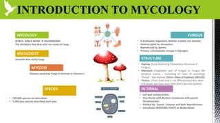
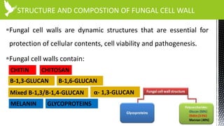
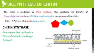
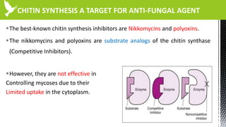
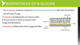
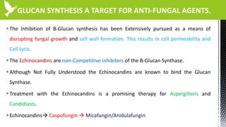
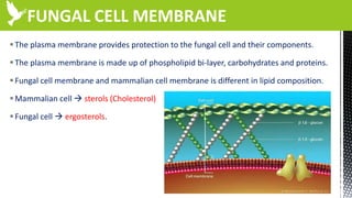
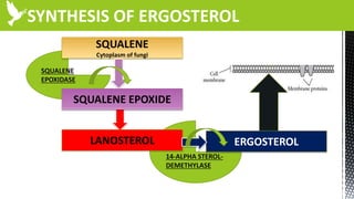
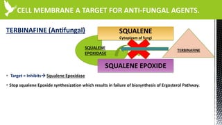
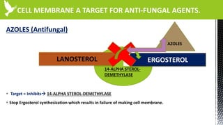
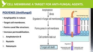
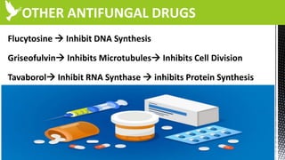
The document discusses anti-fungal drugs that target the fungal cell membrane and cell wall biosynthesis. It provides an overview of fungal cell structure, focusing on the cell wall components of chitin and beta-glucans. It explains how drugs like echinocandins inhibit beta-glucan synthase to disrupt cell wall formation, and how azoles and terbinafine inhibit ergosterol biosynthesis in the cell membrane. The summary highlights the main cellular targets of anti-fungal drugs and how inhibiting key processes like chitin, beta-glucan, and ergosterol synthesis impacts fungal growth and viability.
Mechanism of action of major antibiotic classes including betal lactam agents, aminoglycosides, macrolides, tetracyclines, quinolons, vancomycin, oxazolidionons. Detailed review and illustrations
Hello friends. In this PPT I am talking about Anti-viral drugs drugs. If you like it, please do let me know in the comments section. A single word of appreciation from you will encourage me to make more of such videos. Thanks. Enjoy and welcome to the beautiful world of pharmacology where pharmacology comes to life. This video is intended for MBBS, BDS, paramedical and any person who wishes to have a basic understanding of the subject in the simplest way.
An antifungal medication is a pharmaceutical fungicide used to treat and prevent mycoses such as athlete's foot, ringworm, candidiasis (thrush), serious systemic infections such as cryptococcal meningitis, and others. Such drugs are usually obtained by a doctor's prescription, but a few are available OTC (over-the-counter).
Antifungals work by exploiting differences between mammalian and fungal cells to kill the fungal organism with fewer adverse effects to the host. Unlike bacteria, both fungi and humans are eukaryotes. Thus, fungal and human cells are similar at the biological level. This makes it more difficult to discover drugs that target fungi without affecting human cells. As a consequence, many antifungal drugs cause side-effects. Some of these side-effects can be life-threatening if the drugs are not used properly.
The presentation provide in depth knowledge about two of the most affecting bacteria to human health. They are Neisseria ( causing gonorrhea and Meningitis) and Shigella ( Diarrhea)
Mycotoxins are naturally occurring toxins produced by certain moulds (fungi) and can be found in food.
The moulds grow on a variety of different crops and foodstuffs including cereals, nuts, spices, dried fruits, apples and coffee beans, often under warm and humid conditions.
Mycotoxins can cause a variety of adverse health effects and pose a serious health threat to both humans and livestock.
Observation of Io’s Resurfacing via Plume Deposition Using Ground-based Adapt...Sérgio Sacani
Since volcanic activity was first discovered on Io from Voyager images in 1979, changes
on Io’s surface have been monitored from both spacecraft and ground-based telescopes.
Here, we present the highest spatial resolution images of Io ever obtained from a groundbased telescope. These images, acquired by the SHARK-VIS instrument on the Large
Binocular Telescope, show evidence of a major resurfacing event on Io’s trailing hemisphere. When compared to the most recent spacecraft images, the SHARK-VIS images
show that a plume deposit from a powerful eruption at Pillan Patera has covered part
of the long-lived Pele plume deposit. Although this type of resurfacing event may be common on Io, few have been detected due to the rarity of spacecraft visits and the previously low spatial resolution available from Earth-based telescopes. The SHARK-VIS instrument ushers in a new era of high resolution imaging of Io’s surface using adaptive
optics at visible wavelengths.
Introduction:
RNA interference (RNAi) or Post-Transcriptional Gene Silencing (PTGS) is an important biological process for modulating eukaryotic gene expression.
It is highly conserved process of posttranscriptional gene silencing by which double stranded RNA (dsRNA) causes sequence-specific degradation of mRNA sequences.
dsRNA-induced gene silencing (RNAi) is reported in a wide range of eukaryotes ranging from worms, insects, mammals and plants.
This process mediates resistance to both endogenous parasitic and exogenous pathogenic nucleic acids, and regulates the expression of protein-coding genes.
What are small ncRNAs?
micro RNA (miRNA)
short interfering RNA (siRNA)
Properties of small non-coding RNA:
Involved in silencing mRNA transcripts.
Called “small” because they are usually only about 21-24 nucleotides long.
Synthesized by first cutting up longer precursor sequences (like the 61nt one that Lee discovered).
Silence an mRNA by base pairing with some sequence on the mRNA.
Discovery of siRNA?
The first small RNA:
In 1993 Rosalind Lee (Victor Ambros lab) was studying a non- coding gene in C. elegans, lin-4, that was involved in silencing of another gene, lin-14, at the appropriate time in the
development of the worm C. elegans.
Two small transcripts of lin-4 (22nt and 61nt) were found to be complementary to a sequence in the 3' UTR of lin-14.
Because lin-4 encoded no protein, she deduced that it must be these transcripts that are causing the silencing by RNA-RNA interactions.
Types of RNAi ( non coding RNA)
MiRNA
Length (23-25 nt)
Trans acting
Binds with target MRNA in mismatch
Translation inhibition
Si RNA
Length 21 nt.
Cis acting
Bind with target Mrna in perfect complementary sequence
Piwi-RNA
Length ; 25 to 36 nt.
Expressed in Germ Cells
Regulates trnasposomes activity
MECHANISM OF RNAI:
First the double-stranded RNA teams up with a protein complex named Dicer, which cuts the long RNA into short pieces.
Then another protein complex called RISC (RNA-induced silencing complex) discards one of the two RNA strands.
The RISC-docked, single-stranded RNA then pairs with the homologous mRNA and destroys it.
THE RISC COMPLEX:
RISC is large(>500kD) RNA multi- protein Binding complex which triggers MRNA degradation in response to MRNA
Unwinding of double stranded Si RNA by ATP independent Helicase
Active component of RISC is Ago proteins( ENDONUCLEASE) which cleave target MRNA.
DICER: endonuclease (RNase Family III)
Argonaute: Central Component of the RNA-Induced Silencing Complex (RISC)
One strand of the dsRNA produced by Dicer is retained in the RISC complex in association with Argonaute
ARGONAUTE PROTEIN :
1.PAZ(PIWI/Argonaute/ Zwille)- Recognition of target MRNA
2.PIWI (p-element induced wimpy Testis)- breaks Phosphodiester bond of mRNA.)RNAse H activity.
MiRNA:
The Double-stranded RNAs are naturally produced in eukaryotic cells during development, and they have a key role in regulating gene expression .
Slide 1: Title Slide
Extrachromosomal Inheritance
Slide 2: Introduction to Extrachromosomal Inheritance
Definition: Extrachromosomal inheritance refers to the transmission of genetic material that is not found within the nucleus.
Key Components: Involves genes located in mitochondria, chloroplasts, and plasmids.
Slide 3: Mitochondrial Inheritance
Mitochondria: Organelles responsible for energy production.
Mitochondrial DNA (mtDNA): Circular DNA molecule found in mitochondria.
Inheritance Pattern: Maternally inherited, meaning it is passed from mothers to all their offspring.
Diseases: Examples include Leber’s hereditary optic neuropathy (LHON) and mitochondrial myopathy.
Slide 4: Chloroplast Inheritance
Chloroplasts: Organelles responsible for photosynthesis in plants.
Chloroplast DNA (cpDNA): Circular DNA molecule found in chloroplasts.
Inheritance Pattern: Often maternally inherited in most plants, but can vary in some species.
Examples: Variegation in plants, where leaf color patterns are determined by chloroplast DNA.
Slide 5: Plasmid Inheritance
Plasmids: Small, circular DNA molecules found in bacteria and some eukaryotes.
Features: Can carry antibiotic resistance genes and can be transferred between cells through processes like conjugation.
Significance: Important in biotechnology for gene cloning and genetic engineering.
Slide 6: Mechanisms of Extrachromosomal Inheritance
Non-Mendelian Patterns: Do not follow Mendel’s laws of inheritance.
Cytoplasmic Segregation: During cell division, organelles like mitochondria and chloroplasts are randomly distributed to daughter cells.
Heteroplasmy: Presence of more than one type of organellar genome within a cell, leading to variation in expression.
Slide 7: Examples of Extrachromosomal Inheritance
Four O’clock Plant (Mirabilis jalapa): Shows variegated leaves due to different cpDNA in leaf cells.
Petite Mutants in Yeast: Result from mutations in mitochondrial DNA affecting respiration.
Slide 8: Importance of Extrachromosomal Inheritance
Evolution: Provides insight into the evolution of eukaryotic cells.
Medicine: Understanding mitochondrial inheritance helps in diagnosing and treating mitochondrial diseases.
Agriculture: Chloroplast inheritance can be used in plant breeding and genetic modification.
Slide 9: Recent Research and Advances
Gene Editing: Techniques like CRISPR-Cas9 are being used to edit mitochondrial and chloroplast DNA.
Therapies: Development of mitochondrial replacement therapy (MRT) for preventing mitochondrial diseases.
Slide 10: Conclusion
Summary: Extrachromosomal inheritance involves the transmission of genetic material outside the nucleus and plays a crucial role in genetics, medicine, and biotechnology.
Future Directions: Continued research and technological advancements hold promise for new treatments and applications.
Slide 11: Questions and Discussion
Invite Audience: Open the floor for any questions or further discussion on the topic.
Multi-source connectivity as the driver of solar wind variability in the heli...Sérgio Sacani
The ambient solar wind that flls the heliosphere originates from multiple
sources in the solar corona and is highly structured. It is often described
as high-speed, relatively homogeneous, plasma streams from coronal
holes and slow-speed, highly variable, streams whose source regions are
under debate. A key goal of ESA/NASA’s Solar Orbiter mission is to identify
solar wind sources and understand what drives the complexity seen in the
heliosphere. By combining magnetic feld modelling and spectroscopic
techniques with high-resolution observations and measurements, we show
that the solar wind variability detected in situ by Solar Orbiter in March
2022 is driven by spatio-temporal changes in the magnetic connectivity to
multiple sources in the solar atmosphere. The magnetic feld footpoints
connected to the spacecraft moved from the boundaries of a coronal hole
to one active region (12961) and then across to another region (12957). This
is refected in the in situ measurements, which show the transition from fast
to highly Alfvénic then to slow solar wind that is disrupted by the arrival of
a coronal mass ejection. Our results describe solar wind variability at 0.5 au
but are applicable to near-Earth observatories.
(May 29th, 2024) Advancements in Intravital Microscopy- Insights for Preclini...Scintica Instrumentation
Intravital microscopy (IVM) is a powerful tool utilized to study cellular behavior over time and space in vivo. Much of our understanding of cell biology has been accomplished using various in vitro and ex vivo methods; however, these studies do not necessarily reflect the natural dynamics of biological processes. Unlike traditional cell culture or fixed tissue imaging, IVM allows for the ultra-fast high-resolution imaging of cellular processes over time and space and were studied in its natural environment. Real-time visualization of biological processes in the context of an intact organism helps maintain physiological relevance and provide insights into the progression of disease, response to treatments or developmental processes.
In this webinar we give an overview of advanced applications of the IVM system in preclinical research. IVIM technology is a provider of all-in-one intravital microscopy systems and solutions optimized for in vivo imaging of live animal models at sub-micron resolution. The system’s unique features and user-friendly software enables researchers to probe fast dynamic biological processes such as immune cell tracking, cell-cell interaction as well as vascularization and tumor metastasis with exceptional detail. This webinar will also give an overview of IVM being utilized in drug development, offering a view into the intricate interaction between drugs/nanoparticles and tissues in vivo and allows for the evaluation of therapeutic intervention in a variety of tissues and organs. This interdisciplinary collaboration continues to drive the advancements of novel therapeutic strategies.
Earliest Galaxies in the JADES Origins Field: Luminosity Function and Cosmic ...Sérgio Sacani
We characterize the earliest galaxy population in the JADES Origins Field (JOF), the deepest
imaging field observed with JWST. We make use of the ancillary Hubble optical images (5 filters
spanning 0.4−0.9µm) and novel JWST images with 14 filters spanning 0.8−5µm, including 7 mediumband filters, and reaching total exposure times of up to 46 hours per filter. We combine all our data
at > 2.3µm to construct an ultradeep image, reaching as deep as ≈ 31.4 AB mag in the stack and
30.3-31.0 AB mag (5σ, r = 0.1” circular aperture) in individual filters. We measure photometric
redshifts and use robust selection criteria to identify a sample of eight galaxy candidates at redshifts
z = 11.5 − 15. These objects show compact half-light radii of R1/2 ∼ 50 − 200pc, stellar masses of
M⋆ ∼ 107−108M⊙, and star-formation rates of SFR ∼ 0.1−1 M⊙ yr−1
. Our search finds no candidates
at 15 < z < 20, placing upper limits at these redshifts. We develop a forward modeling approach to
infer the properties of the evolving luminosity function without binning in redshift or luminosity that
marginalizes over the photometric redshift uncertainty of our candidate galaxies and incorporates the
impact of non-detections. We find a z = 12 luminosity function in good agreement with prior results,
and that the luminosity function normalization and UV luminosity density decline by a factor of ∼ 2.5
from z = 12 to z = 14. We discuss the possible implications of our results in the context of theoretical
models for evolution of the dark matter halo mass function.
This pdf is about the Schizophrenia.
For more details visit on YouTube; @SELF-EXPLANATORY;
https://www.youtube.com/channel/UCAiarMZDNhe1A3Rnpr_WkzA/videos
Thanks...!
ANTI FUNGAL DRUGS AFFECTING CELL MEMBRANE AND CELL WALL.
1. ANTI FUNGAL DRUGS AFFECTING
CELL MEMBRANE AND CELL WALL BIOSYNTHESIS
MOHAMMAD BILAL KAKAR
SCHOLAR
DEPARTMENT OF MICROBIOLOGY
MOLECULAR MECHANISM OF DRUG ACTION
UNIVERSITY OF BALOCHISTAN QUETTA.
2. Agenda
Infographic
Overview , General Characteristics
BREIF INTRO TO MYCOLOGY
01
Infections, structure of fungal cell
BRIEF INTRO TO FUNGAL INFECTION
02
ANTI FUNGAL AGENTS AFFECTING CELL WALL
03
.
ANTI FUNGAL AGENTS AFFECTING CELL
MEMBRANE
04
3. INTRODUCTION TO MYCOLOGY
FUNGUS
• A Eukaryotic organisms. Neither a plants nor animals.
• Heterotrophic by absorption
• Reproduced by Spores.
• Primary carbohydrate storage is Glycogen.
MYCOLOGY
MYKES: GREEK WORD MUSHROOMS
The discipline that deal with the study of Fungi.
SPECIES
• 120,000 species are described
• 1,700 new species described each year..
INTERNAL
• Cell wall contain Chitin.
• True Nuclei with Nuclear membrane with paired
Chromosomes.
• Divided By : Sexual , Asexual and Both Reproduction.
• Unicellular (BUDDING YEAST) or Multicellular
MICOLOGIST
Scientist who study Fungi.
MYCOSIS
Disease caused by Fungi in Animals or Humans by Fungi.
STRUCTURE
• Hyphae (Long Branching Filamentous Structure of
Fungus)
• Mycelium (Vegetative part of Fungus or Fungus like
bacterial Colony , Consisting of mass of branching,
Thread – Like Hyphae. (Shiro: Mass of Hyphae) (MOLDS)
• Thallus ( Plant body that is not differentiated Into stem
and leaves and lacks true roots and a vascular system).
4. INTRODUCTION TO FUNGI
FUNGAL DISEASE
• Not Highly contagious.
• Humans acquire from nature.
• Epidermis, hair & nails.
• Lower in case of contagious.
GROWTH
• All are chemoheterotrophs.
• Absorption of Nutrients by power Enzymes.
• Grow at lower PH 5
• Grow in High Salt and Sugar
SPECIES
CHARACTERISTICS
• Non vascular Organisms.
• Reproduce by means of spores.
• Produce both sexual and asexual spores depending on
the species condition.
• Typically non motile. Although a few like chytrids have
motile phase.
HETEROTROPHIC NATURE
• Present in soil that has great economically importance
in decaying dead organic matters
PATHOGENIC IN NATURE
• Only 100 species of fungi are human and animals
pathogens.
• Some are opportunistic pathogens
BASIC STRUCTURE
• Cell wall is rigid. (Chitin , Mannan and polysaccharides)
• Cell Membrane contain ergoterols.
Fungi are found all around the world and grow in a wide range of habitats, including deserts.
Most grow on land (terrestrial) environments, but several species live only in
aquatic habitats. Most fungi live in either soil or dead matter, and many are symbionts of
plants, animals, or other fungi.
6. CLASSIFICATION OF FUNGAL INFECTION
CUTANEOUS &
SUBCUTANEOUS
SUPER
FICIAL
SYSTEMIC OPPORTUNISTIC
• These
are superficial cosmetic
fungal infections of the skin
or hair shaft.
• No living tissue is invaded
and there is no cellular
response from the host.
• Essentially no pathological
changes are elicited.
• Infection of skin and its
appendages (nail & hairs.
• Subcutaneous Infections
produce chronic
inflammation of
subcutaneous tissues
(sporotrichosis)
Systemic mycoses are
fungal infections affecting
internal organs. In the
right circumstances the
fungi enter the body via
the lungs, through the
gut, paranasal sinuses or
skin.
• Do not normally cause
diseases in healthy
people.
• Cause disease in in
immuno-compromised
People.
• Drug that suppress
immune system.
• Cancer therapy
• HIV Infection
• Cancer
8. FUNGAL INFECTION
If We look at the CDC’s web site that lists
diseases and conditions,
36% of the diseases have a bacterial origin,
33% have a viral origin,
6% are fungal,
25% are due to parasites
10. STRUCTURE AND COMPOSTION OF FUNGAL CELL WALL
Fungal cell walls are dynamic structures that are essential for
protection of cellular contents, cell viability and pathogenesis.
Fungal cell walls contain:
CHITIN CHITOSAN
Β-1,3-GLUCAN Β-1,6-GLUCAN
Mixed Β-1,3/B-1,4-GLUCAN
MELANIN GLYCOPROTEINS
α- 1,3-GLUCAN
11. BIOSYNTHESIS OF CHITIN
The chitin is mediated by chitin synthase, that catalyzes the transfer of
N-acetylglucosamine from UDP-N-acetylglucosamine to a growing chitin chain.
Chitin Polymer of N-acetylglucoseamine
CHITIN SYNTHASE
An enzyme that synthesis a
Chain of chitin in the fungal
Cell wall.
12. CHITIN SYNTHESIS A TARGET FOR ANTI-FUNGAL AGENT
The best-known chitin synthesis inhibitors are Nikkomycins and polyoxins.
The nikkomycins and polyoxins are substrate analogs of the chitin synthase
(Competitive Inhibitors).
However, they are not effective in
Controlling mycoses due to their
Limited uptake in the cytoplasm.
13. BIOSYNTHESIS OF B-GLUCAN
The B-Glucans comprise a group of B-D-Glucose Polysaccharides Naturally Occurring in
the cell wall of Fungi.
B-Glucan is synthesized by an Enzyme called
B-Glucan Synthase which is present in the
Fungal Cell Membrane.
Beta-Glucan Makes 50% of the Fungal Cell Wall.
14. GLUCAN SYNTHESIS A TARGET FOR ANTI-FUNGAL AGENTS.
The Inhibition of B-Glucan synthesis has been Extensively pursued as a means of
disrupting fungal growth and cell wall formation. This results in cell permeability and
Cell Lysis.
The Echinocandins are non-Competitive inhibitors of the B-Glucan Synthase.
Although Not Fully Understood the Echinocandins are known to bind the Glucan
Synthase.
Treatment with the Echinocandins is a promising therapy for Aspergillosis and
Candidiasis.
Echinocandins Caspofungin Micafungin/Anidulafungin
15. FUNGAL CELL MEMBRANE
The plasma membrane provides protection to the fungal cell and their components.
The plasma membrane is made up of phospholipid bi-layer, carbohydrates and proteins.
Fungal cell membrane and mammalian cell membrane is different in lipid composition.
Mammalian cell sterols (Cholesterol)
Fungal cell ergosterols.