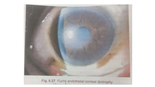
Fuchs endothelial corneal
dystrophy
Presented by:-- Gauri Shankar Saini
MBBS
Onset and course
• Aka endoepithelial corneal dystrophy
• Slow progressive, bilateral conditions
• Females > males
• Inheritance (autosomal dominant or sporadic)
• Genetic locus is 13p.
Clinical features
1. Stage of corneal guttata
2. Oedematous or endothelial decompensation
3. Stage of bullous keratopathy
4. Stage of scaring
Stage of cornea guttata
• Hassall henle type excrescence ( central cornea)
• Beaten mettal appearance
• Asymptomatic stage
Oedematous stage
• Early stromal oedema and epithelial dystrophy
• Blurring of vision
Stage of bullous keratopathy
• Marked epithelial oedema with formation of bullae
• when bulle rupture cause pain, discomfort, irritation, decreased
visual acuity
Stage of scaring
• Bullae replace by scar tissue
• Opaque and vascularized cornea
• Complicated by occurance of infection and glaucoma
Specular microscopy
• Decreased endothelial cell count
• Increased average cell diameter
• Decreased hexagon
• Increased variations in size
Treatment
• 5% NaCl
• Warm air
• 0.5% timolol
• Soft contact lenses
• Penetrating keratoplasty

Fuchs endothelial corneal dystrophy.pptx

  • 1.
    Fuchs endothelial corneal dystrophy Presentedby:-- Gauri Shankar Saini MBBS
  • 2.
    Onset and course •Aka endoepithelial corneal dystrophy • Slow progressive, bilateral conditions • Females > males • Inheritance (autosomal dominant or sporadic) • Genetic locus is 13p.
  • 3.
    Clinical features 1. Stageof corneal guttata 2. Oedematous or endothelial decompensation 3. Stage of bullous keratopathy 4. Stage of scaring
  • 4.
    Stage of corneaguttata • Hassall henle type excrescence ( central cornea) • Beaten mettal appearance • Asymptomatic stage
  • 5.
    Oedematous stage • Earlystromal oedema and epithelial dystrophy • Blurring of vision
  • 7.
    Stage of bullouskeratopathy • Marked epithelial oedema with formation of bullae • when bulle rupture cause pain, discomfort, irritation, decreased visual acuity
  • 8.
    Stage of scaring •Bullae replace by scar tissue • Opaque and vascularized cornea • Complicated by occurance of infection and glaucoma
  • 9.
    Specular microscopy • Decreasedendothelial cell count • Increased average cell diameter • Decreased hexagon • Increased variations in size
  • 10.
    Treatment • 5% NaCl •Warm air • 0.5% timolol • Soft contact lenses • Penetrating keratoplasty