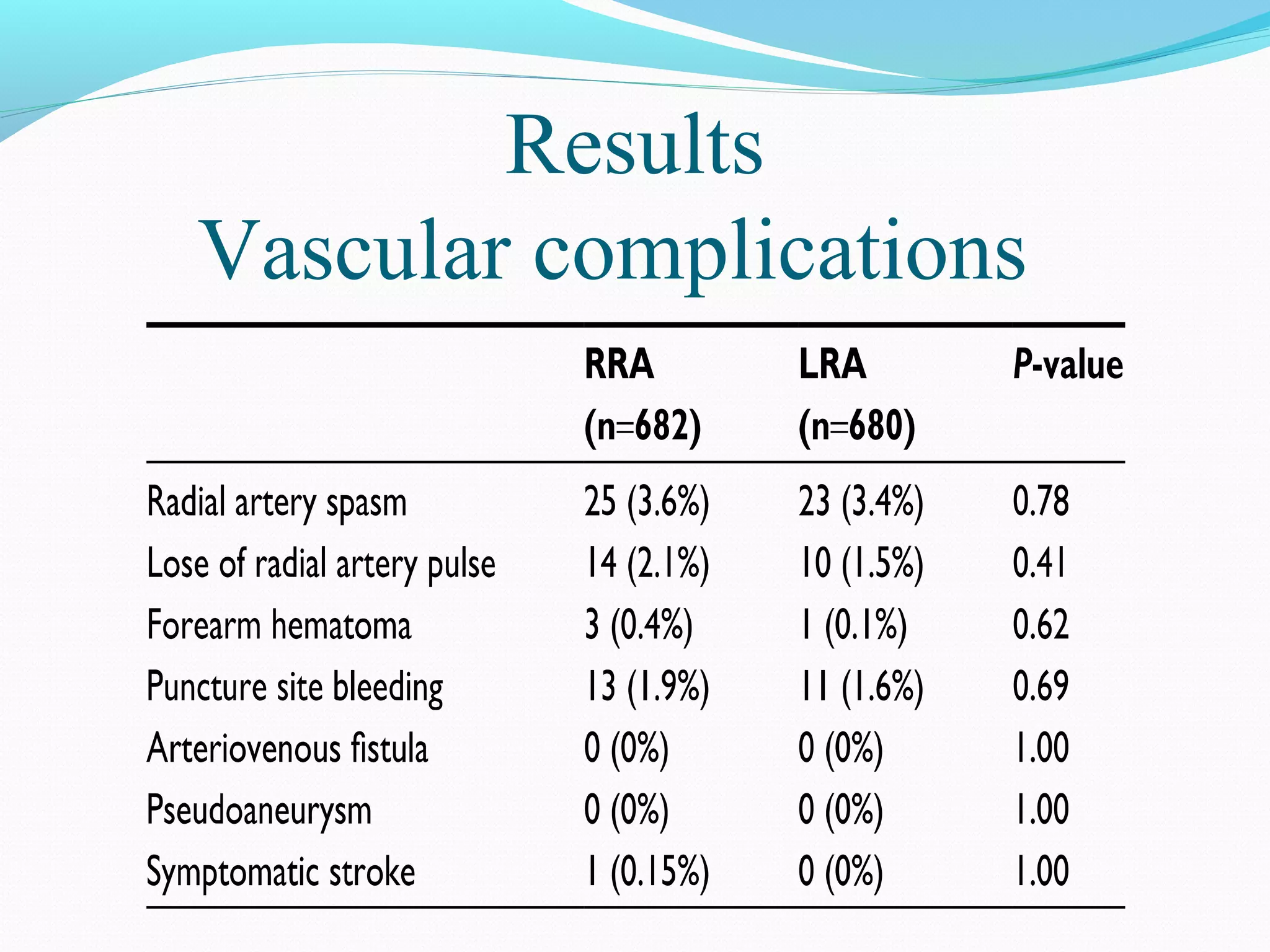
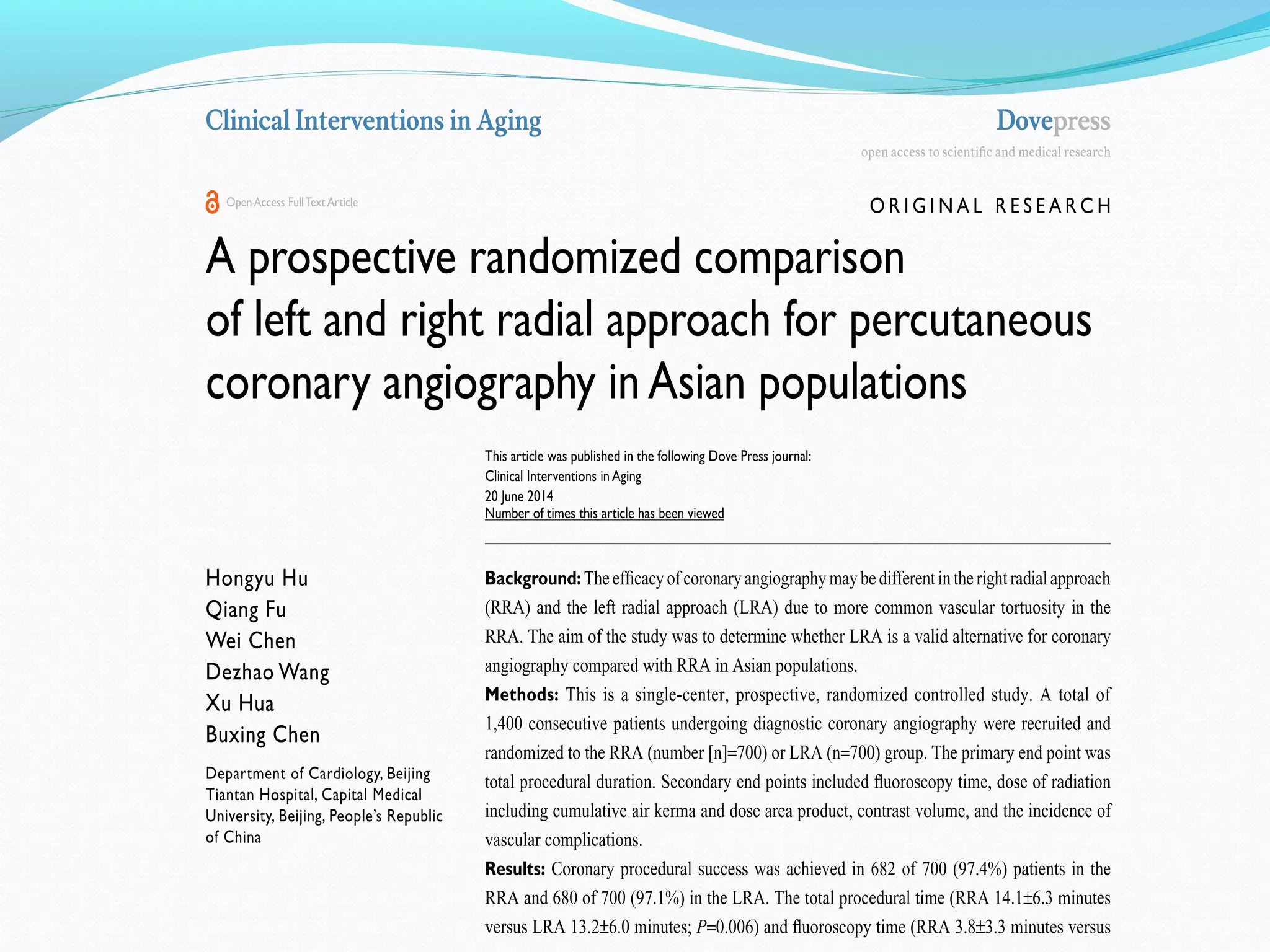
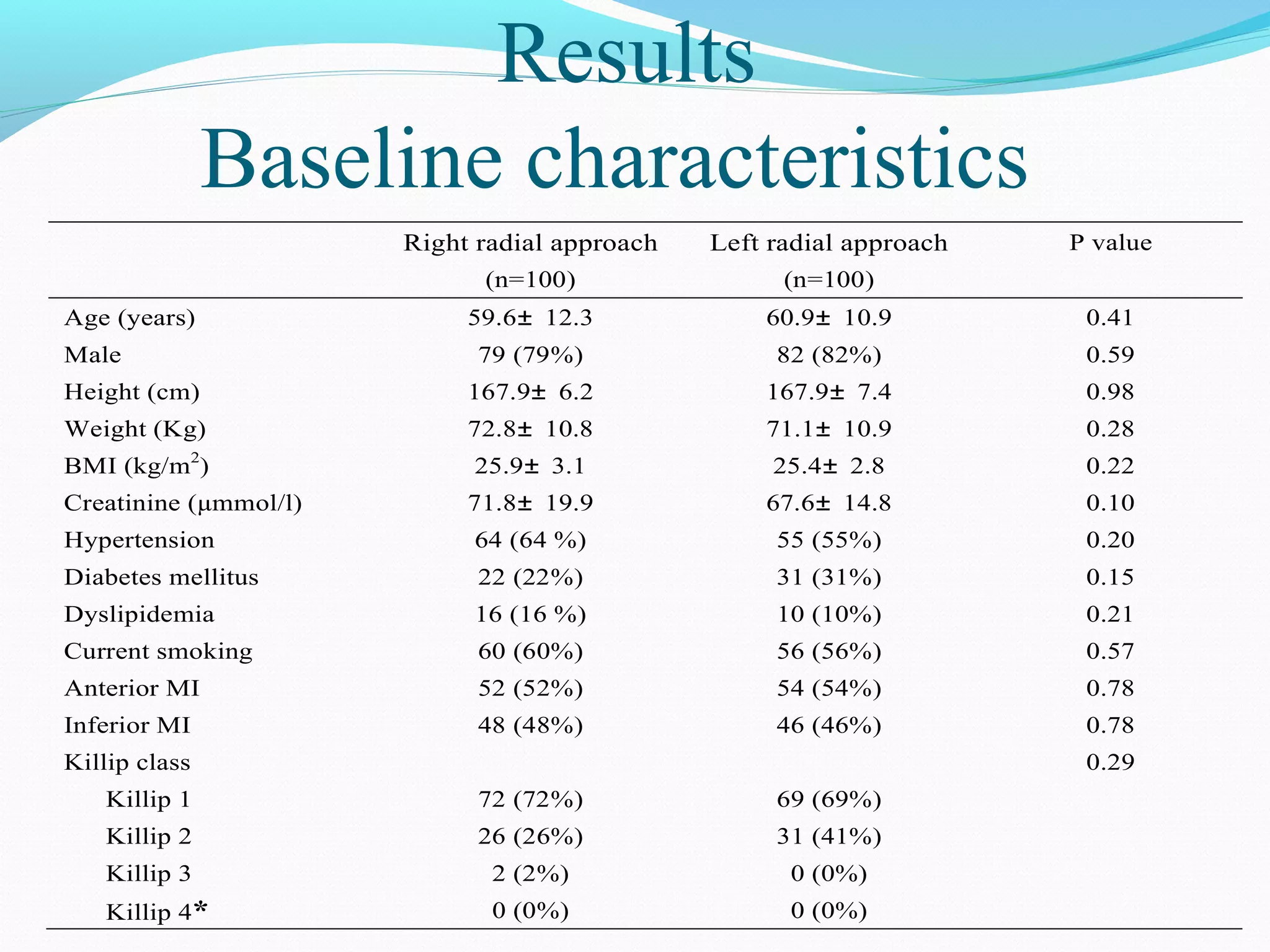
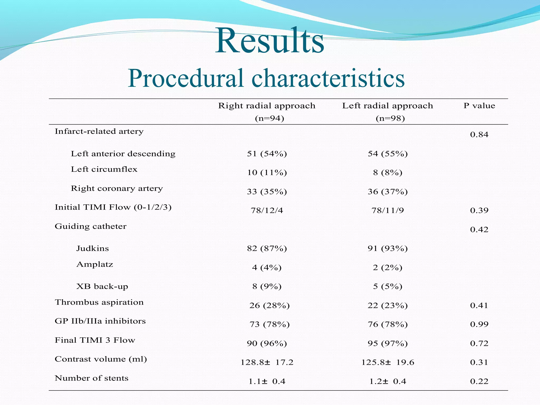
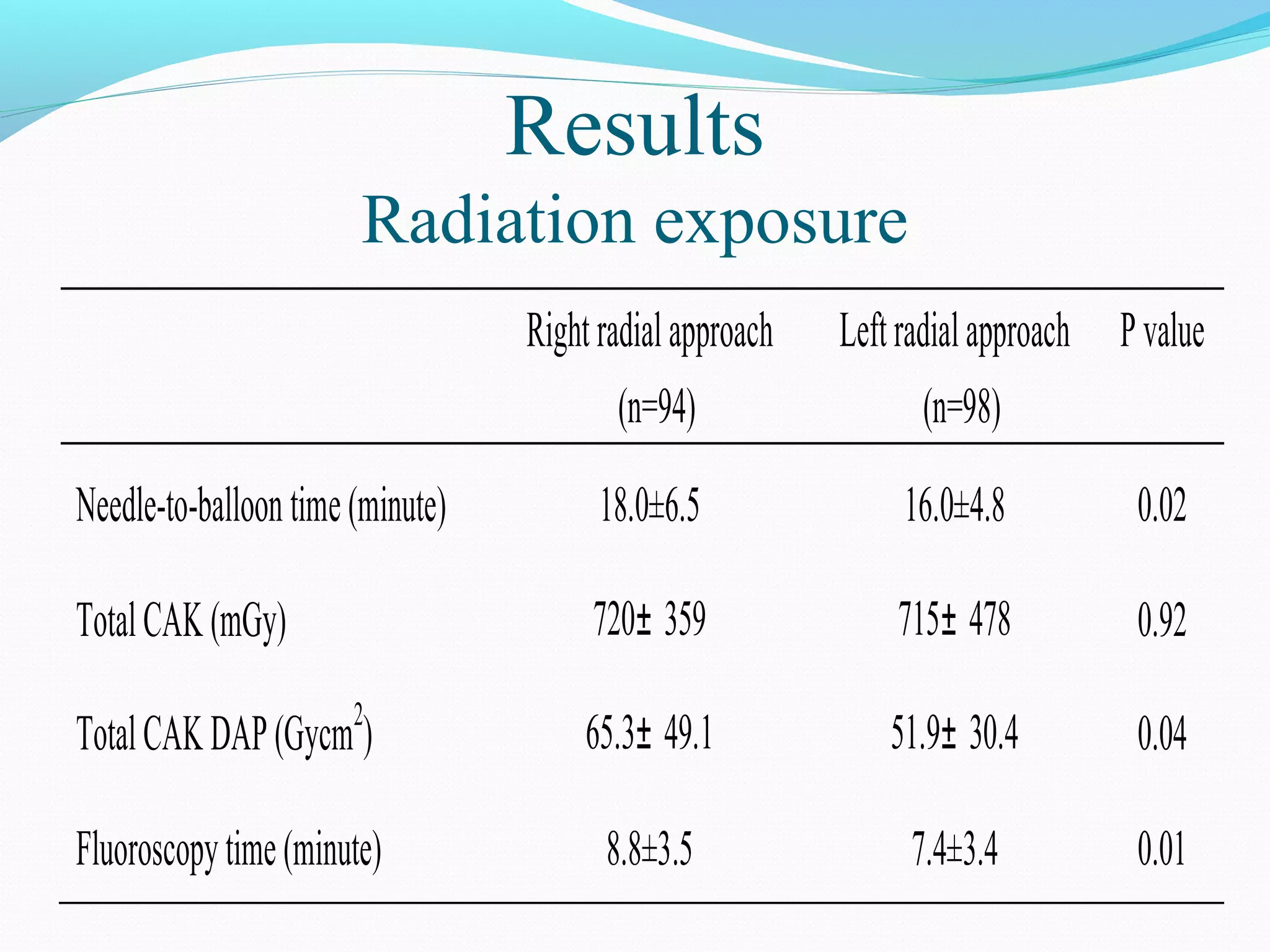
Department of Cardiology, Beijing Tiantan Hospital, 
Capital Medical University, Beijing, China 
Qiang Fu MD,Ph.D
The Temple of Heaven 
New Tiantan Hospital
The feasibility and safety of TRI have been established. 
- Easy hemostasis 
- Less bleeding/neurological complications 
TRI in China: 76%, but the choice of radial access 
depends on the institutions.
Marked vascular tortuosity is more 
common in right radial 
approach(RRA)than that in 
left radial approach 
(LRA)and may lead to 
coronary procedure failure. 
TALENT study showed that LRA for coronary diagnostic 
procedure is associated with lower fluoroscopy time and 
radiation dose adsorbed by patients compared with the 
RRA.
Recent studies indicated that incidence of microemboli was 
higher in coronary procedures performed through RRA, and 
LRA might reduce cerebrovascular complications compared 
with RRA*. *Int J Cardiol. 2012 Oct 6. pii: S0167-5273(12)01130-8. 
Until now, there is still no consensus on optimal radial 
approach for coronary procedure. Left or right ? 
Few data from randomized controlled trials in Asian 
patients undergoing coronary angiography and 
interventions are available.
Study I Comparison of transradial approach 
for coronary angiography. 
Study II Comparison of transradial approach 
for primary PCI.
To determine whether LRA is a valid alternative 
for coronary angiography compared with RRA in 
Chinese populations.
Design: 
Single-centre, prospective, randomized controlled 
study. 
Subjects: 
Consecutive patients who underwent coronary 
diagnostic procedures 
Exclusion criteria: 
Acute ST-elevation myocardial infarction, 
Previous coronary artery bypass graft surgery, 
Hemodynamic instability.
Primary end point 
Coronary procedure success 
Secondary end points 
Total procedure time; 
Fluoroscopy time; 
Dose of radiation 
cumulative air kerma: CAK, 
CAK dose area product: CAK DAP; 
Contrast volume.
Vascular complications 
Radial occlusion, 
Pseudoaneurysm, 
Arteriovenous fistula, 
Major bleeding at access-site.
Flow chart
Results 
Coronary procedural success were 682 of 700 (97.4%) in 
the LRA group and 680 of 488 (97.1%) in the RRA group.
Results 
Baseline characteristics
Results 
Procedural characteristics Table 3 Procedural characteristics in coronary angiography 
RRA 
(n=682) 
LRA 
(n=680) 
P value 
Total procedural time (min) 14.1±6.3 13.2±6.0 0.006 
CAK (mGy) 320±205 302±173 0.09 
CAK DAP (Gycm2) 28±20 26±17 0.13 
Fluoroscopy time (min) 3.8±3.3 3.4±2.8 0.046 
Contrast volume (ml) 63±14 62±17 0.29 
Number of catheters 2.1±0.4 2.1±0.4 0.69 
Data are expressed as the mean±SD or * median (25th percentile and 75th percentile)
Results 
Vascular complications
To investigate the difference between left 
and right radial approach in the setting of 
primary PCI for STEMI.
Subjects: 
All patients with STEMI were screened for 
eligibility. 
Inclusion criteria: 
Patients undergoing primary PCI within 12 
hours of symptom onset. 
Exclusion criteria: 
Cardiogenic shock.
Primary end point 
Needle-to-balloon time, defined as the time from local 
anesthesia infiltration to the first balloon inflation. If a manual 
thrombectomy was conducted before balloon inflation, needle-to-balloon 
time was also considered as the time from local anesthesia 
infiltration to the beginning of thrombus aspiration. 
Secondary end points 
Fluoroscopy time; 
Dose of radiation: CAK and CAK DAP; 
Contrast volume; 
In-hospital MACE: death, reinfarction, urgent 
revascularization, stroke and new congestive heart failure
Results 
Tables 
Baseline characteristics 
Table 1 Baseline characteristics of the study population 
Right radial approach 
(n=100) 
Left radial approach 
(n=100) 
P value 
Age (years) 59.6± 12.3 60.9± 10.9 0.41 
Male 79 (79%) 82 (82%) 0.59 
Height (cm) 167.9± 6.2 167.9± 7.4 0.98 
Weight (Kg) 72.8± 10.8 71.1± 10.9 0.28 
BMI (kg/m2) 25.9± 3.1 25.4± 2.8 0.22 
Creatinine (μmmol/l) 71.8± 19.9 67.6± 14.8 0.10 
Hypertension 64 (64 %) 55 (55%) 0.20 
Diabetes mellitus 22 (22%) 31 (31%) 0.15 
Dyslipidemia 16 (16 %) 10 (10%) 0.21 
Current smoking 60 (60%) 56 (56%) 0.57 
Anterior MI 52 (52%) 54 (54%) 0.78 
Inferior MI 48 (48%) 46 (46%) 0.78 
Killip class 0.29 
Killip 1 72 (72%) 69 (69%) 
Killip 2 26 (26%) 31 (41%) 
Killip 3 2 (2%) 0 (0%) 
Killip 4* 0 (0%) 0 (0%)
Results 
Table 2 AngioPgrarpohicc anedd intuerrveantlio ncalh chaarracatecristtiecsr oif sstutdiyc psop ulation 
Right radial approach 
(n=94) 
Left radial approach 
(n=98) 
P value 
Infarct-related artery 0.84 
Left anterior descending 51 (54%) 54 (55%) 
Left circumflex 10 (11%) 8 (8%) 
Right coronary artery 33 (35%) 36 (37%) 
Initial TIMI Flow (0-1/2/3) 78/12/4 78/11/9 0.39 
Guiding catheter 0.42 
Judkins 82 (87%) 91 (93%) 
Amplatz 4 (4%) 2 (2%) 
XB back-up 8 (9%) 5 (5%) 
Thrombus aspiration 26 (28%) 22 (23%) 0.41 
GP IIb/IIIa inhibitors 73 (78%) 76 (78%) 0.99 
Final TIMI 3 Flow 90 (96%) 95 (97%) 0.72 
Contrast volume (ml) 128.8± 17.2 125.8± 19.6 0.31 
Number of stents 1.1± 0.4 1.2± 0.4 0.22 
Data are expressed as the mean±SD or number (%)
Results 
Table 3 Procedural durations and radiation exposure of study population 
Radiation exposure 
Right radial approach 
(n=94) 
Left radial approach 
(n=98) 
P value 
Needle-to-balloon time (minute) 18.0±6.5 16.0±4.8 0.02 
Total CAK (mGy) 720± 359 715± 478 0.92 
Total CAK DAP (Gycm2) 65.3± 49.1 51.9± 30.4 0.04 
Fluoroscopy time (minute) 8.8±3.5 7.4±3.4 0.01
In-hospital MACE 
Only one patient suffered a severe congestive heart 
failure in right radial approach group. No patient 
experienced death, reinfarction and stroke. No patient 
required re-PCI or bypass surgery.
Similar to TELENT study, the LRA is associated with 
shorter coronary procedure time and fluoroscopy time 
compared with the RRA for Chinese populations in a 
diagnostic coronary angiography. 
Even in STEMI patients, primary PCI can be performed 
via LRA with earlier blood flow restoration in infarct-related 
artery and lower radiation exposure compared 
with RRA.
Possible explanations of our findings: 
1, The LRA has lower subclavian tortuosity and permits 
more direct access to the ascending aorta, whereas the 
RRA may be more difficult to access, owing to its 
tortuosity and to the atherosclerosis of the right 
common brachiocephalic trunk and subclavian artery
2, The engagement of coronary ostia is easier through 
the LRA. However, the catheter must be rotated to 
afford the S-shaped geometry of subclavian-innominate-aorta 
axis through the RRA.
On the basis of these advantages, procedure duration and 
fluoroscopy time could be decreased in the LRA 
compared to the RRA. 
Therefore, in view of lower subclavian tortuosity, easier 
catheter manipulation, and less radiation exposure via the 
LRA, the LRA may be a better and more reasonable 
choice, and it should be recommended in real-world 
cardiac catheterization, especially in urgent cases that 
need a faster coronary procedure.
Left radial approach may become a feasible and 
attractive alternative to perform not only diagnostic 
coronary angiography but also primary PCI for 
STEMI patients.
Fu Q - AIMRADIAL 2014 - Left vs right radial approach

Fu Q - AIMRADIAL 2014 - Left vs right radial approach

  • 1.
    Department of Cardiology,Beijing Tiantan Hospital, Capital Medical University, Beijing, China Qiang Fu MD,Ph.D
  • 2.
    The Temple ofHeaven New Tiantan Hospital
  • 3.
    The feasibility andsafety of TRI have been established. - Easy hemostasis - Less bleeding/neurological complications TRI in China: 76%, but the choice of radial access depends on the institutions.
  • 4.
    Marked vascular tortuosityis more common in right radial approach(RRA)than that in left radial approach (LRA)and may lead to coronary procedure failure. TALENT study showed that LRA for coronary diagnostic procedure is associated with lower fluoroscopy time and radiation dose adsorbed by patients compared with the RRA.
  • 5.
    Recent studies indicatedthat incidence of microemboli was higher in coronary procedures performed through RRA, and LRA might reduce cerebrovascular complications compared with RRA*. *Int J Cardiol. 2012 Oct 6. pii: S0167-5273(12)01130-8. Until now, there is still no consensus on optimal radial approach for coronary procedure. Left or right ? Few data from randomized controlled trials in Asian patients undergoing coronary angiography and interventions are available.
  • 6.
    Study I Comparisonof transradial approach for coronary angiography. Study II Comparison of transradial approach for primary PCI.
  • 7.
    To determine whetherLRA is a valid alternative for coronary angiography compared with RRA in Chinese populations.
  • 8.
    Design: Single-centre, prospective,randomized controlled study. Subjects: Consecutive patients who underwent coronary diagnostic procedures Exclusion criteria: Acute ST-elevation myocardial infarction, Previous coronary artery bypass graft surgery, Hemodynamic instability.
  • 9.
    Primary end point Coronary procedure success Secondary end points Total procedure time; Fluoroscopy time; Dose of radiation cumulative air kerma: CAK, CAK dose area product: CAK DAP; Contrast volume.
  • 10.
    Vascular complications Radialocclusion, Pseudoaneurysm, Arteriovenous fistula, Major bleeding at access-site.
  • 11.
  • 12.
    Results Coronary proceduralsuccess were 682 of 700 (97.4%) in the LRA group and 680 of 488 (97.1%) in the RRA group.
  • 13.
  • 14.
    Results Procedural characteristicsTable 3 Procedural characteristics in coronary angiography RRA (n=682) LRA (n=680) P value Total procedural time (min) 14.1±6.3 13.2±6.0 0.006 CAK (mGy) 320±205 302±173 0.09 CAK DAP (Gycm2) 28±20 26±17 0.13 Fluoroscopy time (min) 3.8±3.3 3.4±2.8 0.046 Contrast volume (ml) 63±14 62±17 0.29 Number of catheters 2.1±0.4 2.1±0.4 0.69 Data are expressed as the mean±SD or * median (25th percentile and 75th percentile)
  • 15.
  • 17.
    To investigate thedifference between left and right radial approach in the setting of primary PCI for STEMI.
  • 18.
    Subjects: All patientswith STEMI were screened for eligibility. Inclusion criteria: Patients undergoing primary PCI within 12 hours of symptom onset. Exclusion criteria: Cardiogenic shock.
  • 19.
    Primary end point Needle-to-balloon time, defined as the time from local anesthesia infiltration to the first balloon inflation. If a manual thrombectomy was conducted before balloon inflation, needle-to-balloon time was also considered as the time from local anesthesia infiltration to the beginning of thrombus aspiration. Secondary end points Fluoroscopy time; Dose of radiation: CAK and CAK DAP; Contrast volume; In-hospital MACE: death, reinfarction, urgent revascularization, stroke and new congestive heart failure
  • 20.
    Results Tables Baselinecharacteristics Table 1 Baseline characteristics of the study population Right radial approach (n=100) Left radial approach (n=100) P value Age (years) 59.6± 12.3 60.9± 10.9 0.41 Male 79 (79%) 82 (82%) 0.59 Height (cm) 167.9± 6.2 167.9± 7.4 0.98 Weight (Kg) 72.8± 10.8 71.1± 10.9 0.28 BMI (kg/m2) 25.9± 3.1 25.4± 2.8 0.22 Creatinine (μmmol/l) 71.8± 19.9 67.6± 14.8 0.10 Hypertension 64 (64 %) 55 (55%) 0.20 Diabetes mellitus 22 (22%) 31 (31%) 0.15 Dyslipidemia 16 (16 %) 10 (10%) 0.21 Current smoking 60 (60%) 56 (56%) 0.57 Anterior MI 52 (52%) 54 (54%) 0.78 Inferior MI 48 (48%) 46 (46%) 0.78 Killip class 0.29 Killip 1 72 (72%) 69 (69%) Killip 2 26 (26%) 31 (41%) Killip 3 2 (2%) 0 (0%) Killip 4* 0 (0%) 0 (0%)
  • 21.
    Results Table 2AngioPgrarpohicc anedd intuerrveantlio ncalh chaarracatecristtiecsr oif sstutdiyc psop ulation Right radial approach (n=94) Left radial approach (n=98) P value Infarct-related artery 0.84 Left anterior descending 51 (54%) 54 (55%) Left circumflex 10 (11%) 8 (8%) Right coronary artery 33 (35%) 36 (37%) Initial TIMI Flow (0-1/2/3) 78/12/4 78/11/9 0.39 Guiding catheter 0.42 Judkins 82 (87%) 91 (93%) Amplatz 4 (4%) 2 (2%) XB back-up 8 (9%) 5 (5%) Thrombus aspiration 26 (28%) 22 (23%) 0.41 GP IIb/IIIa inhibitors 73 (78%) 76 (78%) 0.99 Final TIMI 3 Flow 90 (96%) 95 (97%) 0.72 Contrast volume (ml) 128.8± 17.2 125.8± 19.6 0.31 Number of stents 1.1± 0.4 1.2± 0.4 0.22 Data are expressed as the mean±SD or number (%)
  • 22.
    Results Table 3Procedural durations and radiation exposure of study population Radiation exposure Right radial approach (n=94) Left radial approach (n=98) P value Needle-to-balloon time (minute) 18.0±6.5 16.0±4.8 0.02 Total CAK (mGy) 720± 359 715± 478 0.92 Total CAK DAP (Gycm2) 65.3± 49.1 51.9± 30.4 0.04 Fluoroscopy time (minute) 8.8±3.5 7.4±3.4 0.01
  • 23.
    In-hospital MACE Onlyone patient suffered a severe congestive heart failure in right radial approach group. No patient experienced death, reinfarction and stroke. No patient required re-PCI or bypass surgery.
  • 24.
    Similar to TELENTstudy, the LRA is associated with shorter coronary procedure time and fluoroscopy time compared with the RRA for Chinese populations in a diagnostic coronary angiography. Even in STEMI patients, primary PCI can be performed via LRA with earlier blood flow restoration in infarct-related artery and lower radiation exposure compared with RRA.
  • 25.
    Possible explanations ofour findings: 1, The LRA has lower subclavian tortuosity and permits more direct access to the ascending aorta, whereas the RRA may be more difficult to access, owing to its tortuosity and to the atherosclerosis of the right common brachiocephalic trunk and subclavian artery
  • 26.
    2, The engagementof coronary ostia is easier through the LRA. However, the catheter must be rotated to afford the S-shaped geometry of subclavian-innominate-aorta axis through the RRA.
  • 27.
    On the basisof these advantages, procedure duration and fluoroscopy time could be decreased in the LRA compared to the RRA. Therefore, in view of lower subclavian tortuosity, easier catheter manipulation, and less radiation exposure via the LRA, the LRA may be a better and more reasonable choice, and it should be recommended in real-world cardiac catheterization, especially in urgent cases that need a faster coronary procedure.
  • 28.
    Left radial approachmay become a feasible and attractive alternative to perform not only diagnostic coronary angiography but also primary PCI for STEMI patients.