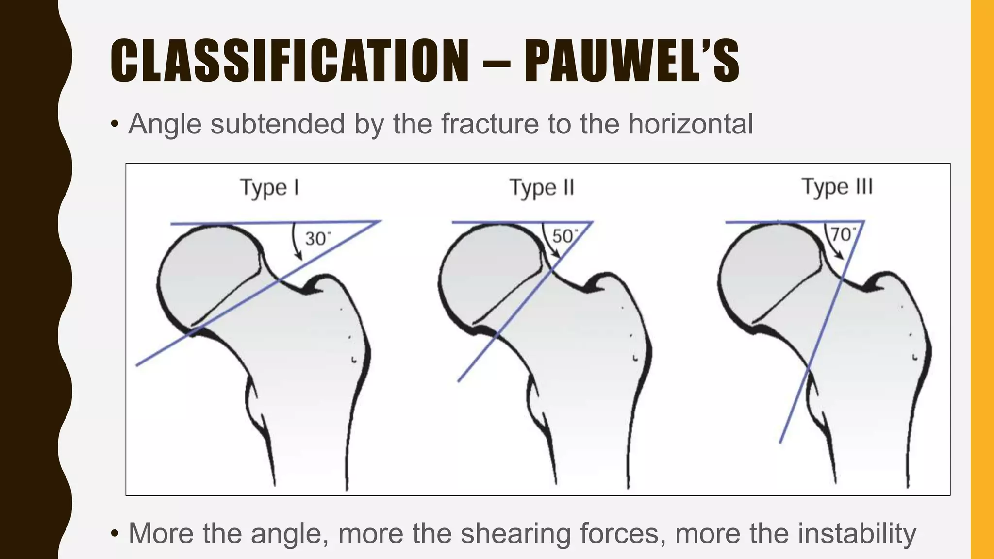
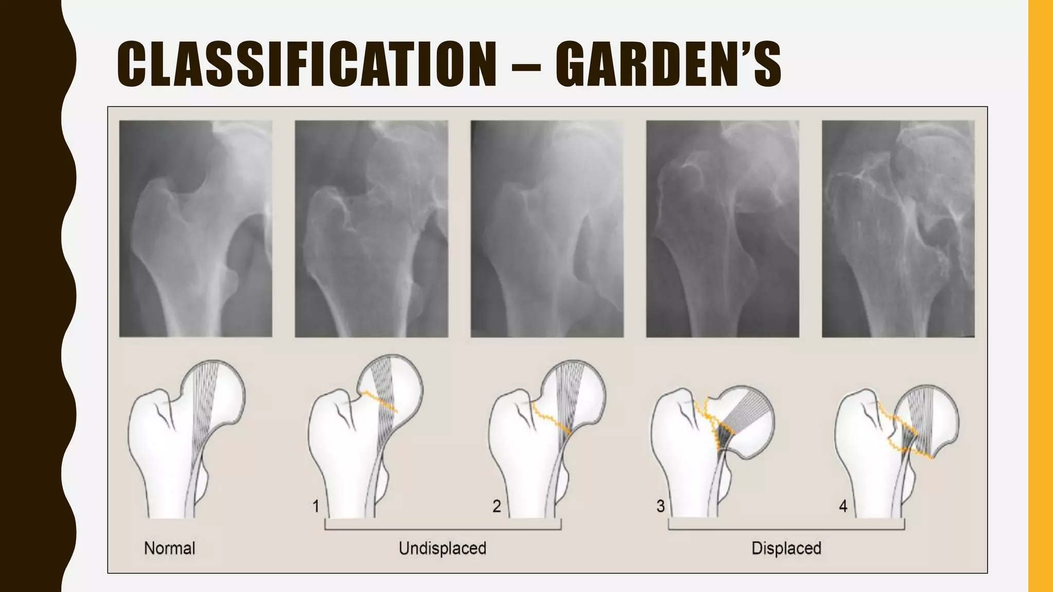
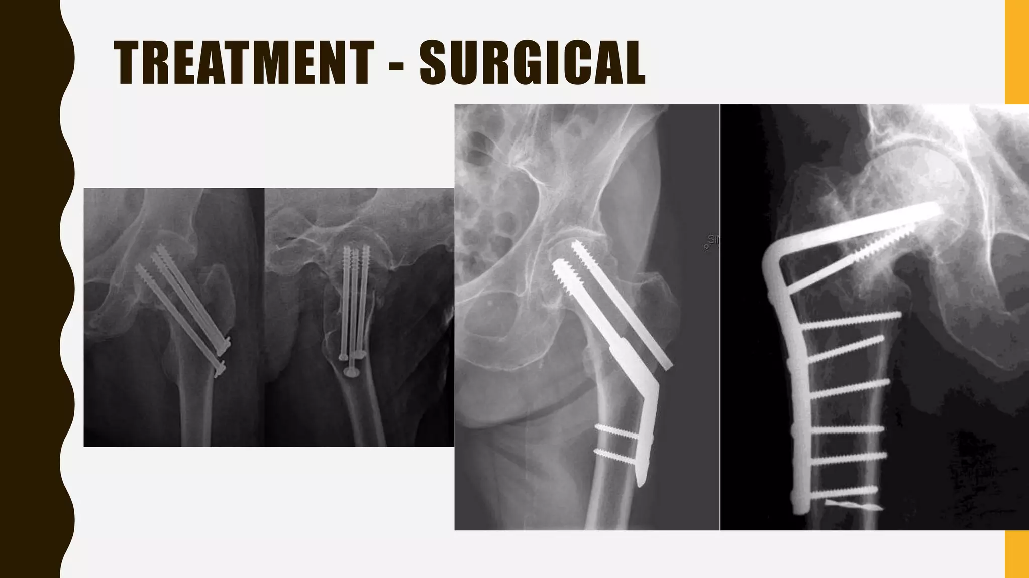
FRACTURE NECK
OF FEMUR
D R . B I P U L B O R T H A K U R
P R O F E S S O R , D E P T O F O R T H O PA E D I C S , S M C H
INTRODUCTION - ANATOMY
INTRODUCTION -ANATOMY
• Normal Neck-shaft angle =130±7degrees
• Femoral anteversion
INTRODUCTION - ANATOMY
INTRODUCTION BLOOD SUPPLY
INTRODUCTION
BLOOD SUPPLY OF NECK OF FEMUR
(1)An extracapsular arterial ring located at the base of femoral
neck
(2)Ascending cervical branches of the extracapsular arterial
ring of the surface of the femoral neck known as retinacular
arteries)
(3)The arteries of the ligamentum teres
INTRODUCTION
BLOOD SUPPLY OF NECK OF FEMUR
• The extracapsular arterial ring is formed posteriorly by a large branch of
the medial femoral circumflex artery and anteriorly by branches of the
lateral femoral circumflex artery
• The ascending cervical arteries can be divided into four groups
(Anterior, Medial, Posterior, and lateral) based on their relationship with
femoral neck.
Lateral group provides most of the blood supply to the femoral head and
neck
• The artery of ligamentum teres is a branch of the Obturator or the
Medial femoral circumflex artery ( Only a small amount of femoral head
INTRODUCTION
FRACTURE NECK OF FEMUR
• Commonly seen in elderly women – low energy falls
• Very low incidence in younger patients – high energy trauma
• Mechanism of injury:
–Low energy trauma – Direct or Indirect
–High energy trauma – MVA or fall from heights
–Cyclical loading-stress fractures
–Insufficiency fractures – osteoporosis or osteopenia
FRACTURE NECK OF FEMUR IS AN
UNSOLVED FRACTURE, WHY??
• Elongated position of femoral neck in joint capsule
• Absence of cambium layer of periosteum, periosteum is replaced by retinaculum,
which is a reflected part of capsule
• Fracture heals without external callus, almost heals entirely from intramedullary
endosteal callus
• Peculiar blood supply of the head of femur
• Intracapsular callus formed will get washed by synovial fluid.
• Intracapsular hemorrhage following fracture neck of femur causes tamponade effect
and obliterate retinacular vessels and fuerther decrease blood supply to head
REVASCULARIZATION OF THE HEAD
OCCURS FROM;
• The areas of the femoral head that remains viable
• Vascular ingrowth from the distal fragment
• Capillary ingrowth from distal fragment( It’s a slow process and occurs
when the fixation is rigid and stable)
CLASSIFICATION
• Anatomical location
SUBCAPITAL BASICERVICALTRANSCERVICAL
CLASSIFICATION – PAUWEL’S
• Angle subtended by the fracture to the horizontal
• More the angle, more the shearing forces, more the instability
CLASSIFICATION – GARDEN’S
CLINICAL FEATURES
• H/o trauma – High or low energy
• Non ambulatory (except in impacted and stress fractures)
• Pain over the groin
• Deformities – external rotation of the limb with shortening
• Tenderness – on hip ROM, axial compression and over the groin,
Scarpa’s triangle
• In low energy fracture – h/o LOC, syncopal attacks, chest pain, prior hip
pain (pathological #)
• Preinjury ambulatory status – determinant of treatment
CLINICAL FEATURES
RADIOLOGICAL EVALUATION
• Plain radiograph of pelvis with both hip joint – AP view, with
15 deg of internal rotation of the limb
• Cross table lateral view of proximal femur of fracture side
• CT scan – undisplaced femoral neck fractures
• MRI – imaging of choice in delineating non-displaced or
occult fractures
– Bone scans and CT scan is reserved for those who have
contraindications for MRI
AP VIEW VARIATIONS
LATERAL VIEW FROG LATERAL
RADIOLOGICAL EVALUATION
SHENTON’S LINE IN AN X-RAY
RADIOLOGICAL EVALUATION
TREATMENT
• Goals of the treatment are:
–Minimize patient discomfort
–Restoration of hip function
–Early stable fixation of fracture or prosthetic replacement
–Allow rapid mobilization
• Early bed to chair mobilization - to avoid risks and
complications of prolonged recumbence (Venous stasis, DVT,
Pulmonary Embolism, poor pulmonary toilet, atelectasis,
pressure sores)
TREATMENT
• Preoperative skin traction – below knee traction
–Immobilization of limb gives pain relief
–Reduce the risk of further soft tissue injury
–Helps to maintain reduction
–Easier intraoperative fracture reduction
• Use is contraversial
TREATMENT
• Non-operative treatment – can be employed in
undisplaced fractures; in patients with severe medical
comorbidities
–Good results have been reported from some studies
–Advantage: surgery can be avoided
–Disadvantage: Greater risk of fracture displacement
–Hence there is no role of non-operative in treatment of fracture
neck of femur
TREATMENT - SURGICAL
• Displaced fractures
• In young patients: urgent reduction and internal fixation and
capsulotomy
–Multiple screw fixation – 3 cannulated cancellous screws in
inverted triangular configuration
–Sliding hip screw or dynamic hip screw
–Angled blade plates
TREATMENT - SURGICAL
• Displaced fractures
• In elderly patients:
– High functional demand and good bone quality: Total hip
replacement
– Low functional demand and poor bone quality: Hemiarthroplasty
– Severely ill, demented, bedridden patients: non-operative
TREATMENT - SURGICAL
COMPLICATIONS
• Non-union: if the fracture has not united by 3months
–5% in undisplaced and 25% in displaced fracture
–In elderly: arthroplasty
–In young patients: proximal femoral osteotomy
• Osteonecrosis: avascular necrosis of femoral head
–10% in undisplaced and 30% in displaced fractures
–Early without x-ray changes: core decompression
–Late with x-ray changes: arthroplasty in elderly;
osteotomy/arthrodesis/arthroplasty in younger patients
COMPLICATIONS
• Fixation failure: poor reduction/fixation; osteoporotic bone
• Prosthetic dislocation and prosthetic loosening
• Prominent hardware: due to fracture collapse and screw
backout
• Others: complications of prolonged recumbence, infection
THANK YOU

Fracture neck of femur

  • 1.
    FRACTURE NECK OF FEMUR DR . B I P U L B O R T H A K U R P R O F E S S O R , D E P T O F O R T H O PA E D I C S , S M C H
  • 2.
  • 4.
    INTRODUCTION -ANATOMY • NormalNeck-shaft angle =130±7degrees • Femoral anteversion
  • 5.
  • 6.
  • 7.
    INTRODUCTION BLOOD SUPPLY OFNECK OF FEMUR (1)An extracapsular arterial ring located at the base of femoral neck (2)Ascending cervical branches of the extracapsular arterial ring of the surface of the femoral neck known as retinacular arteries) (3)The arteries of the ligamentum teres
  • 8.
    INTRODUCTION BLOOD SUPPLY OFNECK OF FEMUR • The extracapsular arterial ring is formed posteriorly by a large branch of the medial femoral circumflex artery and anteriorly by branches of the lateral femoral circumflex artery • The ascending cervical arteries can be divided into four groups (Anterior, Medial, Posterior, and lateral) based on their relationship with femoral neck. Lateral group provides most of the blood supply to the femoral head and neck • The artery of ligamentum teres is a branch of the Obturator or the Medial femoral circumflex artery ( Only a small amount of femoral head
  • 9.
    INTRODUCTION FRACTURE NECK OFFEMUR • Commonly seen in elderly women – low energy falls • Very low incidence in younger patients – high energy trauma • Mechanism of injury: –Low energy trauma – Direct or Indirect –High energy trauma – MVA or fall from heights –Cyclical loading-stress fractures –Insufficiency fractures – osteoporosis or osteopenia
  • 10.
    FRACTURE NECK OFFEMUR IS AN UNSOLVED FRACTURE, WHY?? • Elongated position of femoral neck in joint capsule • Absence of cambium layer of periosteum, periosteum is replaced by retinaculum, which is a reflected part of capsule • Fracture heals without external callus, almost heals entirely from intramedullary endosteal callus • Peculiar blood supply of the head of femur • Intracapsular callus formed will get washed by synovial fluid. • Intracapsular hemorrhage following fracture neck of femur causes tamponade effect and obliterate retinacular vessels and fuerther decrease blood supply to head
  • 11.
    REVASCULARIZATION OF THEHEAD OCCURS FROM; • The areas of the femoral head that remains viable • Vascular ingrowth from the distal fragment • Capillary ingrowth from distal fragment( It’s a slow process and occurs when the fixation is rigid and stable)
  • 12.
  • 13.
    CLASSIFICATION – PAUWEL’S •Angle subtended by the fracture to the horizontal • More the angle, more the shearing forces, more the instability
  • 14.
  • 15.
    CLINICAL FEATURES • H/otrauma – High or low energy • Non ambulatory (except in impacted and stress fractures) • Pain over the groin • Deformities – external rotation of the limb with shortening • Tenderness – on hip ROM, axial compression and over the groin, Scarpa’s triangle • In low energy fracture – h/o LOC, syncopal attacks, chest pain, prior hip pain (pathological #) • Preinjury ambulatory status – determinant of treatment
  • 16.
  • 17.
    RADIOLOGICAL EVALUATION • Plainradiograph of pelvis with both hip joint – AP view, with 15 deg of internal rotation of the limb • Cross table lateral view of proximal femur of fracture side • CT scan – undisplaced femoral neck fractures • MRI – imaging of choice in delineating non-displaced or occult fractures – Bone scans and CT scan is reserved for those who have contraindications for MRI
  • 18.
  • 19.
  • 20.
  • 21.
  • 22.
    TREATMENT • Goals ofthe treatment are: –Minimize patient discomfort –Restoration of hip function –Early stable fixation of fracture or prosthetic replacement –Allow rapid mobilization • Early bed to chair mobilization - to avoid risks and complications of prolonged recumbence (Venous stasis, DVT, Pulmonary Embolism, poor pulmonary toilet, atelectasis, pressure sores)
  • 23.
    TREATMENT • Preoperative skintraction – below knee traction –Immobilization of limb gives pain relief –Reduce the risk of further soft tissue injury –Helps to maintain reduction –Easier intraoperative fracture reduction • Use is contraversial
  • 24.
    TREATMENT • Non-operative treatment– can be employed in undisplaced fractures; in patients with severe medical comorbidities –Good results have been reported from some studies –Advantage: surgery can be avoided –Disadvantage: Greater risk of fracture displacement –Hence there is no role of non-operative in treatment of fracture neck of femur
  • 25.
  • 26.
  • 27.
    TREATMENT - SURGICAL •Displaced fractures • In young patients: urgent reduction and internal fixation and capsulotomy –Multiple screw fixation – 3 cannulated cancellous screws in inverted triangular configuration –Sliding hip screw or dynamic hip screw –Angled blade plates
  • 28.
  • 29.
    TREATMENT - SURGICAL •Displaced fractures • In elderly patients: – High functional demand and good bone quality: Total hip replacement – Low functional demand and poor bone quality: Hemiarthroplasty – Severely ill, demented, bedridden patients: non-operative
  • 30.
  • 31.
    COMPLICATIONS • Non-union: ifthe fracture has not united by 3months –5% in undisplaced and 25% in displaced fracture –In elderly: arthroplasty –In young patients: proximal femoral osteotomy • Osteonecrosis: avascular necrosis of femoral head –10% in undisplaced and 30% in displaced fractures –Early without x-ray changes: core decompression –Late with x-ray changes: arthroplasty in elderly; osteotomy/arthrodesis/arthroplasty in younger patients
  • 32.
    COMPLICATIONS • Fixation failure:poor reduction/fixation; osteoporotic bone • Prosthetic dislocation and prosthetic loosening • Prominent hardware: due to fracture collapse and screw backout • Others: complications of prolonged recumbence, infection
  • 33.