INTRODUCTION
• The studyof repair of maxillofacial skeleton must begin logically
with an understanding of nature of biologic response to osseous
injury and repair.
3.
• Bone isunique structure with several specific functions
• Major reservoir of calcium
• Support to human frame
• Origin and insertion of muscles
• Protects vital soft tissues
• Locomotion
4.
• Bone andliver in the body are only organs capable of undergoing
spontaneous regeneration.
• It is surrounded by a fibrous sheet called as periosteum outer
sheath fibrous layer and inner layer called as cambium layer which
is source of new bone cells.
5.
• Endosteum innerportion of bone marrow cavity is lined with
fibrous sheet called as endosteum.
• Haversian system or osteon is functional unit in mature bone
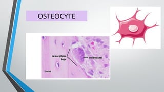
OSTEOBLAST
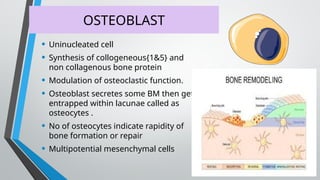
• Uninucleated cell
•Synthesis of collogeneous{1&5} and
non collagenous bone protein
• Modulation of osteoclastic function.
• Osteoblast secretes some BM then get
entrapped within lacunae called as
osteocytes .
• No of osteocytes indicate rapidity of
bone formation or repair
• Multipotential mesenchymal cells
9.
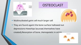
OSTEOCLAST
• Multinucleated gaintcell much larger cell
• They are found againt the bone surface hallowed out
depressions-Howships lacunae( themselves have
created),Resorption of bone ,Hemopoietic in origin.
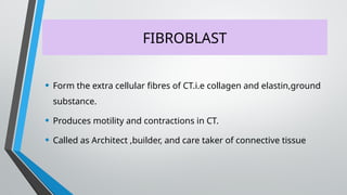
FIBROBLAST
• Form theextra cellular fibres of CT.i.e collagen and elastin,ground
substance.
• Produces motility and contractions in CT.
• Called as Architect ,builder, and care taker of connective tissue
12.
BONE HEALING
• ProcessOf Bone Healing Has Many Features Similar To That Of
Skin Healing Except it involve calcification of connective tissue
matrix.
• Bone heals by means of regeneration rather than repair.
13.
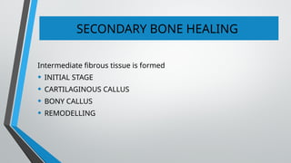
Types of bonehealing
• Direct bone healing [Primary bone healing callus free]
• Indirect bone healing [secondary bone healing ,callus forming
healing]
14.
Primary bone healing
•It occurs in the following condition
• Excellent anatomical reduction
• Minimal or no mobility.
• Good vascular supply
• It can also occur without rigid fixation if there is no gross mobility.
15.
• Osteogenic cellsand capillaries proliferate in the medullary bone
on both sides of fracture, forming new bone along the fracture
site.
PRIMARY HEALING TYPES
- GAP HEALING
- CONTACT HEALING
16.
GAP HEALING
• Evenwith rigid fixation, in some areas of fracture, small gaps
occur between the bone segments.
• Blood vessels from Periosteum, Endosteum or Haversian canals
invade the gaps bringing osteogenic cells into the gap.
• Bone is deposited directly over the fracture ends without
resorption or intermediate cartilage formation.
17.
GAP HEALING
• IfGap is < 0.3mm- lamellar bone forms directly.
• 0.5-1mm Woven bone formation and lamellar bone subsequently
laid down within the trabecular spaces .
• At the end of six weeks lamellar bundles oriented at right angle to
the longitudinal axis of remaining bone . Over several
months ,remodeling then leads to change in this direction.
18.
CONTACT HEALING
• Interfragmentarygap is essentially zero
• Vascular and cellular in growth can not proceed as that occurs in
gap healing
• Bone heals by means through the formation of bone metabolizing
unit(BMU)
• Osteoclast begin to cut away cores on either side of the fracture
progressing towards the fracture site at a rate of 50-80 um /day
19.
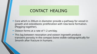
CONTACT HEALING
• Corewhich is 200um in diameter provide a pathway for vessel in
growth and osteoblastic proliferation with new bone formation.
(Pegging together).
• Osteon forms at a rate of 1-2 um/day.
• This lag between resorption and osteon ingrowth produce
transient porosity in the compact bone visible radiographically for
3month after fracture in humans .
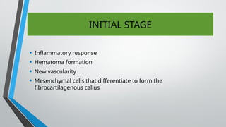
INITIAL STAGE
• Inflammatoryresponse
• Hematoma formation
• New vascularity
• Mesenchymal cells that differentiate to form the
fibrocartilagenous callus
22.
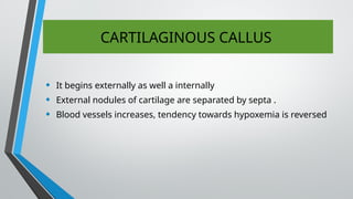
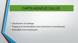
CARTILAGINOUS CALLUS
• Itbegins externally as well a internally
• External nodules of cartilage are separated by septa .
• Blood vessels increases, tendency towards hypoxemia is reversed
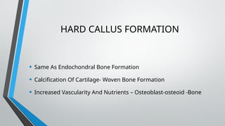
HARD CALLUS FORMATION
•Same As Endochondral Bone Formation
• Calcification Of Cartilage- Woven Bone Formation
• Increased Vascularity And Nutrients – Osteoblast-osteoid -Bone
25.
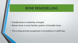
BONE REMODELLING
• Initiallybone is randomly arranged
• Woven bone is more familiar pattern of lamellar bone.
• This is slow process progresses in accordance in wolfs law.
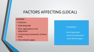
FACTORS AFFECTING (LOCAL)
ADVERSE
•Infection
• Pathological#
• Poor appositions and
alignment
• Continuing movement of bone
ends
• Poor blood supply
FAVORABLE
Good Apposition
Good Immobilisation
Good Blood Supply
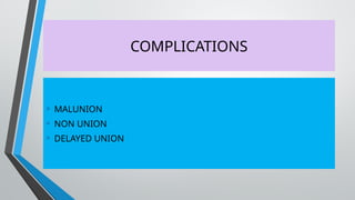
DELAYED UNION
• Fracturethat has not healed in the expected time for type of fracture, patient
and method of repair.
Causes:
• Inadequate blood supply
• Severe soft tissue damage
• Periosteal stripping
• Excessive traction
• Insufficient splintage
• Infection
33.
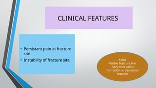
CLINICAL FEATURES
• Persistantpain at fracture
site
• Instability of fracture site X-RAY
-Visible Fracture line
-Very little callus
formation or periosteal
reaction
34.
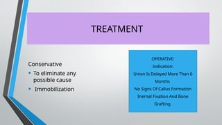
TREATMENT
Conservative
• To eliminateany
possible cause
• Immobilization
OPERATIVE:
Indication:
Union Is Delayed More Than 6
Months
No Signs Of Callus Formation
Inernal Fixation And Bone
Grafting
![Types of bone healing
• Direct bone healing [Primary bone healing callus free]
• Indirect bone healing [secondary bone healing ,callus forming
healing]](https://image.slidesharecdn.com/fracturehealing-250412062521-ed010f70/85/FRACTURE-FRACTURE-HEALING-HEALING-SOCKET-13-320.jpg)


![Fracture Healing (Dr Arinze) [Autosaved].pdf](https://cdn.slidesharecdn.com/ss_thumbnails/fracturehealingdrarinzeautosaved-230520154407-0e798837-thumbnail.jpg?width=640&height=640&fit=bounds)


![2 FRACTURE HEALING reloaded [Autosaved].pptx](https://cdn.slidesharecdn.com/ss_thumbnails/2fracturehealingreloadedautosaved-250810162739-94b56093-thumbnail.jpg?width=640&height=640&fit=bounds)


