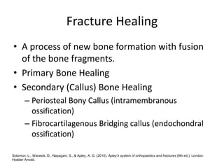
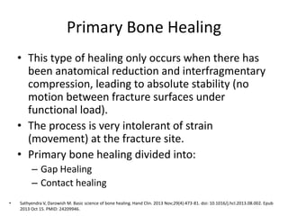
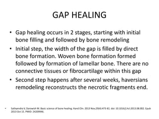
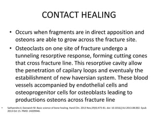
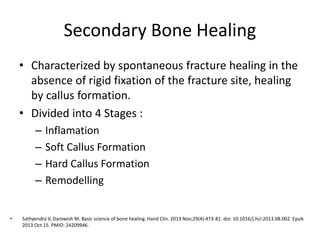
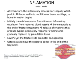
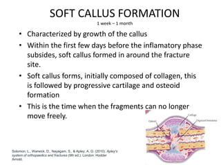
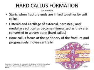
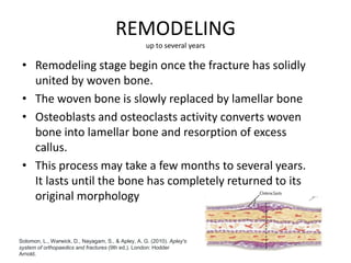
Fracture Healing
MU/BR
Mod: TQ
Supervisor: dr. Benny Murtaza, M. Kes, Sp.OT
Friday, July 29 2022
Fracture Healing
• A process of new bone formation with fusion
of the bone fragments.
• Primary Bone Healing
• Secondary (Callus) Bone Healing
– Periosteal Bony Callus (intramembranous
ossification)
– Fibrocartilagenous Bridging callus (endochondral
ossification)
Solomon, L., Warwick, D., Nayagam, S., & Apley, A. G. (2010). Apley's system of orthopaedics and fractures (9th ed.). London:
Hodder Arnold.
Primary Bone Healing
• This type of healing only occurs when there has
been anatomical reduction and interfragmentary
compression, leading to absolute stability (no
motion between fracture surfaces under
functional load).
• The process is very intolerant of strain
(movement) at the fracture site.
• Primary bone healing divided into:
– Gap Healing
– Contact healing
• Sathyendra V, Darowish M. Basic science of bone healing. Hand Clin. 2013 Nov;29(4):473-81. doi: 10.1016/j.hcl.2013.08.002. Epub
2013 Oct 15. PMID: 24209946.
GAP HEALING
• Gap healing occurs in 2 stages, starting with initial
bone filling and followed by bone remodeling
• Initial step, the width of the gap is filled by direct
bone formation. Woven bone formation formed
followed by formation of lamellar bone. There are no
connective tissues or fibrocartilage within this gap
• Second step happens after several weeks, haversians
remodeling reconstructs the necrotic fragments end.
• Sathyendra V, Darowish M. Basic science of bone healing. Hand Clin. 2013 Nov;29(4):473-81. doi: 10.1016/j.hcl.2013.08.002. Epub
2013 Oct 15. PMID: 24209946.
CONTACT HEALING
• Occurs when fragments are in direct apposition and
osteons are able to grow across the fracture site.
• Osteoclasts on one site of fracture undergo a
tunneling resorptive response, forming cutting cones
that cross fracture line. This resorptive cavity allow
the penetration of capilary loops and eventualy the
estabilishment of new haversian system. These blood
vessels accompanied by endothelial cells and
osteoprogenitor cells for osteoblasts leading to
productions osteons across fracture line
• Sathyendra V, Darowish M. Basic science of bone healing. Hand Clin. 2013 Nov;29(4):473-81. doi: 10.1016/j.hcl.2013.08.002. Epub
2013 Oct 15. PMID: 24209946.
Secondary Bone Healing
• Characterized by spontaneous fracture healing in the
absence of rigid fixation of the fracture site, healing
by callus formation.
• Divided into 4 Stages :
– Inflamation
– Soft Callus Formation
– Hard Callus Formation
– Remodelling
• Sathyendra V, Darowish M. Basic science of bone healing. Hand Clin. 2013 Nov;29(4):473-81. doi: 10.1016/j.hcl.2013.08.002. Epub
2013 Oct 15. PMID: 24209946.
INFLAMATION
up to 1 weeks
• After fracture, the inflamatory process starts rapidly which
peak in 48 hours and lasts until fibrous tissue, cartilage, or
bone formation begins
• Initially there is hematoma formation and inflamatory
exudation from ruptured blood vessels  bone necrosis at
the end of fracture fragments  release of cytokines that
produce typical inflamatory response  hematoma
gradually replaced by granulation tissue
• Low PO 2 at the fracture site promotes angiogenesis
• Osteoclasts remove the necrotic bones in the end of the
fragments
Solomon, L., Warwick, D., Nayagam, S., & Apley, A. G. (2010). Apley's
system of orthopaedics and fractures (9th ed.). London: Hodder
Arnold.
SOFT CALLUS FORMATION
1 week – 1 month
• Characterized by growth of the callus
• Within the first few days before the inflamatory phase
subsides, soft callus formed in around the fracture
site.
• Soft callus forms, initially composed of collagen, this
is followed by progressive cartilage and osteoid
formation
• This is the time when the fragments can no longer
move freely.
Solomon, L., Warwick, D., Nayagam, S., & Apley, A. G. (2010). Apley's
system of orthopaedics and fractures (9th ed.). London: Hodder
Arnold.
HARD CALLUS FORMATION
1-4 months
• Starts when fracture ends are linked together by soft
callus.
• Osteoid and Cartilage of external, perosteal, and
medullary soft callus become mineralized as they are
converted to woven bone (hard callus)
• Bone callus forms at the periphery of the fracture and
progressively moves centrally.
Solomon, L., Warwick, D., Nayagam, S., & Apley, A. G. (2010). Apley's
system of orthopaedics and fractures (9th ed.). London: Hodder
Arnold.
REMODELING
up to several years
• Remodeling stage begin once the fracture has solidly
united by woven bone.
• The woven bone is slowly replaced by lamellar bone
• Osteoblasts and osteoclasts activity converts woven
bone into lamellar bone and resorption of excess
callus.
• This process may take a few months to several years.
It lasts until the bone has completely returned to its
original morphology
Solomon, L., Warwick, D., Nayagam, S., & Apley, A. G. (2010). Apley's
system of orthopaedics and fractures (9th ed.). London: Hodder
Arnold.
VARIABLES INFLUENCE FRACTURE
HEALING
• Internal
• External
• Patient Factors
Augat P, Simon U, Liedert A, Claes L. Mechanics and mechano-biology of fracture healing in normal and osteoporotic bone. Osteoporos Int. 2005
Mar;16 Suppl 2:S36-43. doi: 10.1007/s00198-004-1728-9. Epub 2004 Sep 15. PMID: 15372141.
INTERNAL
• Blood Supply
– initially the blood flow decreases with vascular
disruption
– after few hours to days, the blood flow increases.
This peaks at 2 weeks and normalizes at 3-5
months
• Mechanical factors
bony soft tissue attachments, mechanical stability,
location of injury, degree of bone loss
Augat P, Simon U, Liedert A, Claes L. Mechanics and mechano-biology of fracture healing in normal and osteoporotic bone. Osteoporos Int. 2005
Mar;16 Suppl 2:S36-43. doi: 10.1007/s00198-004-1728-9. Epub 2004 Sep 15. PMID: 15372141.
EXTERNAL
• Bone stimulators
Lead to elevated concentrations of TGF-Beta and
BMP
• COX-2:
promotes fracture healing by causing mesenchymal
stem cells to differentiate into osteoblast
Augat P, Simon U, Liedert A, Claes L. Mechanics and mechano-biology of fracture healing in normal and osteoporotic bone. Osteoporos Int. 2005
Mar;16 Suppl 2:S36-43. doi: 10.1007/s00198-004-1728-9. Epub 2004 Sep 15. PMID: 15372141.
PATIENT FACTORS
• Diet
as high as 84% of patients with nonunion were found to have
metabolic issues
– greater than 66% of these patients had vitamin D deficiencies
• DM
fracture healing takes 1.6 times longer in diabetic patients versus
non-diabetic patients. Because of decreased cellularity of the
fracture callus and delayed endochondral ossification.
• Nicotine
Inhibits growth of new blood vessels as bone is remodeled
increase risk of nonunion
decreased strength of fracture callus
Augat P, Simon U, Liedert A, Claes L. Mechanics and mechano-biology of fracture healing in normal and osteoporotic bone. Osteoporos Int. 2005
Mar;16 Suppl 2:S36-43. doi: 10.1007/s00198-004-1728-9. Epub 2004 Sep 15. PMID: 15372141.
PATIENT FACTORS
• HIV
• Medications
BIPHOSPHONATES
• As a cause of osteoporotic fractures with long term usage
SYSTEMIC CORTICOSTEROIDS
NSAIDs
• Prolonged healing time because of COX enzyme inhibition
QUINOLONES
• Toxic to chondrocytes and diminishes fracture repair
Augat P, Simon U, Liedert A, Claes L. Mechanics and mechano-biology of fracture healing in normal and osteoporotic bone. Osteoporos Int. 2005
Mar;16 Suppl 2:S36-43. doi: 10.1007/s00198-004-1728-9. Epub 2004 Sep 15. PMID: 15372141.
THANK YOU

Fracture Healing.pptx

  • 1.
    Fracture Healing MU/BR Mod: TQ Supervisor:dr. Benny Murtaza, M. Kes, Sp.OT Friday, July 29 2022
  • 2.
    Fracture Healing • Aprocess of new bone formation with fusion of the bone fragments. • Primary Bone Healing • Secondary (Callus) Bone Healing – Periosteal Bony Callus (intramembranous ossification) – Fibrocartilagenous Bridging callus (endochondral ossification) Solomon, L., Warwick, D., Nayagam, S., & Apley, A. G. (2010). Apley's system of orthopaedics and fractures (9th ed.). London: Hodder Arnold.
  • 3.
    Primary Bone Healing •This type of healing only occurs when there has been anatomical reduction and interfragmentary compression, leading to absolute stability (no motion between fracture surfaces under functional load). • The process is very intolerant of strain (movement) at the fracture site. • Primary bone healing divided into: – Gap Healing – Contact healing • Sathyendra V, Darowish M. Basic science of bone healing. Hand Clin. 2013 Nov;29(4):473-81. doi: 10.1016/j.hcl.2013.08.002. Epub 2013 Oct 15. PMID: 24209946.
  • 4.
    GAP HEALING • Gaphealing occurs in 2 stages, starting with initial bone filling and followed by bone remodeling • Initial step, the width of the gap is filled by direct bone formation. Woven bone formation formed followed by formation of lamellar bone. There are no connective tissues or fibrocartilage within this gap • Second step happens after several weeks, haversians remodeling reconstructs the necrotic fragments end. • Sathyendra V, Darowish M. Basic science of bone healing. Hand Clin. 2013 Nov;29(4):473-81. doi: 10.1016/j.hcl.2013.08.002. Epub 2013 Oct 15. PMID: 24209946.
  • 5.
    CONTACT HEALING • Occurswhen fragments are in direct apposition and osteons are able to grow across the fracture site. • Osteoclasts on one site of fracture undergo a tunneling resorptive response, forming cutting cones that cross fracture line. This resorptive cavity allow the penetration of capilary loops and eventualy the estabilishment of new haversian system. These blood vessels accompanied by endothelial cells and osteoprogenitor cells for osteoblasts leading to productions osteons across fracture line • Sathyendra V, Darowish M. Basic science of bone healing. Hand Clin. 2013 Nov;29(4):473-81. doi: 10.1016/j.hcl.2013.08.002. Epub 2013 Oct 15. PMID: 24209946.
  • 6.
    Secondary Bone Healing •Characterized by spontaneous fracture healing in the absence of rigid fixation of the fracture site, healing by callus formation. • Divided into 4 Stages : – Inflamation – Soft Callus Formation – Hard Callus Formation – Remodelling • Sathyendra V, Darowish M. Basic science of bone healing. Hand Clin. 2013 Nov;29(4):473-81. doi: 10.1016/j.hcl.2013.08.002. Epub 2013 Oct 15. PMID: 24209946.
  • 7.
    INFLAMATION up to 1weeks • After fracture, the inflamatory process starts rapidly which peak in 48 hours and lasts until fibrous tissue, cartilage, or bone formation begins • Initially there is hematoma formation and inflamatory exudation from ruptured blood vessels  bone necrosis at the end of fracture fragments  release of cytokines that produce typical inflamatory response  hematoma gradually replaced by granulation tissue • Low PO 2 at the fracture site promotes angiogenesis • Osteoclasts remove the necrotic bones in the end of the fragments Solomon, L., Warwick, D., Nayagam, S., & Apley, A. G. (2010). Apley's system of orthopaedics and fractures (9th ed.). London: Hodder Arnold.
  • 8.
    SOFT CALLUS FORMATION 1week – 1 month • Characterized by growth of the callus • Within the first few days before the inflamatory phase subsides, soft callus formed in around the fracture site. • Soft callus forms, initially composed of collagen, this is followed by progressive cartilage and osteoid formation • This is the time when the fragments can no longer move freely. Solomon, L., Warwick, D., Nayagam, S., & Apley, A. G. (2010). Apley's system of orthopaedics and fractures (9th ed.). London: Hodder Arnold.
  • 9.
    HARD CALLUS FORMATION 1-4months • Starts when fracture ends are linked together by soft callus. • Osteoid and Cartilage of external, perosteal, and medullary soft callus become mineralized as they are converted to woven bone (hard callus) • Bone callus forms at the periphery of the fracture and progressively moves centrally. Solomon, L., Warwick, D., Nayagam, S., & Apley, A. G. (2010). Apley's system of orthopaedics and fractures (9th ed.). London: Hodder Arnold.
  • 10.
    REMODELING up to severalyears • Remodeling stage begin once the fracture has solidly united by woven bone. • The woven bone is slowly replaced by lamellar bone • Osteoblasts and osteoclasts activity converts woven bone into lamellar bone and resorption of excess callus. • This process may take a few months to several years. It lasts until the bone has completely returned to its original morphology Solomon, L., Warwick, D., Nayagam, S., & Apley, A. G. (2010). Apley's system of orthopaedics and fractures (9th ed.). London: Hodder Arnold.
  • 11.
    VARIABLES INFLUENCE FRACTURE HEALING •Internal • External • Patient Factors Augat P, Simon U, Liedert A, Claes L. Mechanics and mechano-biology of fracture healing in normal and osteoporotic bone. Osteoporos Int. 2005 Mar;16 Suppl 2:S36-43. doi: 10.1007/s00198-004-1728-9. Epub 2004 Sep 15. PMID: 15372141.
  • 12.
    INTERNAL • Blood Supply –initially the blood flow decreases with vascular disruption – after few hours to days, the blood flow increases. This peaks at 2 weeks and normalizes at 3-5 months • Mechanical factors bony soft tissue attachments, mechanical stability, location of injury, degree of bone loss Augat P, Simon U, Liedert A, Claes L. Mechanics and mechano-biology of fracture healing in normal and osteoporotic bone. Osteoporos Int. 2005 Mar;16 Suppl 2:S36-43. doi: 10.1007/s00198-004-1728-9. Epub 2004 Sep 15. PMID: 15372141.
  • 13.
    EXTERNAL • Bone stimulators Leadto elevated concentrations of TGF-Beta and BMP • COX-2: promotes fracture healing by causing mesenchymal stem cells to differentiate into osteoblast Augat P, Simon U, Liedert A, Claes L. Mechanics and mechano-biology of fracture healing in normal and osteoporotic bone. Osteoporos Int. 2005 Mar;16 Suppl 2:S36-43. doi: 10.1007/s00198-004-1728-9. Epub 2004 Sep 15. PMID: 15372141.
  • 14.
    PATIENT FACTORS • Diet ashigh as 84% of patients with nonunion were found to have metabolic issues – greater than 66% of these patients had vitamin D deficiencies • DM fracture healing takes 1.6 times longer in diabetic patients versus non-diabetic patients. Because of decreased cellularity of the fracture callus and delayed endochondral ossification. • Nicotine Inhibits growth of new blood vessels as bone is remodeled increase risk of nonunion decreased strength of fracture callus Augat P, Simon U, Liedert A, Claes L. Mechanics and mechano-biology of fracture healing in normal and osteoporotic bone. Osteoporos Int. 2005 Mar;16 Suppl 2:S36-43. doi: 10.1007/s00198-004-1728-9. Epub 2004 Sep 15. PMID: 15372141.
  • 15.
    PATIENT FACTORS • HIV •Medications BIPHOSPHONATES • As a cause of osteoporotic fractures with long term usage SYSTEMIC CORTICOSTEROIDS NSAIDs • Prolonged healing time because of COX enzyme inhibition QUINOLONES • Toxic to chondrocytes and diminishes fracture repair Augat P, Simon U, Liedert A, Claes L. Mechanics and mechano-biology of fracture healing in normal and osteoporotic bone. Osteoporos Int. 2005 Mar;16 Suppl 2:S36-43. doi: 10.1007/s00198-004-1728-9. Epub 2004 Sep 15. PMID: 15372141.
  • 16.