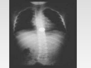
Tips on using my ppt.
1. You can freely download, edit, modify and put your
name etc.
2. Don’t be concerned about number of slides. Half the
slides are blanks except for the title.
3. First show the blank slides (eg. Aetiology ) > Ask
students what they already know about ethology of
today's topic. > Then show next slide which enumerates
aetiologies.
4. At the end rerun the show – show blank> ask questions >
show next slide.
5. This will be an ACTIVE LEARNING SESSION x
three revisions.
6. Good for self study also.
7. See notes for bibliography.
Learning Objectives
Learning Objectives
1. Introduction & History
2. Relevant Anatomy, Physiology
3. Aetiology
4. Pathophysiology
5. Pathology
6. Classification
7. Clinical Features
8. Investigations
9. Management
10. Prevention
11. Guidelines
12. Take home messages
Introduction & History.
•
Introduction & History.
• Foreign bodies in the oesophagus are
usually swallowed, purposefully or
accidentally
• The presentation is usually straightforward
but on occasion can be extremely subtle.
Aetiology
Aetiology
• Idiopathic
• Congenital/ Genetic
• Nutritional Deficiency/excess
• Traumatic
• Infections /Infestation
• Autoimmune
• Neoplastic (Benign/Malignant)
• Degenerative / lifestyle
• Iatrogenic
• Psychosomatic
• Poisoning/ Toxins/ Drug induced
Etiology
• Idiopathic
• Congenital
• Traumatic
• Infections /Infestation
• Autoimmune
• Neoplastic (Benign/Malignant)
• Degenerative
Pathophysiology
Pathophysiology
• The esophagus is a tubular structure
approximately 20-25 cm in length.
• Patients can usually localize foreign bodies
in the upper esophagus but localize them
poorly in the lower two thirds of the
structure.
Pathophysiology
• 3 areas of narrowing :
– the upper esophageal sphincter (UES),
– crossover of the aorta;
– lower esophageal sphincter (LES).
• Structural abnormalities of the esophagus
• strictures
• webs
• Diverticula
• Malignancies
• motor disturbances such as scleroderma,
diffuse esophageal spasm, or achalasia.
Pathophysiology
• Swallowed magnets from toys and
household items have become a serious
health hazard in children.
• food boluses
• coins or marbles
• toothpicks and dentures.
Pathophysiology
• Drug smugglers may swallow multiple
condoms (usually double wrapped) filled
with cocaine or heroin. This is called "body
packing," as opposed to "stuffing," which
occurs when the patient attempts to elude
arrest by swallowing packets of drugs in
their possession.
Clinical Features
•
Clinical Features
• Demography
• Symptoms
• Signs
• Prognosis
• Complications
Demography
Demography
• The site of entrapment
– 75% of children having entrapment at the upper
esophageal sphincter (UES)
– 70% of adults having entrapment at the lower
esophageal sphincter (LES).
Demography
• The site of entrapment
– 75% of children having entrapment at the upper
esophageal sphincter (UES)
– 70% of adults having entrapment at the lower
esophageal sphincter (LES).
Demography
• Incidence of intentionally swallowed
foreign bodies is much higher in men than
in women.
Symptoms
• .
Symptoms:Adults
• History of ingestion
• Foreign body sensation or vague discomfort
in the epigastrium
• Dysphagia
• Inability to handle secretions
Symptoms:Child
• Asymptomatic
• History has seen the child with an object in
his or her mouth and suspects the child
might have swallowed it.
• Gagging, vomiting, and neck or throat pain.
Symptoms:Chronic
• Poor feeding
• Irritability
• Failure to thrive
• Fever
• Stridor
• Pulmonary symptoms, such as repetitive
pneumonias from aspiration
• Large ones ar the UES can cause tracheal
impingement in children, with resultant
stridor or respiratory compromise.
Signs
Signs
• None
• Rarely signs of infections:
– Mediastinitis
– Pyothorax
– Peritonitis
Complications
Complications
• Pressure necrosis,
• fistula,perforation
• infection,
• obstruction.
• Button batteries can rapidly create
– esophageal necrosis.
– oesophageal strictures
Complications
• Mucosal scratches or abrasions, punctures,
and perforations
– Abscesses
– Pneumomediastinum,mediastinitis
– pericarditis/tamponade
– Pneumothorax,pneumomediastinumt
– racheoesophageal fistula
– vascular injuries to
• aorta (aortoesophageal fistulas)
• pulmonary vasculature.
Investigations
Investigations
• Laboratory Studies
– Routine
– Special
• Imaging Studies
• Tissue diagnosis
– Cytology
• FNAC
– Histology
– Germ line Testing and Molecular Analysis
• Diagnostic Laparotomy.
Investigations
• Laboratory Studies
– Routine
– Special
• Imaging Studies
• Tissue diagnosis
– Cytology
• FNAC
– Histlogy
Diagnostic Studies
Diagnostic Studies
Imaging Studies
• X-Ray
• USG
• CT
• Angiography
• MRI
• Endoscopy
• Nuclear scan
Diagnostic Studies
Imaging Studies
• X-Ray
• USG
• CT
• Angiography
• MRI
• Endoscopy
• Nuclear scan
• Metal detectors
Management
•
Management
• Observation
• Endoscopy
• Foley catheter removal
• Magill forceps removal of esophageal
foreign bodies
• Bougienage
Get this ppt in mobile
1. Download Microsoft
PowerPoint from play
store.
2. Open Google assistant
3. Open Google lens.
4. Scan qr code from
next slide.
Get this ppt in mobile
Get my ppt collection
• https://www.slideshare.net/drpradeeppande/
edit_my_uploads
• https://www.dropbox.com/sh/x600md3cvj8
5woy/AACVMHuQtvHvl_K8ehc3ltkEa?dl
=0
• https://www.facebook.com/doctorpradeeppa
nde/?ref=pages_you_manage

Foreign body oesophagus.pptx

  • 1.
    Tips on usingmy ppt. 1. You can freely download, edit, modify and put your name etc. 2. Don’t be concerned about number of slides. Half the slides are blanks except for the title. 3. First show the blank slides (eg. Aetiology ) > Ask students what they already know about ethology of today's topic. > Then show next slide which enumerates aetiologies. 4. At the end rerun the show – show blank> ask questions > show next slide. 5. This will be an ACTIVE LEARNING SESSION x three revisions. 6. Good for self study also. 7. See notes for bibliography.
  • 2.
  • 3.
    Learning Objectives 1. Introduction& History 2. Relevant Anatomy, Physiology 3. Aetiology 4. Pathophysiology 5. Pathology 6. Classification 7. Clinical Features 8. Investigations 9. Management 10. Prevention 11. Guidelines 12. Take home messages
  • 4.
  • 5.
    Introduction & History. •Foreign bodies in the oesophagus are usually swallowed, purposefully or accidentally • The presentation is usually straightforward but on occasion can be extremely subtle.
  • 6.
  • 7.
    Aetiology • Idiopathic • Congenital/Genetic • Nutritional Deficiency/excess • Traumatic • Infections /Infestation • Autoimmune • Neoplastic (Benign/Malignant) • Degenerative / lifestyle • Iatrogenic • Psychosomatic • Poisoning/ Toxins/ Drug induced
  • 8.
    Etiology • Idiopathic • Congenital •Traumatic • Infections /Infestation • Autoimmune • Neoplastic (Benign/Malignant) • Degenerative
  • 9.
  • 10.
    Pathophysiology • The esophagusis a tubular structure approximately 20-25 cm in length. • Patients can usually localize foreign bodies in the upper esophagus but localize them poorly in the lower two thirds of the structure.
  • 11.
    Pathophysiology • 3 areasof narrowing : – the upper esophageal sphincter (UES), – crossover of the aorta; – lower esophageal sphincter (LES). • Structural abnormalities of the esophagus • strictures • webs • Diverticula • Malignancies • motor disturbances such as scleroderma, diffuse esophageal spasm, or achalasia.
  • 12.
    Pathophysiology • Swallowed magnetsfrom toys and household items have become a serious health hazard in children. • food boluses • coins or marbles • toothpicks and dentures.
  • 13.
    Pathophysiology • Drug smugglersmay swallow multiple condoms (usually double wrapped) filled with cocaine or heroin. This is called "body packing," as opposed to "stuffing," which occurs when the patient attempts to elude arrest by swallowing packets of drugs in their possession.
  • 14.
  • 15.
    Clinical Features • Demography •Symptoms • Signs • Prognosis • Complications
  • 16.
  • 17.
    Demography • The siteof entrapment – 75% of children having entrapment at the upper esophageal sphincter (UES) – 70% of adults having entrapment at the lower esophageal sphincter (LES).
  • 18.
    Demography • The siteof entrapment – 75% of children having entrapment at the upper esophageal sphincter (UES) – 70% of adults having entrapment at the lower esophageal sphincter (LES).
  • 19.
    Demography • Incidence ofintentionally swallowed foreign bodies is much higher in men than in women.
  • 20.
  • 21.
    Symptoms:Adults • History ofingestion • Foreign body sensation or vague discomfort in the epigastrium • Dysphagia • Inability to handle secretions
  • 22.
    Symptoms:Child • Asymptomatic • Historyhas seen the child with an object in his or her mouth and suspects the child might have swallowed it. • Gagging, vomiting, and neck or throat pain.
  • 23.
    Symptoms:Chronic • Poor feeding •Irritability • Failure to thrive • Fever • Stridor • Pulmonary symptoms, such as repetitive pneumonias from aspiration • Large ones ar the UES can cause tracheal impingement in children, with resultant stridor or respiratory compromise.
  • 24.
  • 25.
    Signs • None • Rarelysigns of infections: – Mediastinitis – Pyothorax – Peritonitis
  • 26.
  • 27.
    Complications • Pressure necrosis, •fistula,perforation • infection, • obstruction. • Button batteries can rapidly create – esophageal necrosis. – oesophageal strictures
  • 28.
    Complications • Mucosal scratchesor abrasions, punctures, and perforations – Abscesses – Pneumomediastinum,mediastinitis – pericarditis/tamponade – Pneumothorax,pneumomediastinumt – racheoesophageal fistula – vascular injuries to • aorta (aortoesophageal fistulas) • pulmonary vasculature.
  • 29.
  • 30.
    Investigations • Laboratory Studies –Routine – Special • Imaging Studies • Tissue diagnosis – Cytology • FNAC – Histology – Germ line Testing and Molecular Analysis • Diagnostic Laparotomy.
  • 31.
    Investigations • Laboratory Studies –Routine – Special • Imaging Studies • Tissue diagnosis – Cytology • FNAC – Histlogy
  • 32.
  • 33.
    Diagnostic Studies Imaging Studies •X-Ray • USG • CT • Angiography • MRI • Endoscopy • Nuclear scan
  • 34.
    Diagnostic Studies Imaging Studies •X-Ray • USG • CT • Angiography • MRI • Endoscopy • Nuclear scan • Metal detectors
  • 38.
  • 39.
    Management • Observation • Endoscopy •Foley catheter removal • Magill forceps removal of esophageal foreign bodies • Bougienage
  • 40.
    Get this pptin mobile 1. Download Microsoft PowerPoint from play store. 2. Open Google assistant 3. Open Google lens. 4. Scan qr code from next slide.
  • 41.
    Get this pptin mobile
  • 42.
    Get my pptcollection • https://www.slideshare.net/drpradeeppande/ edit_my_uploads • https://www.dropbox.com/sh/x600md3cvj8 5woy/AACVMHuQtvHvl_K8ehc3ltkEa?dl =0 • https://www.facebook.com/doctorpradeeppa nde/?ref=pages_you_manage

Editor's Notes

  • #2 drpradeeppande@gmail.com 7697305442