1. The Fluid Mosaic Model of the Structure of Cell Membranes
Author(s): S. J. Singer and Garth L. Nicolson
Source: Science, New Series, Vol. 175, No. 4023 (Feb. 18, 1972), pp. 720-731
Published by: American Association for the Advancement of Science
Stable URL: http://www.jstor.org/stable/1733071
Accessed: 12/09/2008 12:57
Your use of the JSTOR archive indicates your acceptance of JSTOR's Terms and Conditions of Use, available at
http://www.jstor.org/page/info/about/policies/terms.jsp. JSTOR's Terms and Conditions of Use provides, in part, that unless
you have obtained prior permission, you may not download an entire issue of a journal or multiple copies of articles, and you
may use content in the JSTOR archive only for your personal, non-commercial use.
Please contact the publisher regarding any further use of this work. Publisher contact information may be obtained at
http://www.jstor.org/action/showPublisher?publisherCode=aaas.
Each copy of any part of a JSTOR transmission must contain the same copyright notice that appears on the screen or printed
page of such transmission.
JSTOR is a not-for-profit organization founded in 1995 to build trusted digital archives for scholarship. We work with the
scholarly community to preserve their work and the materials they rely upon, and to build a common research platform that
promotes the discovery and use of these resources. For more information about JSTOR, please contact support@jstor.org.
American Association for the Advancement of Science is collaborating with JSTOR to digitize, preserve and
extend access to Science.
http://www.jstor.org
2. The Fluid Mosaic Model of the
Structure of Cell Membranes
Cell membranesare viewed as two-dimensionalsolutions
of oriented globular proteins and lipids.
S. J. Singer and Garth L. Nicolson
Biological membranesplay a crucial
role in almost all cellular phenomena,
yet our understandingof the molecular
organizationof membranesis still rudi-
mentary.Experiencehas taughtus, how-
ever, thatin orderto achievea satisfac-
tory understandingof how any biologi-
cal system functions, the detailed
molecularcompositionand structureof
that system must be known. While we
are still a long way from such knowl-
edge aboutmembranesin general,prog-
ress at both the theoreticaland experi-
mentallevelsin recentyearshasbrought
us to a stage where at least the gross
aspects of the organizationof the pro-
teins and lipids of membranescan be
discerned.Therearesome investigators,
however,who, impressedwith the great
diversityof membranecompositionsand
functions, do not think there are any
useful generalizationsto be made even
about the gross structureof cell mem-
branes.We do not sharethat view. We
suggest that an analogy exists between
the problemsof the structureof mem-
branes and the structure of proteins.
The latter are tremendouslydiverse in
composition, function, and detailed
structure.Each kind of protein mole-
cule is structurallyunique.Nevertheless,
generalizationsabout protein structure
have been very useful in understanding
the propertiesand functions of protein
molecules. Similarly, valid generaliza-
tions may exist aboutthe waysin which
the proteinsand lipids are organizedin
an intact membrane.The ultimate test
of such generalizations,or models, is
whether they are useful to explain old
experimentsand suggest new ones.
Singer (1) has recently examined in
Dr. Singer is a professor of biology at the Uni-
versity of California at San Diego, La Jolla. Dr.
Nicolson is a research associate at the Armand
Hammer Cancer Center of the Salk Institute for
Biological Studies, La Jolla, California.
720
considerable detail several models of
the gross structural organization of
membranes,in terms of the thermody-
namicsof macromolecularsystems and
in the light of the then available ex-
perimentalevidence.From this analysis,
it was concluded that a mosaic struc-
ture of alternating globular proteins
and phospholipidbilayer was the only
membranemodelamongthoseanalyzed
that was simultaneouslyconsistentwith
thermodynamicrestrictionsandwith all
the experimentaldata available. Since
that articlewas written,much new evi-
dence has been publishedthat strongly
supportsandextendsthismosaicmodel.
In particular,the mosaic appearsto be
a fluid or dynamic one and, for many
purposes,is best thought of as a two-
dimensional oriented viscous solution.
In this article,we thereforepresentand
discuss a fluid mosaic model of mem-
brane structure,and propose that it is
applicable to most biological mem-
branes, such as plasmalemmaland in-
tracellular membranes, including the
membranesof differentcell organelles,
such as mitochondriaand chloroplasts.
These membranes are henceforth re-
ferred to as functional membranes.
There may be some other membrane-
like systems, such as myelin, or the
lipoproteinmembranesof small animal
viruses,which we suggestmay be rigid,
ratherthan fluid,mosaicstructures,but
such membranesystems are not a pri-
mary concern of this article.
Ourobjectivesare(i)to reviewbriefly
some of the thermodynamicsof macro-
molecular, and particularlymembrane,
systems in an aqueous environment;
(ii) to discuss some of the properties
of the proteinsand lipids of functional
membranes;(iii) to describe the fluid
mosaic model in detail; (iv) to analyze
some of the recent and more direct
experimentalevidence in terms of the
model; and (v) to show that the fluid
mosaic model suggests new ways of
thinkingaboutmembranefunctionsand
membranephenomena.
Thermodynamics and
Membrane Structure
The fluid mosaic model has evolved
by a series of stages from earlier ver-
sions (1-4). Thermodynamicconsidera-
tions about membranesand membrane
componentsinitiated,and are still cen-
tral to, these developments.These con-
siderations derived from two decades
of intensivestudies of protein and nu-
cleic acidstructures;the thermodynamic
principles involved, however, are per-
fectly generaland applyto any macro-
molecular system in an aqueous en-
vironment. These principles and their
applicationto membranesystems have
been examinedin detail elsewhere (1)
and are only summarizedhere. For our
present purposes, two kinds of non-
covalent interactions are most impor-
tant, hydrophobic (5) and hydrophilic
(1). By hydrophobic interactions is
meant a set of thermodynamicfactors
that are responsiblefor the sequester-
ing of hydrophobicor nonpolargroups
away from water, as, for example, the
immiscibility of hydrocarbons and
water. To be specific, it requires the
expenditureof 2.6 kilocaloriesof free
energy to transfer a mole of methane
from a nonpolar medium to water at
25?C (5). Free energy contributionsof
this magnitude,summedover the many
nonpolaraminoacid residuesof soluble
proteins, are no doubt of primaryim-
portancein determiningthe conforma-
tions that protein molecules adopt in
aqueoussolution(6), in which the non-
polar residues are predominantlyse-
questeredin the interior of the mole-
cules away from contact with water.
By hydrophilicinteractionsis meant a
set of thermodynamicfactors that are
responsiblefor the preferenceof ionic
and polargroupsfor an aqueousrather
than a nonpolarenvironment.For ex-
ample,the free energyrequiredto trans-
fer a mole of zwitterionicglycine from
water to acetone is about 6.0 kcal at
25?C, showing that ion pairs strongly
prefer to be in water than in a non-
polar medium (1). These and related
free energytermsno doubtprovidethe
reasons why essentially all the ionic
residues of protein molecules are ob-
served to be in contact with water,
SCIENCE, VOL. 175
3. usuallyon the outersurfaceof the mol-
ecule, according to x-ray crystallo-
graphicstudies.Similarthermodynamic
argumentsapply to saccharideresidues
(1). It requiresthe expenditureof sub-
stantialfree energyto transfera simple
saccharide from water to a nonpolar
solvent,andsuch residueswill therefore
be in a lower free energy state in con-
tact with water than in a less polar
environment.
There are other noncovalent inter-
actions, such as hydrogenbonding and
electrostatic interactions, which also
contributeto determinemacromolecular
structure.However,withrespectto gross
structure, with which we are now
concerned,these are very likely of sec-
ondary magnitudecomparedto hydro-
phobic and hydrophilicinteractions.
The familiar phospholipid bilayer
structureillustratesthe combinedeffects
of hydrophobicand hydrophilic inter-
actions. In this structure (Fig. 1) the
nonpolarfatty acid chains of the phos-
pholipidsare sequesteredtogetheraway
from contactwith water, therebymaxi-
mizing hydrophobicinteractions. Fur-
thermore, the ionic and zwitterionic
groups are in direct contact with the
aqueous phase at the exterior surfaces
of the bilayer, thereby maximizinghy-
drophilic interactions. In the case of
zwitterionicphospholipidssuch as phos-
phatidylcholine, dipole-dipole interac-
tions between ion pairs at the surface
of the bilayer may also contribute to
the stabilizationof the,bilayerstructure.
In applying these thermodynamic
principlesto membranes,we recognize
first that of the three majorclasses of
membrane components-proteins, lip-
ids, and oligosaccharides-the proteins
are predominant.The ratio by weight
of proteinsto lipids rangesfrom about
1.5 to 4 for thosefunctionalmembranes
which have been well characterized
[compare (7)]. A substantial frac-
tion of this proteinmost probablyplays
an importantrole in determiningthe
structureof membranes,and the struc-
tural properties of these proteins are
therefore of first-order importance.
Membraneproteins are considered in
some detailin the followingsection. At
this juncture, the significant point is
that if hydrophobicand hydrophilicin-
teractionsare to be maximizedand the
lowest free energy state is to be at-
tained for the intact membranein an
aqueous environment, the nonpolar
amino acid residues of the proteins-
along with the fatty acid chains of the
phospholipids-should be sequestered
18 FEBRUARY 1972
Fig. 1. A phospho-
lipid bilayer: sche-
maticcross-sectional
view.The filledcir-
cles represent the
ionicandpolarhead
groupsof the phos-
pholipid molecules,
whichmakecontact
withwater;thewavy
lines representthe
fatty acid chains.
(to the maximumextent feasible) from
contactwith water,while the ionic and
polar groups of the proteins-along
withthoseof thelipidsandthe oligosac-
charides-should be in contactwith the
aqueous solvent. These requirements
place restrictionson models of mem-
branestructure;in particular,they ren-
der highly unlikely the classical model
of a trilaminararrangementof a con-
tinuous lipid bilayer sandwiched be-
tween two monolayersof protein. The
latter model is thermodynamicallyun-
stable because not only are the non-
polar amino acid residuesof the mem-
brane proteins in this model perforce
largely exposed to water but the ionic
and polar groups of the lipid are se-
questered by a layer of protein from
contact with water. Therefore, neither
hydrophobic nor hydrophilic interac-
tions are maximized in the classical
model.
Some Propertiesof
Membrane Components
Peripheral and integral proteins. It
seems both reasonable and important
to discriminatebetween two categories
of proteinsboundto membranes,which
we have termed peripheral and integral
proteins(1). Peripheralproteinsmay be
characterizedby the following criteria.
(i) They require only mild treatments,
such as an increasein the ionic strength
of the medium or the addition of a
chelatingagent, to dissociatethem mo-
lecularly intact from the membrane;
(ii) they dissociate free of lipids; and
(iii) in the dissociated state they are
relatively soluble in neutral aqueous
buffers. These criteria suggest that a
peripheralprotein is held to the mem-
braneonly by ratherweak noncovalent
(perhaps mainly electrostatic) interac-
tions and is not strongly associated
with membranelipid. The cytochrome
c of mitochondrialmembranes,which
can be dissociatedfree of lipidsby high
salt concentrations, and the protein
spectrin(8) of erythrocytemembranes,
which can be removed by chelating
agents under mild conditions, are ex-
amples of membraneproteins that sat-
isfy the criteriafor peripheralproteins.
On the other hand, the major portion
(> 70 percent) of the proteinsof most
membraneshave different characteris-
tics, which may be assignedto integral
proteins: (i) they require much more
drastic treatments,with reagents such
as detergents,bile acids, protein dena-
turants,or organicsolvents,to dissociate
themfrom membranes;(ii) in many in-
stances, they remain associated with
lipids when isolated; (iii) if completely
freed of lipids, they are usuallyhighly
insolubleor aggregatedin neutralaque-
ous buffers(9).
The distinction between peripheral
and integralproteinsmay be useful in
severalregards.It is assumedthat only
the integralproteins are critical to the
structural integrity of membranes.
Therefore, the properties and interac-
tions of peripheralproteins, while in-
terestingin their own right,may not be
directly relevant to the central prob-
lems of membranestructure.The prop-
erties of cytochrome c, for example,
may not be typical of mitochondrial
membrane proteins. Furthermore,the
biosynthesisof peripheraland integral
proteins and their attachment to the
membranemay be very differentproc-
esses. This is not the appropriateoc-
casion to discuss membranebiogenesis
in any detail, but it may be significant
that, althoughcytochromec is a mito-
chondrialprotein, it is synthesized on
cytoplasmic rather than mitochondrial
ribosomes;in fact only a smallfraction
of the total mitochondrialprotein(per-
haps only the integral proteins of the
inner mitochondrial membrane?) ap-
pears to be synthesized on mitochon-
drial ribosomes (10). In any event,
because of the relatively unimportant
membrane structural role assigned to
the peripheralproteins,they are not a
primaryconcernof this article.
Properties of integral proteins. Since
the proteins we have classified as in-
tegral, according to the criteria speci-
fied, constitute the major fraction of
membraneproteins,we assumethat the
propertiesto be discussedapply to the
integralproteins.
1) For several well-characterized
membrane systems, including erythro-
cyte and otherplasmamembranes,and
mitochondrialmembranes,the proteins
have been shown to be grossly hetero-
geneous with respect to molecular
721
Ita?
4. weights (11). There is no convincing
evidence that there exists one predom-
inant type of membrane protein that is
specifically a structural protein; recent
reports to the contrary have been with-
drawn. We consider this heterogeneity
to be more significant for a general
model of membrane structure than the
fact that in a few specialized instances,
as in the case of disk membranes of
retinal rod outer segments (12, 13), a
single protein species predominates. A
satisfactory membrane model must be
capable of explaining the heterogeneity
of the integral membrane proteins.
2) The proteins of a variety of intact
membranes, on the average, show ap-
preciable amounts of the a-helical con-
formation, as was first shown iby Ke
(14), Wallach and Zahler (4), and
Lenard and Singer (3). For example,
circular dichroism measurements of
aqueous suspensions of intact and me-
chanically fragmented human erythro-
cyte membranes (provided that we take
into account certain optical anomalies
of these measurements) reveal that
about 40 percent of the protein is in
the right-handed a-helical conformation
(15). Most soluble globular proteins
whose circular dichroism spectra have
been obtained exhibit a smaller fraction
of a-helix in their native structures.
This suggests that the integral proteins
in intact membranes are largely globu-
lar in shape rather than spread out as
monolayers. On the other hand, a
membrane model in which such globu-
lar proteins are attached to the outer
surfaces of a lipid bilayer (16) would
not be satisfactory because, among
other reasons, it would require mem-
brane thicknesses much larger than the
75 to 90 angstroms generally observed.
A model in which globular protein
molecules are intercalated within the
membrane would, however, meet these
restrictions.
The phospholipids of membranes.
There is now substantial evidence that
the major portion of the phospholipids
is in bilayer form in a variety of intact
membranes. For example, differential
calorimetry of intact mycoplasma mem-
branes shows that they undergo a phase
transition in a temperature range very
similar to that of aqueous dispersions of
the phospholipids extracted from the
membranes (16, 17). Thus the structures
of the lipid in the membrane and of the
lipid in isolated aqueous dispersion are
closely similar; presumably the latter is
the bilayer form. This conclusion is sup-
ported iby x-ray diffraction '(18) and
spir-label studies (19) on similar mem-
brane preparations.
The bilayer character of membrane
lipids rules out models such as that of
Benson (20) in which the proteins and
lipids form a single-phase lipoprotein
subunit that is repeated indefinitely in
two dimensions to constitute the mem-
brane. In such a model, most of the
lipids would be expected to have dis-
tinctly different properties from those
of a bilayer.
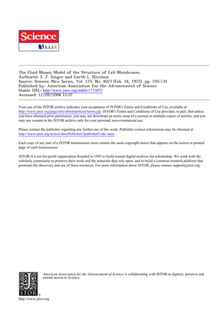
4.
Fig. 2. The lipid-globular protein mosaic model of membrane structure: schematic
cross-sectionalview. The phospholipids are depicted as in Fig. 1, and are arrangedas
a discontinuousbilayer with their ionic and polar heads in contact with water. Some
lipid may be structurallydifferentiatedfrom the bulk of the lipid (see text), but this
is not explicitly shown in the figure. The integral proteins, with the heavy lines repre-
senting the folded polypeptide chains, are shown as globular molecules partially em-
bedded in, and partially protruding from, the membrane. The protruding parts have
on their surfaces the ionic residues (- and +) of the protein, while the nonpolar
residues are largely in the embedded parts; accordingly,the protein molecules are am-
phipathic. The degree to which the integral proteins are embedded and, in particular,
whether they span the entire membrane thickness depend on the size and structure of
the molecules. The arrow marks the plane of cleavage to be expected in freeze-etching
experiments (see text). [From Lenard and Singer (3) and Singer (1)]
722
Two qualifications should be stressed,
however, concerning the bilayer form
of membrane lipids. (i) None of the
evidence so far obtained for the bilayer
form permits us to say whether the
bilayer is continuous or interrupted (1).
The calorimetrically observed phase
transitions, for example, occur over a
broad temperature interval, allowing the
possibility that the cooperative unit in-
volved in the phase transition is quite
small, consisting perhaps of only 100
lipid molecules on the average. (ii) None
of the experiments mentioned above is
sufficiently sensitive and quantitative to
prove whether 100 percent of the phos-
pholipid is in the bilayer form. It is
therefore not excluded that some signifi-
cant fraction of the phospholipid (per-
haps as much as 30 percent) is physi-
cally in a different state from the rest
of the lipid.
Protein-lipid interactions in mem-
branes. Several kinds of experiments
indicate that protein-lipid interactions
play a direct role in a variety of
membrane functions. Many membrane-
bound enzymes and antigens require
lipids, often specific phospholipids, for
the expression of their activities [see
table 2 in (21)]. Furthermore, the
nature of the fatty acids incorporated
into phospholipids affects the function
of certain membrane-bound proteins in
bacterial membranes (22).
On the other hand, the calorimetric
data discussed above give no significant
indication that the association of pro-
teins with the phospholipids of intact
membranes affects the phase transitions
of the phospholipids themselves. Ex-
periments with phospholipase C and
membranes have shown that the en-
zymic release of 70 percent of the
phosphorylated amines from intact
erythrocyte membranes profoundly
perturbs the physical state of the resid-
ual fatty acid chains, but has no detect-
able effect (as measured by circular
dichroism spectra) on the average con-
formation of the membrane proteins
(2). Such results therefore suggest that
the phospholipids and proteins of
membranes do not interact strongly; in
fact, they appear to be largely inde-
pendent.
This paradox, that different types of
experiments suggest strong protein-lipid
interactions on the one hand, and weak
or no interactions on the other, can be
resolved in a manner consistent with
all the data if it is proposed that, while
the largest portion of the phospholipid
is in bilayer form and not strongly
coupled to proteins in the membrane,
SCIENCE, VOL. 175
5. a small fraction of the lipid is more
tightly coupled to protein. With any
one membrane protein, the tightly
coupledlipid might be specific;that is,
the interaction might require that the
phospholipidcontain specificfatty acid
chains or particularpolar head groups.
There is at present, however, no satis-
factory direct evidence for such a dis-
tinctive lipid fraction. This problem is
consideredagain in connection with a
discussionof the experimentsof Wilson
and Fox (23).
Fluid Mosaic Model
Mosaic structure of the proteins and
lipids of membranes. The thermody-
namic considerationsand experimental
results so far discussedfit in with the
idea of a mosaic structurefor mem-
branes(1-3, 24) in whichglobularmol-
ecules of the integralproteins(perhaps
in particularinstances attachedto oli-
gosaccharides to form glycoproteins,
or interactingstronglywith specificlip-
ids to form lipoproteins)alternatewith
sections of phospholipidbilayer in the
cross sectionof the membrane(Fig. 2).
The globularproteinmoleculesarepos-
tulatedto be amphipathic(3, 4) as are
the phospholipids. That is, they are
structurallyasymmetric,withone highly
polar end and one nonpolar end. The
highlypolar regionis one in which the
ionic amino acid residuesand any co-
valently bound saccharideresiduesare
clustered,and which is in contactwith
the aqueous phase in the intact mem-
brane;the nonpolarregion is devoid of
ionic and saccharideresidues,contains
many of the nonpolarresidues,and is
embeddedin the hydrophobicinterior
of the membrane. The amphipathic
structure adopted by a particularin-
tegralprotein(or lipoprotein)molecule,
and thereforethe extent to which it is
embeddedin the membrane,are under
thermodynamiccontrol; that is, they
are determinedby the amino acid se-
quence and covalent structureof the
protein,and by its interactionswith its
molecularenvironment,so thatthe free
energyof the systemas a whole is at a
minimum.An integralproteinmolecule
with the appropriatesize and structure,
or a suitableaggregateof integralpro-
teins (below) may transversethe entire
membrane (3); that is, they have re-
gions in contact with the aqueous sol-
vent on both sides of the membrane.
It is clear from these considerations
that differentproteins,if they have the
appropriate amino acid sequence to
18 FEBRUARY 1972
adopt an amphipathicstructure,can be
integralproteinsof membranes;in this
manner, the heterogeneityof the pro-
teinsof most functionalmembranescan
be rationalized.
The sameconsiderationsmay also ex-
plainwhy someproteinsaremembrane-
bound and others are freely soluble in
the cytoplasm. The differencemay be
that either the amino acid sequenceof
the particularproteinallowsit to adopt
an amphipathic structure or, on the
contrary,to adopt a structurein which
the distributionof ionic groupsis nearly
spherically symmetrical,in the lowest
free energy state of the system. If the
ionic distributionon the protein sur-
face were symmetrical, the protein
wouldbe capableof interactingstrongly
with water all over its exteriorsurface,
thatis, it wouldbe a monodispersesol-
uble protein.
The mosaic structurecan be readily
diversified in several ways. Although
the nature of this diversificationis a
matterof speculation,it is importantto
recognizethatthe mosaicstructureneed
not be restrictedby the schematicrep-
resentation in Fig. 2. Protein-protein
interactionsthat are not explicitlycon-
sideredin Fig. 2 may be importantin
determiningthe propertiesof the mem-
brane. Such interactions may result
either in the specific binding of a
peripheralprotein to the exterior ex-
posed surface of a particularintegral
protein or in the associationof two or
more integralprotein subunitsto form
a specific aggregate within the mem-
brane. These features can be accom-
modatedin Fig. 2 without any changes
in thebasicstructure.
The phospholipids of the mosaic
structurearepredominantlyarrangedas
an interruptedbilayer,with their ionic
and polar head groups in contact with
the aqueous phase. As has been dis-
cussed, however,a small portionof the
lipid may be moreintimatelyassociated
with the integralproteins.This feature
is not explicitlyindicatedin Fig. 2. The
thicknessof a mosaic membranewould
vary alongthe surfacefrom that across
a phospholipid bilayer region to that
acrossa proteinregion,with an average
value that could be expected to corre-
spondreasonablywell to experimentally
measuredmembranethicknesses.
Matrix of the mosaic: lipid or pro-
tein?In the cross section of the mosaic
structurerepresentedin Fig. 2, it is not
indicatedwhetherit is the proteinor the
phospholipidthatprovidesthe matrixof
the mosaic.In otherwords,which com-
ponentis the mortar,which the bricks?
This question must be answeredwhen
the thirddimensionof the mosaic struc-
ture is specified. Trhesetwo types of
mosaic structure may be expected to
have very differentstructuraland func-
tional properties, and the question is
therefore a critical one. It is our hy-
Fig. 3. The lipid-globularproteinmosaicmodelwith a lipid matrix(the fluidmosaic
model); schematic three-dimensionaland cross-sectional views. The solid bodies with
stippled surfaces represent the globular integral proteins, which at long range are
randomly distributedin the plane of the membrane. At short range, some may form
specific aggregates, as shown. In cross section and in other details, the legend of
Fig. 2 applies.
723
6. pothesis that functional cell membranes
have a long-range mosaic structure with
the lipids constituting the matrix, as is
shown in Fig. 3. Supporting evidence is
discussed later. At this point, let us
consider some of the consequences of
this hypothesis.
1) There should generally be no long-
range order in a mosaic membrane with
a lipid matrix. By long range, we mean
over distances of the order of a few
tenths of a micrometer and greater.
Suppose we have a membrane prepara-
tion containing many different protein
species, and suppose further that 10,000
molecules of protein A are present in
the membrane of a single cell or or-
ganelle. How is protein A distributed
over the membrane surface? If the
membrane proteins formed the matrix
of the mosaic, defined by specific con-
tacts between the molecules of different
integral proteins, protein A might be
distributed in a highly ordered, two-
dimensional array on the surface. On
the other hand, if lipid formed the
matrix of the mosaic, there would be no
long-range interactions intrinsic to the
membrane influencing the distribution
of A molecules, and they should there-
fore be distributed in an aperiodic ran-
dom arrangement on the membrane
surface.
The absence of long-range order
should not be taken to imply an ab-
sence of short-range order in the mem-
brane. It is very likely that such short-
range order does exist, as, for example,
among at least some components of the
electron transport chain in the mito-
chondrial inner membrane. Such short-
range order is probably mediated by
specific protein (and perhaps protein-
lipid) interactions leading to the forma-
tion of stoichiometrically defined ag-
gregates within the membrane. How-
ever, in a mosaic membrane with a
lipid matrix, the long-range distribu-
tion of such aggregates would be ex-
pected to be random over the entire
surface of the membrane.
The objection may immediately be
raised that long-range order clearly
exists in certain cases where differen-
tiated structures (for example, synapses)
are found within a membrane. We sug-
gest, in such special cases, either that
short-range specific interactions among
integral proteins result in the formation
of an unusually large two-dimensional
aggregateor that some agent extrinsic
to the membrane (either inside or out-
side the cell) interacts multiply with
specific integral proteins to produce a
clustering of those proteins in a limited
724
area of the membrane surface. In
other words, we suggest that long-range
random arrangements in membranes are
the norm; wherever nonrandom distri-
butions are found, mechanisms must
exist which are responsible for them.
2) It has been shown that, under
physiological conditions, the lipids of
functional cell membranes are in a
fluid rather than a crystalline state.
(This is not true of myelin, however.)
This evidence comes from a variety of
sources, such as spin-labeling experi-
ments (25), x-ray diffraction studies
(18), and differential calorimetry (16,
17). If a membrane consisted of integral
proteins dispersed in a fluid lipid matrix,
the membrane would in effect be a two-
dimensional liquid-like solution of mon-
omeric or aggregated integral proteins
(or lipoproteins) dissolved in the lipid
bilayer. The mosaic structure would be
a dynamic rather than a static one. The
integral proteins would be expected to
undergo translational diffusion within
the membrane, at rates determined in
part by the effective viscosity of the
lipid, unless they were tied down by
some specific interactions intrinsic or
extrinsic to the membrane. However,
because of their amphipathic structures,
the integral proteins would maintain
their molecular orientation and their
degree of intercalation in the membrane
while undergoing translational diffusion
in the plane of the membrane (as dis-
cussed below).
In contrast, if the matrix of the mo-
saic were constituted of integral pro-
teins, the long-range structure of the
membrane would be essentially static.
Large energies of activation would be
required for a protein component to
diffuse in the plane of the membrane
from one region to a distant one be-
cause of the many noncovalent bonds
between the proteins that would have
to be simultaneously broken for ex-
change to take place. Therefore, a
mosaic membrane with a protein ma-
trix should make for a relatively rigid
structure with essentially no transla-
tional diffusion of its protein compo-
nents within the membrane.
From the discussion in this and the
previous section, it is clear that the
fluid mosaic model suggests a set of
structural properties for functional
membranes at least some of which can
be tested experimentally. In an earlier
article (1), a large body of experimen-
tal evidence was examined for its rele-
vance to models of membrane structure.
It was concluded that a mosaic struc-
ture was most consistent with the avail-
able evidence. Some more recent re-
sults, however, bear even more directly
on the problem, and only this evidence
is discussed below.
Some Recent Experimental Evidence
Evidence for proteins embedded in
membranes. One proposal of the fluid
mosaic model is that an integral pro-
tein is a globular molecule having a
significant fraction of its volume em-
bedded in the membrane. The results
of recent freeze-etching experiments
with membranes strongly suggest that a
substantial amount of protein is deeply
embedded in many functional mem-
branes. In this technique (26) a frozen
specimen is fractured with a microtome
knife; some of the frozen water is sub-
limed (etched) from the fractured sur-
face if desired; the surface is then
shadow cast with metal, and the surface
replica is examined in the electron mi-
croscope. By this method the topog-
raphy of the cleaved surface is re-
vealed. A characteristic feature of the
exposed surface of most functional
membranes examined by this technique,
including plasmalemmal, vacuolar, nu-
clear, chloroplast, mitochondrial, and
bacterial membranes (27, 28), is a
mosaic-like structure consisting of a
smooth matrix interrupted by a large
number of particles. These particles
have a fairly characteristic uniform
size for a particular membrane, for
example, about 85-A diameter for eryth-
rocyte membranes. Such surfaces re-
sult from the cleavage of a membrane
along its interior hydrophobic face
(29). This interior face (Fig. 2) corre-
sponds to the plane indicated by the
arrow. If cleavage were to occur
smoothly between the two layers of
phospholipid in the bilayer regions, but
were to circumvent the protein mole-
cules penetrating the mid-plane of the
membrane, then the alternating smooth
and particulate regions observed on the
freeze-etch surfaces can be readily ex-
plained by a mosaic structure for the
membrane (Fig. 2), provided that the
particles can be shown to be protein
in nature. That the particles are indeed
protein has been suggested by recent
experiments (30).
Another consequence of the mosaic
model, suggested from its inception
(3), is that certain integral proteins pos-
sessing the appropriate size and struc-
ture may span the entire thickness of
the membrane and be exposed at both
membrane surfaces. Chemical evidence
SCIENCE, VOL. 175
7. that a trans-membraneprotein, whose
molecular weight is about 100,000, is
present in large amountsin the human
erythrocyte membrane has been ob-
tained by two independent methods-
one involving proteolysis of normal
compared to everted membranes(31),
and the other specificchemicallabeling
of themembraneproteins!(32).
Distribution of components in the
plane of the membrane.A prediction
of the fluid mosaic model is that the
two-dimensionallong-rangedistribution
of any integralprotein in the plane of
the membrane is essentially random.
To test this prediction,we have devel-
oped and applied electron microscopic
techniquesto visualize the distribution
of specific membrane antigens over
large areas of their membranesurfaces
(33) and have so far studied the dis-
tribution *of the Rhd(D) antigen on
humanerythrocytemembranes(34),and
of H-2 histocompatibilityalloantigens
on mouse erythrocytemembranes(35).
In the case of the Rho(D) antigen,
for example, cells of 0, Rh-positive
type were reacted with a saturating
amount of 125I-labeledpurifiedhuman
antibodyto Rho(D) [anti-Rlho(D)],and
the treated(sensitized)cells were lysed
at an air-waterinterface, so that the
cell membraneswere spread out flat.
The flattened membranes, after being
picked up on an electron microscope
grid, were treatedwith the specific"in-
direct stain," ferritin-conjugatedgoat
antibodiesspecific for human y-globu-
lin. Thus,whereverthe humananti-Rho
(D) molecules were bound to the Rho
(D) antigen on the membranesurface,
the ferritin-labeledgoat antibodiesbe-
came specifically attached. In other
words, the human y-globulin antibody
now functioned as an antigen for the
goat antibodies (Fig. 4). The ferritin
was distributedin discreteclusters,each
containing two to eight ferritin mole-
clues within a circle of radius about
300 A. The numblersof such clusters
per unit area of the membranesurface
correspondedto the number of 125I-
labeled human anti-Rho(D)molecules
boundper unit area.This indicatesthat
each ferritin cluster was bound to a
singleanti-Rho(D)molecule,and a clus-
ter represents the number of goat
antibody molecules bound to a single
humany-globulinmolecule. Each clus-
ter therefore corresponds to a single
Rho(D) antigen site (36) on the mem-
brane. Since the clusters were distrib-
uted in a random array, we conclude
that the Rho(D)antigen,which exhibits
propertiesof an integral protein (37),
is molecularlydispersedand is distrib-
utedin a random,two-dimensionalarray
on the human erythrocytemembrane.
Similarexperimentswere carriedout
with the H-2 alloantigenic sites on
mouse erythrocytemembranes.In this
case (Fig. 5) the clusters -of ferritin
moleculesof the indirectstainwere not
isolated, as in the case of the Rho(D)
antigen,butinsteadoccurredin patches.
The patchy distribution of the H-2
histocompatibilityalloantigenicsiteshad
earlierbeen observedby differenttech-
niques (38), but the two-dimensional
distributionof the patchescould not be
ascertained. In our experiments, the
patches contained variablenumbersof
clusters, and were arrangedin an ir-
regular two-dimensionalarray on the
membrane surface. The histocompati-
bility antigen appears to be glycopro-
teinin nature(39). The long-rangedis-
trilbutionof both the Rho(D) and H-2
histocompatibilityantigenson their re-
spective membranesurfaces, therefore,
Fig. 4 (left). The outer membranesurface of an Rh-positive humanerythrocytesensitizedwith human anti-Rho(D) and stainedwith
ferritin-conjugatedgoat antibody to human y-globulin.The cells were first labeled to saturationwith purifiedlIL-labeledhuman anti-
body to Rho(D) and then lysed at an air-water interface. The erythrocyte membrane ghosts, flattened by surface forces (inset,
low magnification) were picked up on a coated, electron microscopegrid and indirectly stained with ferritin-conjugatedgoat anti-
bodies to human y-globulin.The ferritin appearsbound to the membranein discreteclusters of two to eight ferritin-conjugates;each
clusteris circumscribedby a circle of radius300 A. The numberof such clustersper cell (9300) is equal within experimentalerror to
the number of "2I-labeledhuman antibody to Rho(D) molecules bound per cell (10,200). Each cluster -thereforecorresponds
to an individual Rho(D) antigenic site. Scale is 0.1 tum;inset scale is i ,tm. [From Nicolson, Masouredis,and Singer (34)] Fig.
5 (right). The outer membrane surface on a mouse erythrocyte (H-2b) sensitized with alloantibodies against H-2b histocompatibil-
ity antigens and stained with ferritin-conjugatedantibodies against 7S mouse r-globulin. The procedures are the same as listed in
the legend to Fig. 4. The ferritin-antibodyclusters are present in randomly spaced "patches"of variable size on the membrane sur-
face. Scale is 0.1 ,um.[From Nicolson, Hyman, and Singer (35)]
18 FEBRUARY 1972 725
8. are in accordwith the predictionof the
fluid mosaic model that the integral
proteins of membranes are randomly
arrangedin two dimensions.
The particleson the innermembrane
faces revealedby freeze-etchingexperi-
ments, which (as discussed above) are
probablyproteinin nature,aregenerally
also relatively randomly distributedin
two dimensions.
Evidence that proteins are in a
fluid state in intact membranes. An im-
portant series of experimentshas been
carried out (12, 40-44) with receptor
disk membranesfrom the retinaof the
frog. This membranesystemis unusual
in that it contains as its predominant,
if not only, proteincomponentthe pig-
ment rhodopsin.In electronmicroscopy
of the negativelystainedsurfacesof the
dried membranes,a somewhat tightly
packed and ordered array of par-
ticles (about40 A) was observed.These
particles are the individual rhodopsin
molecules. Although the earlierstudies
suggested that there was a long-range
orderin the distributionof the particles
(40), more recentx-ray diffractiondata
(42) on pellets of wet, receptor disk
membranes showed that only a few
ordersof reflectionwere observedcor-
respondingto the spacings of the rho-
dopsin molecules in the plane of the
membrane.This indicated that a non-
crystalline, aperiodic arrangement of
the particlesexisted in the plane of the
membrane.Furthermore,the tempera-
ture dependence of the characteristics
of the x-ray diffractionmaxima were
consistent with the suggestionthat the
particles were in a planar liquid-like
statein theintactmembrane.,Additional
supportfor the existenceof this liquid-
like state was the observationthat the
absorptionof a foreign protein (bovine
serumalbumin)to the membranecould
definitely alter the x-ray spacings due
to the rhodopsinparticles;that is, the
distributionof the rhodopsinmolecules
in the plane of the membranewas rad-
ically altered by the weak binding of
the albumin.This alterationwould not
be expected if a rigid lattice structure
of Itherhodopsin molecules, or aggre-
gates, were presentin the plane of the
membrane,
These studies are particularlynote-
worthy because they involved a mem-
brane which, by conventionalelectron
microscopictechniques,appearsto show
long-rangeperiodicity over its surface.
Otherspecializedmembraneshave also
exhibitedorderedelectronmicrographic
imagesof theirsurfaces[compare(43)].
However,it is likelythat a very concen-
726
tratedtwo-dimensionalfluid solution of
identicalproteinmoleculeswill appear,
when dried, to be arrangedin an or-
dered array, particularlywhen optical
tricksare used to enhancethe apparent
order(43). Whatis really a fluidphase
may thereforeartifactuallybe made 'to
appear as a crystallinesolid. This ap-
pears to be the situationwith the reti-
nal receptordisk membranes.
A major contributionto membrane
studies has been made by Frye and
Edidin(44), who investigatedthe mem-
brane properties of some cell fusion
heterokaryons.Human and mouse cells
in culture were induced to fuse with
one another, with Sendai virus as the
fusingagent.The distributionof human
and mouse antigeniccomponentsof the
fused cell membraneswas then deter-
minedby immunofluorescence,with the
use of rabbitantibodiesdirectedto the
whole human cells, mouse antibodies
directedagainstthe H-2 alloantigenon
the mouse cell membranes,and, as in-
direct stains, goat antiserumto rabbit
y-glolbulinand goat antiserumto mouse
y-globulin labeled with two different
fluorescentdyes. Shortly after cell fu-
sion, the mouse and human antigenic
componentswere largely segregatedin
differenthalves of the fused cell mem-
branes;but after about 40 minutes at
37?C the componentswere essentially
completely intermixed. Inhibitors of
protein synthesis,of adenosinetriphos-
phate (ATP) formation, and of gluta-
mine-dependentsyntheticpathways,ap-
plied before or aftercell fusion, had no
effect on the rate of this intermixing
process, but lowering the temperature
below 15?C sharplydecreasedit.
Frye and Edidin (44) suggest that
the intermixingof membrane compo-
nents is due to diffusionof these com-
ponents within the membrane, rather
than to their removal and reinsertion,
or to the synthesis and insertion of
new copies of these components, into
the heterokaryonmembrane.An unex-
plainedfindingof theseexperimentswas
the fairly frequentoccurrence,at early
andintermediatetimesaftercell fusion,
of heterokaryonmembranesin which
the human antigeniccomponentswere
uniformly distributed over the mem-
brane surface but the mouse compo-
nents were still largely segregated to
about half the membrane (Ml/2-H1
cells). On the other hand, the reverse
situation, with the mouse antigenic
componentsuniformlyspreadout over
the membraneand the human compo-
nents segregated (M1-H1/2), was only
rarelyobserved.This resultcan now be
explained by a diffusion mechanism for
the intermixingprocess,as follows. The
antibodiesto the humancell membrane
were no doubt directedto a heteroge-
neous set of antigens,whereasthe anti-
bodies itothe mouse cell were directed
specifically to the histocompatibility
alloantigen.However,the histocompati-
bility antigensoccur as largeaggregates
in the membrane (Fig. 5), and might
therefore be expected to diffuse more
slowlythana complexmixtureof largely
unaggregated human antigens in the
membrane.Thus, at appropriateinter-
mediate times after cell fusion, signifi-
cant numbersof (M1/2-HI)but not of
(Ml-HI/2) fused cells might appear,to
be converted at longer times to cells
withcompletelyintermixedcomponents.
A rough estimate may be made of
the averageeffective diffusionconstant
requiredof the membranecomponents
to accountfor the kineticsof intermix-
ing in the Frye-Edidin experiments.
Taking the average distance of migra-
tion, x, to have been about 5 micro-
metersin a time, t, of 40 minutesgives
an apparentdiffusionconstant,D=x2/
2t, of 5 X 10-11 cm2/sec. For com-
parison,the diffusionconstantof hemo-
glob,in in aqueous solutions is about
7 X 10⁻⁷ cm2/sec. The apparent effec-
tive viscosity of the membrane fluid
phase is therefore about 103 to 104
times that of water.
The Frye-Edidinexperimentscan be
rationalizedby the fluid mosaic model
of membranestructureas being the re-
sultof thefree diffusionandintermixing
of the lipids and the proteins (or lipo-
proteins) within the fluid lipid matrix.
Some experiments,however, appear
to suggestthat the lipids of membranes
are not readily interchangeablewithin
the membrane and are therefore not
free to diffuse independently.For ex-
ample, Wilson and Fox (23) have
studied the induction of /l-galactoside
and 8/-glucoside transport systems in
mutants of Escherichia coli that cannot
synthesizeunsaturatedfatty acids. Such
fatty acids can be lincorporatedinto
phospholipids,however,if they are sup-
plied in the growthmedium.Whencells
weregrownin particularfatty acid sup-
plements and induced for the synthesis
of the transportsystems, the effect of
temperatureon the transportrate was
characteristicof thatfatty acid.If, then,
the cells were first grown in medium
containingoleic acid and thenshiftedto
growthin a mediumsupplementedwith
linoleic acid during a brief period of
inductionof either of the transportsys-
tems,theeffectof temperatureon trans-
SCIENCE, VOL. 175
9. port was said to be characteristic of cells
grown continually in the linoleic acid
medium. In other words, although most
of the phopholipids of the membrane
contained oleic acid chains, these did
not appear to exchange with the newly
synthesized small amounts of phospholi-
pids containing linoleic acid chains.
These experiments, however, do not
necessarily contradict the thesis that
most of the phospholipids of membranes
are freely diffusible and, hence, ex-
changeable. For example, each of the
two transport systems might be or-
ganized in the membrane as a specific
protein aggregate containing intercal-
ated and strongly bound phospholipid
components. If such lipoprotein aggre-
gates had first to be assembled in order
to be incorporated into the bulk lipid
matrix of the membrane, the results of
Wilson and Fox would be anticipated.
In particular, the small fraction of
the membrane phospholipid that was
strongly bound, and perhaps segregated
in such aggregates from the bulk of the
membrane lipid, might not exchange
rapidly with the bulk lipid. The Wilson-
Fox experiments therefore do not re-
quire that the major part of the mem-
brane phospholipid be static, but only
that a small fraction of the lipids be
structurally differentiated from the rest.
The structural differentiation of some
of the membrane lipid by strong bind-
ing to integral proteins is a possibility
that was discussed above.
The observations of Wilson and Fox,
that there is a significant coupling of
lipid and protein incorporation into
membranes, appear to be a special case.
The experiments of Mindich (45) dem-
onstrate that more generally lipid and
protein incorporation into bacterial
membranes can occur independently,
and 'thatquite wide variations in the ratio
of lipids and proteins in the membrane
can be produced in vivo, as might be
expected from the fluid mosaic model
of membrane structure.
The asymmetry of membranes. A
substantial amount of evidence has ac-
cumulated showing that the two sur-
faces of membranes are not identical
in composition or structure. One aspect
of this asymmetry is the distribution of
oligosaccharides on the two surfaces of
membranes. There exist plant proteins,
called lectins or plant agglutinins, which
bind to specific sugar residues, and, as
a result, can cause the agglutination of
cells bearing the sugar residues on their
surfaces. By conjugating several such
agglutinins to ferritin, we have been able
to visualize the distribution of oligosac-
18 FEBRUARY 1972
charides on membranes in the electron
microscope (33). For example, the fer-
ritin conjugate of concanavalin A, a
protein agglutinin that binds specifically
to terminal a-D-glucopyranosyl or a-D-
mannopyranosyl residues (46), attaches
specifically to the outer surface of eryth-
rocyte membranes and not at all to
the inner cytoplasmic surface (33). A
similar, completely asymmetric distri-
bution of ferritin conjugates of ricin (a
protein agglutinin) on the membranes
of rabbit erythrocytes is shown in Fig.
6. Ricin binds specifically to terminal
f/-D-galactopyranosyl and sterically re-
lated sugar residues (47). Such asym-
metry has now been observed with
several ferritin-conjugated agglutinins
and a number of different mammalian
cell plasma membranes (48). These find-
ings extend earlier results obtained by
different methods (49).
The foregoing observations bear on
many problems, including cell-cell inter-
actions and membrane biogenesis (50).
In the context of this article, however,
the absence of oligosaccharides on in-
ner membrane surfaces indicates that
rotational transitions of the glycopro-
teins of erythrocyte and other plasma
membranes from the outer to the inner
surfaces must occur at only negligibly
slow rates. This conclusion probably
applies to membrane proteins other
than glycoproteins; for example, the
Na,K-dependent and Mg-dependent
adenosine triphosphatase activities of
erythrocyte membranes are exclusively
localized to the inner cytoplasmic sur-
faces (51). Individual molecules of spin-
labeled zwitterionic and anionic phos-
pholipids also exhibit very slow inside-
outside transitions in synthetic vesicles
of phospholipid bilayers (52). The very
slow or negligible rates of such transi-
tions can be explained by the mosaic
model and the thermodynamic argu-
ments already discussed. If the integral
proteins (including the glycoproteins)
in intact membranes have, like the phos-
pholipids, an amphipathic structure, a
large free energy of activation would
be required to rotate the ionic and polar
regions of the proteins through the
hydrophobic interior of the membrane
to the other side.
To accommodate the fluid mosaic
model to these conclusions concerning
asymmetry, we specify that, while the
two-dimensional translational diffusion
of the integral proteins and the phos-
pholipids of membranes occurs freely,
Fig. 6. The inner (i) and outer (o) membrane surfaces of a rabbit erythrocyte mem-
branethat has been stained with ferritin-conjugatedricin. In preparingmembranespeci-
mens such as are shown in Figs. 4 and 5, occasionally a cell lyses with membrane
rupturesuch that both inner and outer surfaces of the membrane are exposed. In this
case the mounted membranewas stainedwith ferritin conjugatedto ricin, a plant agglu-
tinin that specifically binds to terminal p-D-galactopyranosyland sterically related
terminalsugarresidues in oligosaccharides.The ferritin-agglutininis found on the outer
membrane surface only. The scale is equivalent to 0.1 ,m; the insert scale is equiva-
lent to 1 Am.
727
10. the rotational diffusion of these com-
ponents is generally restricted to axes
perpendicular to the plane of the mem-
brane; that is, in general, molecular
tumbling does not occur at significant
rates within the membrane. The asym-
metry of the membrane introduces
another factor into the problem of
translational diffusion of membrane
components. In the experiments of Frye
and Edidin (44) only those membrane
antigens exposed at the outer surface of
the membrane were labeled by fluores-
cent antibodies, and the conclusion that
these particular antigens were mobile in
the plane of the membranes therefore,
strictly speaking, applies only to those
components accessible at the outer sur-
face. Whether components confined to
the inner surfaces also intermix and
diffuse should be separately established.
Thus, recent evidence obtained with
many experimental methods and differ-
ent kinds of functional membrane sys-
tems is entirely consistent with the pre-
dictions of the fluid mosaic model of
membrane structure and provides strong
support for the model. It seems amply
justified, therefore, to speculate about
how a fluid mosaic structure might
carry out various membrane functions,
and to suggest specific mechanisms for
various functions that can be subjected
to experimental tests.
The Fluid Mosaic Model and
Membrane Functions
The hypothesis that a membrane is
an oriented, two-dimensional, viscous
solution of amphipathic proteins (or
lipoproteins) and lipids in instantaneous
thermodynamic equilibrum, leads to
many specific predictions about the
mechanisms of membrane functions.
Rather than catalog a large number of
these, we suggest some directions that
such speculations may usefully take.
Among these problems are nerve im-
pulse transmission, transport through
membranes, and the effects of specific
drugs and hormones on membranes (1).
The fluidity of the mosaic structure,
which introduces a new factor into such
speculations, is emphasized here. This
new factor may be stated in general
form as follows. The physical or chem-
ical perturbation of a membrane may
affect or alter a particular membrane
component or set of components; a re-
distribution of membrane components
can then occur by translational diffu-
sion through the viscous two-dimen-
728
sional solution, thereby allowing new
thermodynamic interactions among the
altered components to take effect. This
general mechanism may play an im-
portant role in various membrane-me-
diated cellular phenomena that occur
on a time scale of minutes or longer.
Much more rapidly occurring phenom-
ena, such as nerve impulse transmission,
would find the mosaic structure to be a
static one, insofar as translational diffu-
sion of the membrane components is
concerned. In order to illustrate the
concepts involved, we discuss two spe-
cific membrane phenomena.
Malignant transformation of cells and
the "exposure of cryptic sites." Normal
mammalian cells grown in monolayer
culture generally exhibit "contact in-
hibition"; that is, they divide until they
form a confluent monolayer and they
then stop dividing. Cells that have be-
come transformed to malignancy by
oncogenic viruses or by chemical car-
cinogens lose the property of contact
inhibition; that is, they overgrow the
monolayer. For some time, this experi-
mental finding has been thought to re-
flect the difference between the normal
and the malignant states in vivo, and
to be due to differences in the surface
properties of normal and malignant
cells. Much excitement and investiga-
tive activity therefore attended the dem-
onstration (53, 54) that malignant trans-
formation is closely correlated with a
greatly increased capacity for the trans-
formed cells to be agglutinated by sev-
eral saccharide-binding plant aggluti-
nins. Furthermore, mild treatment of
normal cells with proteolytic enzymes
can render them also more readily ag-
glutinable by these protein agglutinins.
Burger (54) has suggested, therefore,
that the agglutinin-binding sites are pres-
ent on the membrane surfaces of nor-
mal cells but are "icryptic" (Fig. 7A)
(that is, they are shielded by some other
membrane components from effectively
participating in the agglutination proc-
ess), and that proteolytic digestion of
normal cells or the processes of malig-
nant transformation "exposes" these
cryptic sites on the membrane surface.
In some cases, quantitative binding
studies have indeed indicated that no
significant change in the numbers of
agglutinin-binding sites on the mem-
brane accompanies either mild pro-
teolysis of normal cells or malignant
transformation (55).
An alternative explanation of these
phenomena (Fig. 7B), based on the
fluid mosaic model of membrane struc-
ture, may be proposed. Consider first
the proteolysis experiments with nor-
mal cells. Suppose that the integral gly-
coproteins in the normal cell mem-
brane are molecularly dispersed in the
fluid mosaic structure. It is likely that
mild proteolysis would preferentially
release a small amount of glycopeptides
and other polar peptides from these
proteins because these are the most
exposed portions of the integral pro-
teins at the outer surface of the mem-
brane (Figs. 2 and 3). The remaining
portions of these proteins may still
contain a large fraction of their original
oligosaccharide chains after the limited
proteolysis, but the release of some of
the more polar structures would make
the remaining portions more hydro-
phobic. As these more hydrophobic
glycoproteins diffused in the membrane,
they might then aggregate in the plane
of the membrane. The result would be
a clustering of the agglutinin-binding
sites on the enzyme-treated cell sur-
face, as compared to the normal un-
treated surface. Such clustering (with
no increase, or perhaps even a decrease
in the total numbers of sites because
of digestion) could enhance the agglu-
tination of the treated cells, as com-
pared to that of normal cells, because
it would increase the probability of
agglutinin bridges forming between the
surfaces of two cells.
In malignant transformation, distinct
chemical changes in the glycolipids and
the glycoproteins of the cell membrane
are known to occur (56), and the en-
hanced agglutinability of the transform-
ed cells may be much more complicated
than is the case in the proteolysis of
normal cells. If, however, the two phe-
nomena do have a basic feature in com-
mon, it could be a similar clustering of
saccharide-binding sites on the trans-
formed and the enzyme-treated normal
cells. In malignant transformation, such
clustering could be the result of the
chemical changes in the membrane
mentioned above; or some virus-induced
gene product (57) may be incorporated
into the cell membrane and serve as a
nucleus for the aggregation of the ag-
glutinin-binding glycoproteins within the
membrane.
These suggestions can be tested ex-
perimentally by the use of ferritin-con-
jugated agglutinins (33) as already dis-
cussed (Fig. 6). The prediction is that
with normal cells subjected to mild
proteolysis, and also with malignant
transformed cells, the total number of
ferritin-agglutinin particles specifically
SCIENCE, VOL. 175
11. boundto the outersurfacesof the cells
mightnotbe greatlydifferentfromthose
of normal cells, but larger clusters of
ferritin particles would be found.
Cooperative phenomena in mem-
branes. By a cooperativephenomenon
we mean an effect which is initiated
at one site on a complex structureand
transmittedto another remote site by
some structuralcoupling between the
two sites. A number of important
membrane phenomena may fall into
this category. However, before enum-
eratingthem, we should first discrimi-
nate between two types of cooperative
effects that may occur. These can be
termed trans and cis. Trans effects refer
to cooperative(allosteric) changesthat
havebeenpostulatedto operateat some
localized region on the membranesur-
face, to transmitaneffectfrom one side
of the membraneto the other (58). For
example,fanintegralproteinmayexistin
themembraneasanaggregateof two (or
more)subunits,one of whichis expc,sed
to the aqueoussolutionat the outersur-
face of the membrane,and the other
is exposedto the cytoplasmat the inner
surface.The specificbindingof a drug
or hormone molecule to the active site
of the outward-orientedsubunit may
induce a conformationalrearrangement
withintheaggregate,andtherebychange
some functionalpropertyof the aggre-
gate or of its inward-orientedsubunit.
By cis effects, on the other hand, we
refer to cooperativechanges that may
be producedover the entiremembrane,
or at least large areasof it, as a conse-
quence of some event or events occur-
ring at only one or a few localized
points on the membranesurface. For
example, the killing effects of certain
bacteriocinson bacteria(59), the lysis
of the cortical granules of egg cells
upon fertilization of eggs by sperm
(60), and the interaction of growth
hormone with erythrocyte membranes
(61) are cases which may involve
transmissionand amplificationof local-
ized events over the entire surface of
a membrane. These phenomena may
not all occur by the same or related
mechanisms,but in at least two experi-
mentalstudies,that involvingthe inter-
action of colicin E1 with intact Esche-
richiacoli cells (62), andthatof human
growth hormone and isolated human
erythrocytemembranes (61), there is
substantialevidencethatlong-rangecis-
type cooperativeeffects intrinsicto the
membranesare involved.
The questionwe now addressis, How
might such cis effectswork?Changeux
18 FEBRUARY 1972
and his co-workers (63) have pro-
posed an extension to membranesof
the Monod-Wyman-Changeuxallosteric
model of protein cooperativephenom-
ena, using as a model of membrane
structure an infinite two-dimensional
aggregateof identical lipoprotein sub-
units [as, for example, the model de-
scribedby Benson (20)]. In this theo-
reticaltreatment,the individualsubunits
are capableof existingin eitherof two
conformational states, one of which
has a much larger binding affinityfor
a specific ligand than does the other.
The bindingof a singleligandmolecule
to any one subunit then triggers the
cooperativeconversionof many of the
subunits to the ligand-bound confor-
mation,in orderto maximizethe inter-
actions among the subunits.
This theory as presented relies on
the membranemodel used.If, however,
the membraneis not a two-dimensional
aggregateof lipoprotein subunits, but
is instead a fluid mosaic of proteins
and lipids, the physicalsituationwould
be quite different.The basic theory of
Changeuex et al. (63) might still be
formally applicable, but with impor-
tant changesin physicalsignificance.It
is possible,for example,that a particu-
lar integralprotein can exist in either
of two conformationalstates, one of
which is favored by ligand binding;in
its normal unbound conformationthe
integral protein is monomolecularly
dispersedwithin the membrane,but in
the conformationpromoted by ligand
binding, its aggregation is thermody-
namically favored. The binding of a
ligand molecule at one integralprotein
site, followed by diffusionof the non-
ligandedprotein molecules to it, might
then lead to an aggregationand simul-
taneouschange in conformationof the
aggregated protein within the mem-
brane.This mechanismcould result in
a long-rangecis-type cooperativephe-
nomenon, if the eventual aggregate
size was very large and if its presence
produced local perturbations in the
propertiesof the membrane.However,
A
B
w v
Fig. 7. Two differentmechanismsto explainthe findingsthateithermalignantlytrans-
formedcells or normalcells that are subjectedto mild proteolysisbecome much
morereadilyagglutinableby severalplantagglutinins.(A) The mechanismof Burger
(54): agglutinin-bindingsitesthatarepresenton the surfacesof normalcells, but are
obstructed("crypticsites"),are exposedby proteolysisor the processesof malignant
transformation.(B) The redistributionmechanism(see text): the agglutininsites on
normalcell surfacesare largelymonomolecularlydispersedin the fluidmosaicstruc-
ture, but on proteolysisor malignanttransformation,they diffuseand aggregatein
clusters.The probabilityof agglutinationof two such modifiedcells is enhancedby
the clusteringof bindingsites.
729
12. the transitionwould occur at a rate
and over a time period determinedby
the rate of diffusion of the molecules
of the integral protein in the fluid
mosaic membrane.This time period is
likely to be relativelylong, of the order
of minutes (44), as alreadymentioned.
On the other hand, if cis-type cooper-
ative effects occurred in a lipoprotein
subunitmodel accordingto the mecha-
nism postulated by Changeux et al.
(63), one would expect the coopera-
tive change to be much faster. Con-
formation changes in the soluble allo-
steric protein aspartyltranscarbamylase,
for example, have half-times of the
order of 10 milliseconds (64). It is
therefore of some interest that in the
studies of the interaction of colicin
E1 andE. coli the fluorescencechanges
that marked the apparentcis-type co-
operative transitionsin the cell mem-
brane occurred over intervals of one
to several minutes (62). If this sug-
gested mechanismfor the colicin effect
is valid, one would predict that (i)
freeze-etchingexperimentson the coli-
cin-treatedbacteria (28) might reveal
an aggregationof normally dispersed
particles at the inner membraneface,
or (ii) changes in membranefluidity,
such as would be producedby suitable
changes in temperatureor by different
compositions of membrane phospho-
lipids (65), might markedlyaffect the
kinetics of the fluorescence changes
that are observed on addition of the
colicin to the bacteria.
In this discussionof membranefunc-
tions, some detailed mechanisms to
accountfor two membranephenomena
have been presented.It may well turn
out that these mechanisms are incor-
rect. Our objecthas been not so much
to arguefor these specificmechanisms,
as to illustrate that the fluid mosaic
model of membranestructurecan sug-
gest novel ways of thinking about
membrane functions-ways that are
amenable to experimentaltests. Other
membrane phenomena may be influ-
encedbysimilardiffusionalmechanisms:
for example, cell-cell and cell-sub-
strateinteractions,wherethe apposition
of intense local electric fields to a cell
membranemay affect the distribution
of electricallychargedintegralproteins
within the membranes;or the specific
bindingof multivalentantibodyto cell
surface antigens, where the simultane-
ous binding of one antibodymolecule
to several molecules of the antigen
may induce rearrangementsof the dis-
tributionof the antigenin the plane of
730
the membrane,an effect that may be
involved in the phenomenon of anti-
genic modulation(66). Thereareother
specific examples as well.
It may well be that a number of
critical metabolic functions performed
by cell membranes may require the
translationalmobility of some impor-
tant integral proteins. This could be
the ultimate significance of the long-
standing observation (67) that the
membranelipidsof poikilothermicorga-
nisms contain a larger fraction of un-
saturated fatty acids the lower their
temperature of growth. Appropriate
enzymes apparentlycarry out the nec-
essary biochemical adjustment (68)
that keeps the membrane lipids in a
fluidstate at the particulartemperature
of growth; if these enzymes are not
functional, for example, because of
mutations, the organism-to grow at
the lower temperature(65)-must be
suppliedwith the unsaturatedfatty acid
exogenously. While it has been sug-
gested before that the maintenanceof
lipid fluidity may be important to
carriermechanismsoperatingacross a
functionalmembrane,it is also possible
that the real purpose of fluidity is to
permit some critical integral proteins
to retaintheir translationalmobility in
the plane of the membrane, as an
obligatory step in their function.
Summary
A fluid mosaic model is presented
for the grossorganizationand structure
of the proteinsand lipids of biological
membranes. The model is consistent
with the restrictionsimposed by ther-
modynamics. In this model, the pro-
teins that are integralto the membrane
are a heterogeneous set of globular
molecules,each arrangedin an amphi-
pathic structure,that is, with the ionic
and highly polar groups protruding
from the membrane into the aqueous
phase, and the nonpolargroupslargely
buried in the hydrophobicinterior of
themembrane.Theseglobularmolecules
are partiallyembeddedin a matrix of
phospholipid.The bulk of the phosplho-
lipid is organized as a discontinuous,
fluid bilayer, althougha small fraction
of the lipid may interact specifically
with the membraneproteins.The fluid
mosaic structureis therefore formally
analogous to a two-dimensional ori-
ented solution of integralproteins (or
lipoproteins) in the viscous phospho-
lipid bilayer solvent. Recent experi-
ments with a wide varietyof techniques
and several different membrane sys-
tems are described, all of which are
consistent with, and add much detail
to, the fluidmosaic model. It therefore
seems appropriateto suggest possible
mechanisms for various membrane
functions and membrane-mediated
phenomenain the light of the model.
As examples, experimentally testable
mechanismsare suggestedfor cell sur-
face changes in malignanttransforma-
tion, and for cooperative effects ex-
hibitedin the interactionsof membranes
with some specific ligands.
Note added in proof: Since this ar-
ticle was written, we have obtained
electronmicroscopicevidence (69) that
the concanavalin A binding sites on
the membranes of SV40 virus-trans-
formed mouse fibroblasts (3T3 cells)
are more clusteredthan the sites on the
membranesof normalcells, as predicted
by the hypothesis representedin Fig.
7B. Therehas also appeareda studyby
Taylor et al. (70) showing the re-
markableeffects produced on lympho-
cytes by the addition of antibodiesdi-
rected to their surface immunoglobulin
molecules. The antibodiesinduce a re-
distribution and pinocytosis of these
surfaceimmunoglobulins,so thatwithin
about 30 minutes at 37?C the surface
immunoglobulinsare completely swept
out of the membrane.These effects do
not occur,however,if the bivalentanti-
bodies are replaced by their univalent
Fab fragments or if the antibody ex-
periments are carried out at 0?C in-
steadof 37?C. These andrelatedresults
stronglyindicatethat the bivalentanti-
bodies produce an aggregationof the
surface immunoglobulin molecules in
the plane of the membrane,which can
occuronly if the immunoglobulinmole-
cules are free to diffuse in the mem-
brane.This aggregationthen appearsto
triggeroff the pinocytosisof the mem-
brane components by some unknown
mechanism. Such membrane transfor-
mations may be of crucial importance
in the induction of an antibody re-
sponse to an antigen, as well as in
otherprocessesof cell differentiation.
References and Notes
1. S. J. Singer, in Structure and Function of
Biological Membranes, L. I. Rothfield, Ed.
(Academic Press, New York, 1971), p. 145.
2. M. Glaser, H. Simpkins, S. J. Singer, M.
Sheetz, S. I. Chan, Proc. Nat. Acad. Sci. U.S.
65, 721 (1970).
3. J. Lenard and S. J. Singer, ibid. 56, 1828
(1966).
4. D. F. H. Wallach and P. H. Zahler, ibid., p.
1552.
5. W. Kauzmann, Advan. Protein Chem. 14, 1
(1959).
SCIENCE, VOL. 175
13. 6. C. Tanford, J. Amer. Chem. Soc. 84, 4240
(1962).
7. E. D. Korn, Annu. Rev. Biochem. 38, 263
(1969).
8. V. T. Marchesi and E. Steers, Jr., Science
159, 203 (1968).
9. S. H. Richardson, H. 0. Hultin, D. E. Green,
Proc. Nat. Acad. Sci. U.S. 50, 821 (1963).
10. M. Ashwell and T. S. Work, Annu. Rev. Bio-
chem. 39, 251 (1970).
11. D. Haldar, K. Freeman, T. S. Work, Nature
211, 9 (1966); E. D. Kiehn and J. J. Holland,
Proc. Nat. Acad. Sci. U.S. 61, 1370 (1968);
D. E. Green, N. F. Haard, G. Lenaz, H. I.
Silman, ibid. 60, 277 (1968); S. A. Rosenberg
and G. Guidotti, in Red Cell Membrane, G.
A. Jamieson and T. J. Greenwalt, Eds. (Lip-
pincott, Philadelphia, 1969), p. 93; J. Lenard,
Biochemistry 9, 1129 (1970).
12. J. K. Blasie, C. R. Worthington, M. M.
Dewey, J. Mol. Biol. 39, 407 (1969).
13. D. Bownds and A. C. Gaide-Huguenin, Na-
ture 225, 870 (1970).
14. B. Ke, Arch. Biochem. Biophys. 112, 554
(1965).
15. M. Glaser and S. J. Singer, Biochemistry 10,
1780 (1971).
16. J. M. Steim, M. E. Tourtellotte, J. C. Reinert,
R. N. McElhaney, R. L. Rader, Proc. Nat.
Acad. Sci. U.S. 63, 104 (1969).
17. D. L. Melchoir, H. J. Morowitz, J. M. Sturte-
vant, T. Y. Tsong, Biochim. Biophys. Acta
219, 114 (1970).
18. D. M. Engelman, J. Mol. Biol. 47, 115 (1970);
M. H. F. Wilkins, A. E. Blaurock, D. M.
Engelman, Nature 230, 72 (1971).
19. M. E. Tourtellotte, D. Branton, A. Keith,
Proc. Nat. Acad. Sci. U.S. 66, 909 (1970).
20. A. A. Benson, J. Amer. Oil Chem. Soc. 43,
265 (1966).
21. D. Triggle, Recent Progr. Surface Sci. 3, 273
(1970).
22. H. U. Schairer and P. Overath, J. Mol. Biol.
44, 209 (1969).
23. G. Wilson and C. F. Fox, ibid. 55, 49 (1971).
24. J. Lenard and S. J. Singer, Science 159, 738
(1968); G. Vanderkooi and D. E. Green,
Proc. Nat. Acad. Sci. U.S. 66, 615 (1970).
25. W. L. Hubbell and H M. McConnell, Proc.
Nat. Acad. Sci. U.S. 61, 12 (1968); A. D.
Kieth, A. S. Waggoner, 0. H. Griffith, ibid.,
p. 819.
26. R. L. Steere, J. Biophys. Biochem. Cytol. 3,
45 (1957); H. Moor, K. Miihlethaler, H. Wald-
ner, A. Frey-Wyssling, ibid. 10, 1 (1961).
27. D. Branton, Annu. Rev. Plant Physiol. 20, 209
(1969). J. M. Wrigglesworth, L. Packer, D.
6. C. Tanford, J. Amer. Chem. Soc. 84, 4240
(1962).
7. E. D. Korn, Annu. Rev. Biochem. 38, 263
(1969).
8. V. T. Marchesi and E. Steers, Jr., Science
159, 203 (1968).
9. S. H. Richardson, H. 0. Hultin, D. E. Green,
Proc. Nat. Acad. Sci. U.S. 50, 821 (1963).
10. M. Ashwell and T. S. Work, Annu. Rev. Bio-
chem. 39, 251 (1970).
11. D. Haldar, K. Freeman, T. S. Work, Nature
211, 9 (1966); E. D. Kiehn and J. J. Holland,
Proc. Nat. Acad. Sci. U.S. 61, 1370 (1968);
D. E. Green, N. F. Haard, G. Lenaz, H. I.
Silman, ibid. 60, 277 (1968); S. A. Rosenberg
and G. Guidotti, in Red Cell Membrane, G.
A. Jamieson and T. J. Greenwalt, Eds. (Lip-
pincott, Philadelphia, 1969), p. 93; J. Lenard,
Biochemistry 9, 1129 (1970).
12. J. K. Blasie, C. R. Worthington, M. M.
Dewey, J. Mol. Biol. 39, 407 (1969).
13. D. Bownds and A. C. Gaide-Huguenin, Na-
ture 225, 870 (1970).
14. B. Ke, Arch. Biochem. Biophys. 112, 554
(1965).
15. M. Glaser and S. J. Singer, Biochemistry 10,
1780 (1971).
16. J. M. Steim, M. E. Tourtellotte, J. C. Reinert,
R. N. McElhaney, R. L. Rader, Proc. Nat.
Acad. Sci. U.S. 63, 104 (1969).
17. D. L. Melchoir, H. J. Morowitz, J. M. Sturte-
vant, T. Y. Tsong, Biochim. Biophys. Acta
219, 114 (1970).
18. D. M. Engelman, J. Mol. Biol. 47, 115 (1970);
M. H. F. Wilkins, A. E. Blaurock, D. M.
Engelman, Nature 230, 72 (1971).
19. M. E. Tourtellotte, D. Branton, A. Keith,
Proc. Nat. Acad. Sci. U.S. 66, 909 (1970).
20. A. A. Benson, J. Amer. Oil Chem. Soc. 43,
265 (1966).
21. D. Triggle, Recent Progr. Surface Sci. 3, 273
(1970).
22. H. U. Schairer and P. Overath, J. Mol. Biol.
44, 209 (1969).
23. G. Wilson and C. F. Fox, ibid. 55, 49 (1971).
24. J. Lenard and S. J. Singer, Science 159, 738
(1968); G. Vanderkooi and D. E. Green,
Proc. Nat. Acad. Sci. U.S. 66, 615 (1970).
25. W. L. Hubbell and H M. McConnell, Proc.
Nat. Acad. Sci. U.S. 61, 12 (1968); A. D.
Kieth, A. S. Waggoner, 0. H. Griffith, ibid.,
p. 819.
26. R. L. Steere, J. Biophys. Biochem. Cytol. 3,
45 (1957); H. Moor, K. Miihlethaler, H. Wald-
ner, A. Frey-Wyssling, ibid. 10, 1 (1961).
27. D. Branton, Annu. Rev. Plant Physiol. 20, 209
(1969). J. M. Wrigglesworth, L. Packer, D.
Branton [Biochim. Biophys, Acta 205, 125
(1970)] find no evidence by freeze-etching for
the existence of the stalked-knobs on mito-
chondrial inner membranes that are observed
with negatively stained preparations.
28. M. E. Bayer and C. C. Remsen, J. Bacteriol.
101, 304 (1970).
29. P. Pinto da Silva and D. Branton, J. Cell
Biol. 45, 598 (1970); T. W. Tillack and V. T.
Marchesi, ibid., p. 649.
30. P. Pinto da Silva, S. D. Douglas, D. Branton,
Abstracts of the 10th Meeting of the American
Society for Cell Biology, San Diego, Calif.,
November 1970 (1970), p. 159; T. W. Tillack,
R. E. Scott, V. T. Marchesi, ibid., p. 213.
31. T. L. Steck, G. Fairbanks, D. F. H. Wallach,
Biochemistry 10, 2617 (1971).
32. M. S. Bretscher, Nature 231, 225 (1971); J.
Mol. Biol. 59, 351 (1971).
33. G. L. Nicolson and S. J. Singer, Proc. Nat.
Acad. Sci. U.S. 68, 942 (1971).
34. G. L. Nicolson, S. P. Masouredis, S. J. Singer,
ibid., p. 1416.
35. G. L. Nicolson, R. Hyman, S. J. Singer, J.
Cell Biol. 50, 905 (1971).
36. R. E. Lee and J. D. Feldman, ibid. 23, 396
(1964).
37. F. A. Green, Immunochemistry 4, 247 (1967);
J. Biol. Chem. 243, 5519 (1968).
38. W. C. Davis and L. Silverman, Transplanta-
tion 6, 536 (1968); T. Aoki, U. Hammerling,
E. de Harven, E. A. Boyse, L. J. Old, J. Exp.
Med. 130, 979 (1969).
39. A. Shimada and S. G. Nathenson, Biochemis-
try 8, 4048 (1969); T. Muramatsu and S. G.
Nathenson, ibid. 9, 4875 (1970).
40. J. K. Blasie, M. M. Dewey, A. E. Blaurock,
C. R. Worthington, J. Mol. Biol. 14, 143 (1965).
41. M. M. Dewey, P. K. Davis, J. K. Blasie, L.
Barr, ibid. 39, 395 (1969).
42. J. K. Blasie and C. R. Worthington, ibid.,
p. 417.
43. R. C. Warren and R. M. Hicks, Nature 227,
280 (1970).
44. C. D. Frye and M. Edidin, J. Cell Sci. 7, 313
(1970).
45. L. Mindich, J. Mol. Biol, 49, 415, 433 (1971).
46. R. D. Poretz and I. J. Goldstein, Biochemis-
try 9, 2870 (1970).
47. R. G. Drysdale, P. R. Herrick, D. Franks,
Vox Sang. 15, 194 (1968).
48. G. L. Nicolson and S. J. Singer, in prepara-
tion.
49. E. H. Eylar, M. A. Madoff, 0. V. Brody, J.
L. Oncley, J. Biol. Chem. 237, 1962 (1962);
E. L. Benedetti and P. Emmelot, J. Cell Sci.
2, 499 (1967).
50. G. L. Nicolson and S. J. Singer, in prepara-
tion.
51. V. T. Marchesi and G. E. Palade, J. Cell
Biol. 35, 385 (1967).
52. R. K. Kornberg and H. M. McConnell, Bio-
chemistry 10, 1111 (1971).
53. M. M. Burger and A. R. Goldberg, Proc. Nat.
Acad. Sci. U.S. 57, 359 (1967); M. Inbar and
L. Sachs, ibid. 63, 1418 (1969).
54. M. M. Burger, ibid. 62, 994 (1969).
55. B. Sela, H. Lis, N. Sharon, L. Sachs, Bio-
chim. Biophys. Acta, in press; B. Ozanne and
J. Sambrook, Nature 232, 156 (1971).
56. S. Hakamori and W. T. Murakami, Proc. Nat.
Acad. Sci. U.S. 59, 254 (1968); P. T. Mora,
R. 0. Brady, R. M. Bradley, V. M. McFar-
land, ibid. 63, 1290 (1969); C. A. Buck, M.
C. Glick, L. Warren, Biochemistry 9, 4567
(1970); H. C. Wu, E. Meezan, P. H. Black,
P. W. Robbins, ibid. 8, 2509 (1969).
57. T. L. Benjamin and M. M. Burger, Proc. Nat.
Acad. Sci. U.S. 67, 929 (1970).
58. T. R. Podleski and J.-P. Changeux, in Funda-
mental Concepts in Drug-Receptor Interac-
tions, D. J. Triggle, J. F. Danielli, J. F.
Moran, Eds. (Academic Press, New York,
1969), p. 93.
59. M. Nomura, Proc. Nat. Acdd. Sci. U.S. 52,
1514 (1964).
60. D. Epel, B. C. Pressman, S. Elsaesser, A. M.
Weaver, in The Cell Cycle: Gene-Enzyme In-
teractions, G. N. Padilla, G. L. Whitson, I.
L. Camerson, Eds. (Academic Press, New
York, 1969), p. 279.
61. M. Sonenberg, Biochem. Biophys. Res. Com-
mun. 36, 450 (1969); Proc. Nat. Acad. Sci.
U.S. 68, 1051 (1971).
62. W. A. Cramer and S. K. Phillips, J. Bacteriol.
104, 819 (1970).
63. J. P. Changeux, J. Thi6ry, Y. Tung, C. Kittel,
Proc. Nat. Acad. Sci. U.S. 57, 335 (1967).
64. J. Eckfeldt, G. G. Hammes, S. C. Mohr, C.
W. Wu, Biochemistry 9, 3353 (1970).
65. D. F. Silbert and P. R. Vagelos, Proc. Nat.
Acad. Sci. U.S. 58, 1579 (1967).
66. E. A. Boyse and L. J. Old, Annu. Rev. Genet.
3, 269 (1969).
67. E. F. Terroine, C. Hofferer, P. Roehrig, Bull.
Soc. Chim. Biol. 12, 657 (1930); G. Frankel
and A. S. Hopf, Biochem. J. 34, 1085 (1940).
68. M. Sinensky, J. Bacteriol. 106, 449 (1971).
69. G. L. Nicolson, Nature 233, 244 (1971).
70. R. B. Taylor, W. P. H. Duffus, M. C. Raff,
S. dePetris, ibid., p. 225.
71. The original studies reported in this article
were supported by grant GM 15971 from the
National Institutes of Health (to S.J.S.).
NEWS AND COMMENTNEWS AND COMMENT
The Soviet Space Program:Effort
Said to Surpass Peak U.S. Level
The Soviet Space Program:Effort
Said to Surpass Peak U.S. Level
A new and authoritativestudyof the
Soviet space program indicates that,
while American space efforts continue
winding down toward the last Apollo
flight this year, the overallSoviet space
programremains"a strongandgrowing
enterprise,"its ambitionsunhinderedby
budgetarystrainand undimmedby the
A new and authoritativestudyof the
Soviet space program indicates that,
while American space efforts continue
winding down toward the last Apollo
flight this year, the overallSoviet space
programremains"a strongandgrowing
enterprise,"its ambitionsunhinderedby
budgetarystrainand undimmedby the
deaths of three cosmonauts last year.
The study,* producedfor the Senate
Committeeon Aeronauticaland Space
Sciences by analystsin three divisions
of the Libraryof Congress, concludes
that the current level of Soviet space
activity exceeds that of the United
States at its peak in 1966. The space
deaths of three cosmonauts last year.
The study,* producedfor the Senate
Committeeon Aeronauticaland Space
Sciences by analystsin three divisions
of the Libraryof Congress, concludes
that the current level of Soviet space
activity exceeds that of the United
States at its peak in 1966. The space
study also indicates that the Soviet
Unionis almostcertainlypressingahead
-cautiously but intently-with a
mannedlunarprogramthat may be ex-
pected to put cosmonautson the moon
in the mid-1970'sand possibly as early
as 1973. A relatedconclusion,perhaps
the most surprising of the 670-page
study, is that the Russiansmay end up
spending the equivalent of $49 billion
to land men on the moon, far more
than the cost of the Apollo program.
Whether or not the Soviets actually
carry through with their evidentinten-
tions, the study goes on, "it is not pos-
sible to establishthat the Russianshave
investedsmallertotal resourcesin lunar
explorationthanthe UnitedStates"even
though the Soviet effort "has not pro-
duced the visible result in this regard
which the United States has achieved."
These and otherfindingsstandin direct
contradiction of assertions by Soviet
731
* " Soviet Space Programs, 1966-70" Report of the Committee on Aeronautical and Space Sciences,
prepared by the Science Policy Research Division, Foreign Affairs Division, and the European Law
Division, Library of Congress; available from the Government Printing Office, Washington, D.C.
20402, $3; stock number 5271-0263.
18 FEBRUARY 1972