Pathology cptr3 regeneration & healing
•Download as PPTX, PDF•
35 likes•12,743 views

This document discusses regeneration and healing through cell proliferation, differentiation, and tissue remodeling. It defines regeneration as growth of cells to replace lost tissues, and healing as the reparative response to wounds and inflammation, often resulting in fibrosis. The roles of growth factors, the extracellular matrix, angiogenesis and granulation tissue formation in healing are described. First and second intention wound healing processes are also differentiated.

Recommended
More Related Content
What's hot
What's hot (20)
Similar to Pathology cptr3 regeneration & healing
Similar to Pathology cptr3 regeneration & healing (20)
More from MBBS IMS MSU
More from MBBS IMS MSU (20)
Recently uploaded
Recently uploaded (20)
Pathology cptr3 regeneration & healing
- 2. LEARNING OBJECTIVES • Review the normal physiology and concepts of cell proliferation, cell growth, cell “cycle”, and cell differentiation • Understand the basic factors of tissue regeneration • Understand the relationships between cells and their ExtraCellular Matrix (ECM) • Understand the roles of the major players of healing---angiogenesis, growth factors (GFs), and fibrosis • Differentiate 1st & 2nd intention healing
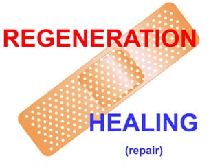
- 3. DEFINITIONS: •REGENERATION: Growth of cells to replace lost tissues •HEALING:A reparative tissue response to a wound, inflammation or necrosis, often leads to fibrosis • GRANULATION TISSUE • “ORGANIZING” INFLAMATION
- 4. REGENERATION • Replacement of lost structures • Is dependent on the type of normal turnover the original tissue has • Can be differentiated from “compensatory” growth
- 5. HEALING (repair) • Needs a wound, inflammatory process, or necrosis • Many disease appearances anatomically are the result of “healing” such as atherosclerosis • Often ends with a scar • Fibrosis, as one of the 3 possible outcomes of inflammation, follows “healing” • Requires a connective tissue “scaffold” • Fibrosis occurs in proportion to the damage of the ECM
- 7. Cell Population Fates • PROLIFERATION – Hormonal, especially steroid hormones – eg., EPO, CSF • DIFFERENTIATION* – UNIDIRECTIONAL, GAIN and LOSS • APOPTOSIS *One of the most KEY concepts in neoplasia
- 10. CELL CYCLE • G0 – Quiescent (not a very long or dominent phase) • G1 – PRE-synthetic, but cell GROWTH taking place • S – Cells which have continuous “turnover” have longer, or larger S-phases, i.e., DNA synthesis – S-phase of TUMOR CELLS can be prognostic • G2 – PRE-mitotic • M (Mitotic:, P,M,A,T, Cytokinesis)
- 11. CELL TYPES • Labile: eg., marrow, GI • Quiescent: liver, kidney • NON-mitotic: neuron, striated muscle
- 13. EMBRYONIC STEM CELLS• DIFFERENTIATION • KNOCKOUT MICE ( mice raised with specific gene defects) • REPOPULATION OF DAMAGED TISSUES, in research
- 14. ADULT STEM CELLS • MARROW (HEMOCYTOBLAST) (hematopoetic stem cells) • NON-MARROW (RESERVE)
- 16. ADULT TISSUE DIFFERENTIATION and REGENERATION PARALLELS EMBRYONIC DEVELOPMENT
- 17. Growth Factors (GFs) • Polypeptides • Cytokines • LOCOMOTION • CONTRACTILITY • DIFFERENTIATION • ANGIOGENESIS
- 18. Growth Factors (GFs) • Epidermal • Transforming (alpha, beta) • Hepatocyte • Vascular Endothelial • Platelet Derived • Fibroblast • Keratinocyte • Cytokines (TNF, IL-1, Interferons)
- 20. CELL PLAYERS (source AND targets) • Lymphocytes, especially T-cells • Macrophages • Platelets • Endothelial cells • Fibroblasts • Keratinocytes • “Mesenchymal” cells • Smooth muscle cells
- 21. E(Epidermal) GF • Made in platelets, macrophages • Present in saliva, milk, urine, plasma • Acts on keratinocytes to migrate, divide • Acts on fibroblasts to produce “granulation” tissue
- 22. T(Transforming) GF-alpha • Made in macrophages, T-cells, keratinocytes • Similar to EGF, also effect on hepatocytes
- 23. H(Hepatocyte) GF • Made in “mesenchymal” cells • Proliferation of epithelium, endothelium, hepatocytes • Effect on cell “motility”
- 24. VE(Vascular Endothelial) GF • Made in mesenchymal cells • Triggered by HYPOXIA • Increases vascular permeability • Mitogenic for endothelial cells • KEY substance in promoting “granulation” tissue
- 25. PD(Platelet Derived) GF • Made in platelets, but also MANY other cell types • Chemotactic for MANY cells • Mitogen for fibroblasts • Angiogenesis • Another KEY player in granulation tissue
- 26. F(Fibroblast) GF • Made in MANY cells • Chemotactic and mitogenic, for fibroblasts and keratinocytes • Re-epithelialization • Angiogenesis, wound contraction • Hematopoesis • Cardiac/Skeletal (striated) muscle
- 27. T(Transforming) GF-beta • Made in MANY CELLS • Chemotactic for PMNs and MANY other types of cells • Inhibits epithelial cells • Fibrogenic • Anti-Inflammatory
- 28. K(Keratinocyte) GF • Made in fibroblasts • Stimulates keratinocytes: –Migration –Proliferation –Differentiation
- 29. I(Insulin-like) GF-1 • Made in macrophages, fibroblasts • Stimulates: –Sulfated proteoglycans –Collagen –Keratinocyte migration –Fibroblast proliferation • Action similar to GH (Pituitary Growth Hormone)
- 30. TNF (Tumor Necrosis Factor) • Made in macrophages, mast cells, T-cells • Activates macrophages • KEY influence on other cytokines
- 31. Interleukins • Made in macrophages, mast cells, T-cells, but also MANY other cells • MANY functions: –Chemotaxis –Angiogenesis –REGULATION of other cytokines
- 32. INTERFERONS • Made by lymphocytes, fibroblasts • Activates MACROPHAGES • Inhibits FIBROBLASTS • REGULATES other cytokines
- 33. SIGNALING • Autocrine (same cell) • Paracrine (next door neighbor) (many GFs) • Endocrine (far away, delivered by blood, steroid hormones)
- 36. ExtraCellular Matrix (ECM) • Collagen(s) I-XVIII • Elastin • Fibrillin • CAMs (Cell Adhesion Molecules) – Immunoglobulins, cadherins, integrins, selectins • Proteoglycans • Hyaluronic Acid
- 37. ECM • Maintain cell differentiation • “Scaffolding” • Establish microenvironment • Storage of GF’s
- 38. Collagen One - bONE (main component of bone) Collagen Two - carTWOlage (main component of cartilage) Collagen Three - reTHREEculate (main component of reticular fibers) Collagen Four - FLOOR - forms the basement membrane
- 39. GENETIC COLLAGEN DISORDERS • I OSTEOGENESIS IMPERFECTA, E-D • II ACHONDROGENESIS TYPE II • III VASCULAR EHLERS-DANLOS • V CLASSICAL E-D • IX STICKLER SYNDROME • IV ALPORT SYNDROME • VI BETHLEM MYOPATHY • VII DYSTROPHIC EPIDERMOLYSIS BULLOS. • IX EPIPHYSEAL DYSPLASIAS • XVII GEN. EPIDERMOLYSYS BULLOSA • XV, XVIII KNOBLOCH SYNDROME
- 40. DEFINITIONS: •REGENERATION: Growth of cells to replace lost tissues •HEALING:A reparative tissue response to a wound, inflammation or necrosis
- 41. HEALING• FOLLOWS INFLAMMATION • PROLIFERATION and MIGRATION of connective tissue cells • ANGIOGENESIS (Neovascularization) • Collagen, other ECM protein synthesis • Tissue Remodeling • Wound contraction • Increase in wound strength (scar = fibrosis)
- 42. ANGIOGENESIS (NEOVASCULARIZATION) • From endothelial precursor cells • From PRE-existing vessels • Stimulated/Regulated by GF’s, especially VEGF • Also regulated by ECM proteins • aka, “GRANULATION”, “GRANULATION TISSUE”, “ORGANIZATION”, “ORGANIZING INFLAMMATION”
- 46. WOUND HEALING • 1st INTENTION • Edges lined up • 2nd INTENTION • Edges NOT lined up • Ergo…. • More granulation • More epithelialization • MORE FIBROSIS
- 49. FIBROSIS/SCARRING • DEPOSITION OF COLLAGEN by FIBROBLASTS • With time (weeks, months, years?) the collagen becomes more dense, ergo, the tissue becomes “STRONGER”
- 50. Wound RETARDING factors (LOCAL) • DECREASED Blood supply • Denervation • Local Infection • FB • Hematoma • Mechanical stress • Necrotic tissue
- 51. Wound RETARDING factors (SYSTEMIC) • DECREASED Blood supply • Age • Anemia • Malignancy • Malnutrition • Obesity • Infection • Organ failure
Editor's Notes
- An example of compensatory growth is when one kidney becomes larger after a nephrectomy, or the left portion on the right lobe of the liver “enlarges” after a left lobectomy.
- Healing (repair), like inflammation, can be thought of as a predictable sequence of events, just like in the Cecil B. DeMille “Inflammation” epic!
- There isn’t a single day in the life of a pathologist when he does not think of the concept of “differentiation” a lot, particularly in reference of neoplasms!
- Typical protein (polypeptide) configurations of GF’s
- The fact that the GF’s are made by the cells involved in inflammation and healing shows the PARACRINE nature of their behavior.
- You can this that this GF works on both ectodermally as well as mesodermally (mesenchymal) derived cells.
- KEY interplay between mesoderm and ectoderm, like embryonic “induction”
- It is tempting to estimate the actual times of events in tissue injury and repair. Another way to describe, in three words, the three phases of “repair”. In which phase would you see “fibrin”? Ans: Inflam. In which phase would you see a dense “scar”? Ans: Maturation Which phase is characterized by prominence of “budding” blood vessels? Ans: Prolif.