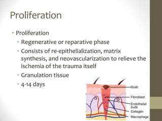
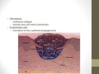
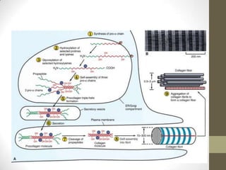
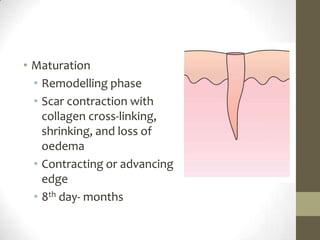
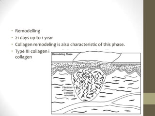
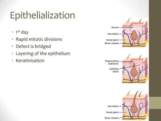
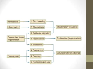
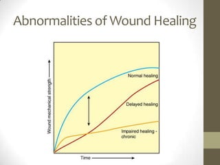
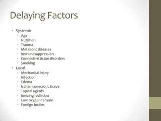
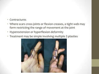
Wound HealingDr. Ranjeet Patil
DefinitionRegenerationReplacement of dead or damaged cells by functioning cells of same structure and functionBones, LiverRepairReplacement of dead or damaged cells by granulation tissue which matures to form scarFunction is lost-Lower order cellsSkinNoneBrain, Skeletal muscle, Cardiac muscle
Wound closure types PrimaryFirst-intentionClosure, the wounds are sealed immediately with simple suturing, skin graft placement, or flap closureClosure at end of a surgical procedure.SecondarySpontaneous-intention, no active intent to seal the woundCloses by re-epithelialization, contraction of the woundHighly contaminated woundTertiary Initially treated by repeated debridement, systemic or topical antibiotics, or negative pressure wound therapy for several days to control infectionSurgical intervention, such as suturing, skin graft placement, or flap design, is performed
WOUND-HEALING PHASESInflammationReactive phaseThe body's defences are aimed at limiting the amount of damage and preventing further injuryEschar or fibrinous exudateUpto 6 days
InflammationHaemostasisplatelet aggregation, degranulation, and activation of the coagulation cascadeplatelet-derived growth factor (PDGF), transforming growth factor beta (TGF), platelet-activating factor, fibronectin, and serotoninIncreased Vascular Permeability
InflammationInflammationPMN 24 to 48 hoursProstaglandins, complement factors, interleukin-1 (IL-1), tumour necrosis factor alpha (TNF-), TGF, platelet factor 4, bacterial productsprimary role-phagocytosis of bacteria and tissue debrisangiogenesis and collagen synthesis
Macrophages 48 to 96 hours – complete healingPhagocytosis Reactive oxygen species, Nitric oxide Debridement Collagenase, elastase Cell recruitment and activation Growth factors: PDGF, TGF, EGF, IGF Cytokines: TNF-, IL-1, IL-6, Fibronectin Matrix synthesis Growth factors: TGF, EGF, PDGF, Cytokines: TNF-, IL-1, Enzymes: arginase, collagenase, Prostaglandins, Nitric oxide Angiogenesis Growth factors: FGF, VEGF , Cytokines: TNF, Nitric oxide
T lymphocytes Less numerous than macrophagesPeak at about 1 week post injury role in wound healing is not fully defined
ProliferationProliferationRegenerative or reparative phaseConsists of re-epithelialization, matrix synthesis, and neovascularization to relieve the ischemia of the trauma itselfGranulation tissue4-14 days
Fibroblastssynthesize collagenactively carry out matrix contractionEndothelial cellsFormation of new capillaries (angiogenesis)
Maturation Remodelling phase Scar contraction with collagen cross-linking, shrinking, and loss of oedemaContracting or advancing edge8th day- months
Remodelling21 days up to 1 yearCollagen remodeling is also characteristic of this phase.Type III collagen is initially laid down, replaced by type I collagen
Epithelialization1st dayRapid mitotic divisionsDefect is bridgedLayering of the epitheliumKeratinisation
Abnormalities of Wound Healing
Delaying FactorsSystemic  Age Nutrition Trauma Metabolic diseases Immunosuppression Connective tissue disorders Smoking Local  Mechanical injury Infection EdemaIschemia/necrotic tissue Topical agents Ionizing radiation Low oxygen tension Foreign bodies
Excess HealingHepatic cirrhosis, Pulmonary Fibrosis, Scleroderma, Retrolental Fibroplasia, Diabetic Retinopathy, OsteoarthritisHypertrophic scarDefined as excessive scar tissue that does not extend beyond the boundary of the original incision or woundProlonged inflammatory phaseKeloidDefined as excessive scar tissue that extends beyond the boundaries of the original incision or woundAetiology is unknownBoth hypertrophic and keloid scars shows excess collagen with hypervascularity, but this is more marked in keloids where there is more type B collagen
TreatmentTreatment of hypertrophic and keloid scarsPressure – local moulds or elasticated garmentsSilicone gel sheeting (mechanism unknown)Intralesionalsteroid injection (triamcinolone)Excision and steroid injectionExcision and postoperative radiation (external beam or brachytherapy)Intralesionalexcision (keloids only)Laser – to reduce redness (which may resolve in any event)Vitamin E or palm oil massage (unproven)
ContracturesWhere scars cross joints or flexion creases, a tight web may form restricting the range of movement at the jointHyperextension or hyperflexion deformityTreatment may be simple involving multiple Z-plasties
Thankyou

Wound healing

  • 1.
  • 2.
    DefinitionRegenerationReplacement of deador damaged cells by functioning cells of same structure and functionBones, LiverRepairReplacement of dead or damaged cells by granulation tissue which matures to form scarFunction is lost-Lower order cellsSkinNoneBrain, Skeletal muscle, Cardiac muscle
  • 3.
    Wound closure typesPrimaryFirst-intentionClosure, the wounds are sealed immediately with simple suturing, skin graft placement, or flap closureClosure at end of a surgical procedure.SecondarySpontaneous-intention, no active intent to seal the woundCloses by re-epithelialization, contraction of the woundHighly contaminated woundTertiary Initially treated by repeated debridement, systemic or topical antibiotics, or negative pressure wound therapy for several days to control infectionSurgical intervention, such as suturing, skin graft placement, or flap design, is performed
  • 5.
    WOUND-HEALING PHASESInflammationReactive phaseThebody's defences are aimed at limiting the amount of damage and preventing further injuryEschar or fibrinous exudateUpto 6 days
  • 6.
    InflammationHaemostasisplatelet aggregation, degranulation,and activation of the coagulation cascadeplatelet-derived growth factor (PDGF), transforming growth factor beta (TGF), platelet-activating factor, fibronectin, and serotoninIncreased Vascular Permeability
  • 7.
    InflammationInflammationPMN 24 to48 hoursProstaglandins, complement factors, interleukin-1 (IL-1), tumour necrosis factor alpha (TNF-), TGF, platelet factor 4, bacterial productsprimary role-phagocytosis of bacteria and tissue debrisangiogenesis and collagen synthesis
  • 8.
    Macrophages 48 to96 hours – complete healingPhagocytosis Reactive oxygen species, Nitric oxide Debridement Collagenase, elastase Cell recruitment and activation Growth factors: PDGF, TGF, EGF, IGF Cytokines: TNF-, IL-1, IL-6, Fibronectin Matrix synthesis Growth factors: TGF, EGF, PDGF, Cytokines: TNF-, IL-1, Enzymes: arginase, collagenase, Prostaglandins, Nitric oxide Angiogenesis Growth factors: FGF, VEGF , Cytokines: TNF, Nitric oxide
  • 9.
    T lymphocytes Lessnumerous than macrophagesPeak at about 1 week post injury role in wound healing is not fully defined
  • 10.
    ProliferationProliferationRegenerative or reparativephaseConsists of re-epithelialization, matrix synthesis, and neovascularization to relieve the ischemia of the trauma itselfGranulation tissue4-14 days
  • 11.
    Fibroblastssynthesize collagenactively carryout matrix contractionEndothelial cellsFormation of new capillaries (angiogenesis)
  • 13.
    Maturation Remodelling phaseScar contraction with collagen cross-linking, shrinking, and loss of oedemaContracting or advancing edge8th day- months
  • 14.
    Remodelling21 days upto 1 yearCollagen remodeling is also characteristic of this phase.Type III collagen is initially laid down, replaced by type I collagen
  • 15.
    Epithelialization1st dayRapid mitoticdivisionsDefect is bridgedLayering of the epitheliumKeratinisation
  • 19.
  • 20.
    Delaying FactorsSystemic Age Nutrition Trauma Metabolic diseases Immunosuppression Connective tissue disorders Smoking Local Mechanical injury Infection EdemaIschemia/necrotic tissue Topical agents Ionizing radiation Low oxygen tension Foreign bodies
  • 21.
    Excess HealingHepatic cirrhosis,Pulmonary Fibrosis, Scleroderma, Retrolental Fibroplasia, Diabetic Retinopathy, OsteoarthritisHypertrophic scarDefined as excessive scar tissue that does not extend beyond the boundary of the original incision or woundProlonged inflammatory phaseKeloidDefined as excessive scar tissue that extends beyond the boundaries of the original incision or woundAetiology is unknownBoth hypertrophic and keloid scars shows excess collagen with hypervascularity, but this is more marked in keloids where there is more type B collagen
  • 22.
    TreatmentTreatment of hypertrophicand keloid scarsPressure – local moulds or elasticated garmentsSilicone gel sheeting (mechanism unknown)Intralesionalsteroid injection (triamcinolone)Excision and steroid injectionExcision and postoperative radiation (external beam or brachytherapy)Intralesionalexcision (keloids only)Laser – to reduce redness (which may resolve in any event)Vitamin E or palm oil massage (unproven)
  • 23.
    ContracturesWhere scars crossjoints or flexion creases, a tight web may form restricting the range of movement at the jointHyperextension or hyperflexion deformityTreatment may be simple involving multiple Z-plasties
  • 25.

Editor's Notes

  • #6 The three phases of wound healing are inflammation, proliferation, and maturation. The immediate response to injury is the inflammatory (also called reactive) phase. The body's defences are aimed at limiting the amount of damage and preventing further injury. The proliferative (also called regenerative or reparative) phase is the reparative process and consists of re-epithelialization, matrix synthesis, and neovascularization to relieve the ischemia of the trauma itself. The final maturational (or remodelling) phase is the period of scar contraction with collagen cross-linking, shrinking, and loss of enema. In a large wound such as a pressure sore, the eschar or fibrinous exudate reflects the inflammatory phase, the granulation tissue is part of the proliferative phase, and the contracting or advancing edge is part of the maturational phase. All three phases may occur simultaneously, and the phases with their individual processes may overlap (
  • #7 Exposure of sub endothelial collagen to platelets results in platelet aggregation, degranulation, and activation of the coagulation cascade. Platelet -granules release a number of wound-active substances, such as platelet-derived growth factor (PDGF), transforming growth factor beta (TGF), platelet-activating factor, fibronectin, and serotonin. In addition to achieving haemostasis, the fibrin clot serves as scaffolding for the migration into the wound of inflammatory cells such as polymorphonuclear leukocytes (PMNs, neutrophils) and monocytes.
  • #8 Cellular infiltration after injury follows a characteristic, predetermined sequence (see Fig. 9-1). PMNs are the first infiltrating cells to enter the wound site, peaking at 24 to 48 hours. Increased vascular permeability, local prostaglandin release, and the presence of chemotactic substances, such as complement factors, interleukin-1 (IL-1), tumour necrosis factor alpha (TNF-), TGF, platelet factor 4, or bacterial products, all stimulate neutrophil migrationmacrophages achieve significant numbers in the wound by 48 to 96 hours post injury and remain present until wound healing is complete.
  • #9 Phagocytosis Reactive oxygen species Nitric oxide Debridement Collagenase, elastase Cell recruitment and activation Growth factors: PDGF, TGF, EGF, IGF Cytokines: TNF-, IL-1, IL-6 Fibronectin Matrix synthesis Growth factors: TGF, EGF, PDGF Cytokines: TNF-, IL-1, IFN- Enzymes: arginase, collagenase Prostaglandins Nitric oxide Angiogenesis Growth factors: FGF, VEGF Cytokines: TNF- Nitric oxide
  • #10 T lymphocytes comprise another population of inflammatory/immune cells that routinely invades the wound. Less numerous than macrophages, T-lymphocyte numbers peak at about 1 week post injury and truly bridge the transition from the inflammatory to the proliferative phase of healing. Although known to be essential to wound healing, the lymphocytes' role in wound healing is not fully defined
  • #16 Fixed basal cells in a zone near the cut edge undergo a series of rapid mitotic divisions, and these cells appear to migrate by moving over one another in a leapfrog fashion until the defect is covered.22 Once the defect is bridged, the migrating epithelial cells lose their flattened appearance, become more columnar in shape, and increase their mitotic activity. Layering of the epithelium is re-established, and the surface layer eventually keratinizes.23