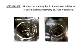
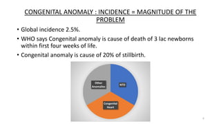
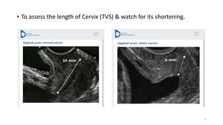
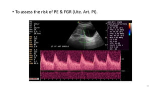
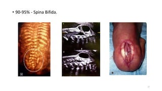
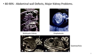
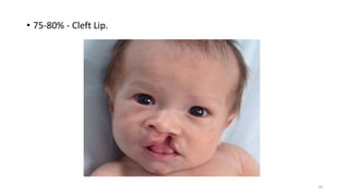
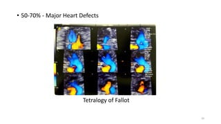
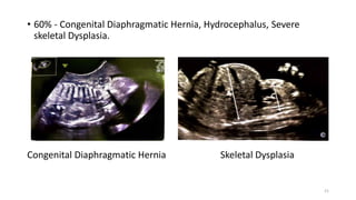
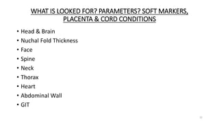
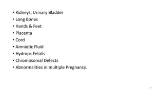
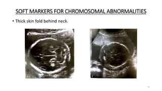
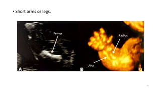
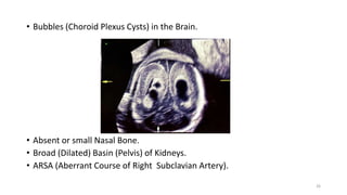
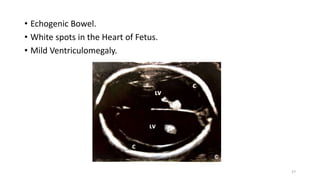
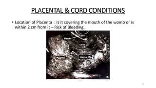
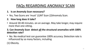
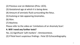
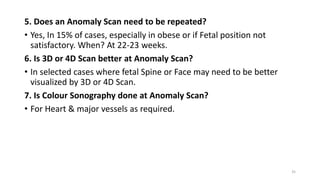
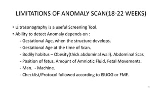
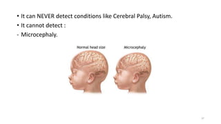
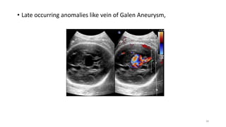
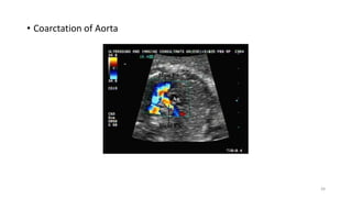
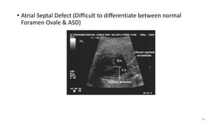
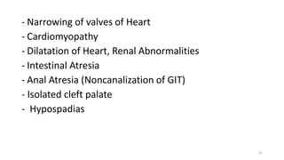
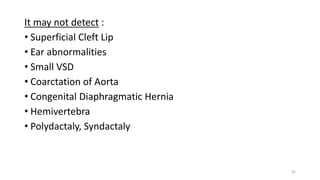
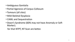
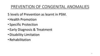
18-22 WEEKS ANOMALY SCAN
TIFFA – TARGETED IMAGING FOR FETAL ANOMALIES
Dr. Nisheeth M. Oza
1
Dr. Nisheeth M. Oza
M.D., D.G.O., F.C.P.S., D.N.B., (M.N.A.M.S.) (OBGYN)
D.A., M.B.B.S., Dip. YAN
Obstetrician, Gynaecologist, Infertility Consultant,
FMF Certified Ultrasonographer (FMF ID 145907)
Licensed for Aneuploidy Screening NT, NB, DV, Cervix & PIH Screening.
Diploma in Yoga, Ayurveda & Naturopathy
Dr. Oza’s Hospital 87/7, Panvel 410206, Maharashtra India.
Email : drnmozas@gmail.com
Website : drozashospital.com
YouTube : DrOzaConnects
2
VIDEO 01 : INTRODUCTION
UNDERSTANDING FEW COMMONLY USED TERMS
CONGENITAL : Present when in womb and at birth
ANOMALY : A structural which is not normal.
MALFORMATION: Abnormality in development. Synonymous with
Anomaly.
3
LETHAL ANOMALY: Cannot survive after birth, not > 3 months eg.
Anencephaly.
MINOR ANOMALY: Does not cause significant health issues eg. Ear tag.
MAJOR ANOMALY: Causes significant health issues which require
surgery or medical assistance eg. Cleft lip, Spina
Bifida.
TERATOGEN : An agent which may be capable of causing a Birth Defect.
4
SOFT MARKER : Not itself an anomaly, but indicates increased chance
of Chromosomal abnormality eg. Thick Nuchal Fold.
5
CONGENITAL ANOMALY : INCIDENCE = MAGNITUDE OF THE
PROBLEM
• Global incidence 2.5%.
• WHO says Congenital anomaly is cause of death of 3 lac newborns
within first four weeks of life.
• Congenital anomaly is cause of 20% of stillbirth.
6
NTD
Congenital
Heart
Other
Anomalies
WHO SHOULD BE SCREENED?
HIGH RISK CASES
• Pregnant woman is > 35 years old.
• Pregnant woman is Diabetic.
• In previous pregnancy there was Congenital Anomaly.
• Family history of Congenital Anomaly.
• History of Fever, Radiation, Drug intake between 4 & 10 weeks from
LMP.
Please note : Majority occur in without risk factor, therefore ANOMALY
SCAN SHOULD BE OFFERED to ALL Pregnant Women.
7
WHY? WHEN ? HOW?
WHY? PURPOSE? (ROLE/AIMS AND OBJECTIVES)
• In 97.5% of cases, the report would be ‘ALL GOOD’ so it helps in
(IUGR).
• Rule out major structural abnormality.
• Rule out minor structural abnormality.
• Look for Soft Markers for Chromosomal Abnormality.
• Evaluate Placenta, Umbilical cord & Amniotic fluid.
• Look at Growth for future comparison in case of FGR.
• Psychological Assurance of the pregnant women & help in bonding.
8
• To assess the length of Cervix (TVS) & watch for its shortening.
9
• To assess the risk of PE & FGR (Ute. Art. PI).
10
WHEN?
• As a Screening Tool, offer to ALL pregnant women at 18-22 weeks.
• Not before 18 weeks - because fetal organs are of small size & may
miss an Anomaly.
• Not after 22 weeks - because if abnormality found, more time
required for second opinion, Counseling, Decision Making, so it
should not go beyond legal limit of MTP, which is 24 weeks at present
in India.
11
HOW?
• First complete formalities as per Government rules.
• Room is dimly lit to allow better visualization.
• Lie down on a couch.
• Expose part of abdomen.
• Tissue paper is tuck around clothing.
• Gel is applied on tummy (the gel makes good contact between probe &
skin.
• The Sonographer moves a handheld device called a probe over your skin.
• Probe is moved over skin of abdomen.
• Image may visible on monitor screen.
• May take 30 minutes or longer, depending on position of Fetus and
visibility of all the structures.
• Spine is seen better when Fetus is lying with tummy down, Face is seen
better when Fetus is lying on its back
12
CHECKLIST AND METHOD FOLLOWED
• Checklist is followed so that one does not forget to look at any
structure.
• ISUOG recommends Six Step Approach.
• ISUOG recommends 22+2 planes to cover all the structures.
• FMF recommends 18 views.
• All the images are recorded.
• Report and films are prepared.
• Opportunity is provided to discuss the report and offer Counseling as
per the result.
13
PRETEST COUNSELING
• Global incidence of in general population is 2.5%.
• Ultrasonography can detect many, though not all.
• Risk factors for Congenital Anomaly – Maternal Diabetes Mellitus,
Infection, Radiation, Drugs.
• Remember 80-90% of Congenital Anomalies occur in fetus without
risk factors.
• Systematic examination for structural anomaly and/or soft marker is
carried out.
14
FACTORS WHICH INFLUENCE DETECTION OF ANOMALIES
• Gestational age at ultrasound examination.
• Natural history of the disease during Fetal life.
• Man - Operator’s training, skill, experience.
• Machine - Resolution of equipment.
• Malformation – Major vs Minor, Single vs Multiple.
• Proper follow up after birth.
15
DETECTION RATE OF DIFFERENT ANOMALIES
• Almost 100% - Anencephaly.
16
• 90-95% - Spina Bifida.
17
• 80-90% - Abdominal wall Defects, Major Kidney Problems.
Multicystic Kidney Omphalocele
Gastroschisis
18
• 75-80% - Cleft Lip.
19
• 50-70% - Major Heart Defects
Tetralogy of Fallot
20
• 60% - Congenital Diaphragmatic Hernia, Hydrocephalus, Severe
skeletal Dysplasia.
Congenital Diaphragmatic Hernia Skeletal Dysplasia
21
WHAT IS LOOKED FOR? PARAMETERS? SOFT MARKERS,
PLACENTA & CORD CONDITIONS
• Head & Brain
• Nuchal Fold Thickness
• Face
• Spine
• Neck
• Thorax
• Heart
• Abdominal Wall
• GIT
22
• Kidneys, Urinary Bladder
• Long Bones
• Hands & Feet
• Placenta
• Cord
• Amniotic Fluid
• Hydrops Fetalis
• Chromosomal Defects
• Abnormalities in multiple Pregnancy.
23
SOFT MARKERS FOR CHROMOSOMAL ABNORMALITIES
• Thick skin fold behind neck.
24
• Short arms or legs.
25
• Bubbles (Choroid Plexus Cysts) in the Brain.
• Absent or small Nasal Bone.
• Broad (Dilated) Basin (Pelvis) of Kidneys.
• ARSA (Aberrant Course of Right Subclavian Artery).
26
• Echogenic Bowel.
• White spots in the Heart of Fetus.
• Mild Ventriculomegaly.
27
• LR – Likelihood Ratio for the Soft Markers, meaning How likely is that
particular Marker to be indicative of a Chromosomal Abnormality?
• In simple words, is the soft marker really significant & how much?
28
PLACENTAL & CORD CONDITIONS
• Location of Placenta : Is it covering the mouth of the womb or is
within 2 cm from it – Risk of Bleeding.
29
• Features of Morbidly adherent placenta : Excessive bleeding at time
of Caesarean Operation.
• Extra Separate lobe of Placenta : Specifically remember to remove it
also, at time of Delivery & check for it at delivery.
• Abnormal insertion of Cord or Vessel crossing over mouth of womb :
Risk of bleeding.
• No. of vessels in Cord : If 2 instead of 3 look at Fetal Heart in detail
Fetal 2D Echo.
30
POST TEST COUNSELING AFTER ANOMALY SCAN
1. What if Structural Anomaly is found? Is it lethal?
Explain. Will it require surgery or special care after birth? Long term
Prognosis – Outcome.
Team of Doctors for Counseling. Decision Making. Recurrence Risk.
2. What if Soft Marker is found?
LR – If high : offer Amniocentesis – If confirms Chromosomal
abnormality, Counsel accordingly.
If LR is low – follow up.
3. What if no abnormality or soft marker is found?
Reassuring at present. There are few anomalies show later in
pregnancy. There are few abnormalities which may be found only after
birth.
31
POST TEST COUNSELING
Scan Normal
Reassure
Follow up scans
28-30 weeks(Growth)
34-36 weeks(Fetal well
being)
Major Anomaly
Prenatal investigations
and treatment
High Morbidity & Guarded
Long Term Outcome
Recurrence Risk
Reasonably good
outcome
Recurrence risk
Minor Anomaly
Counseling
Decision making
32
FAQs REGARDING ANOMALY SCAN
1. Is an Anomaly Scan necessary?
• Yes, Two Scans are ‘must’ (1)NT Scan (2)Anomaly Scan.
2. How long does it take?
• Around 30-40 minutes. on an average. May take longer, may require
more than one sitting.
3. Can Anomaly Scan detect all the structural anomalies with 100%
detection rate?
• No. No medical test can guarantee 100% accuracy. Detection rate is
influenced by so many factors, including
(1) Obesity.
33
(2) Previous scar on Abdomen (Prev. LSCS).
(3) Gestational age at which it is being done.
(4) Amount of amniotic fluid surrounding the fetus.
(5) Evolving or late appearing Anomalies.
(6) Man.
(7) Machine.
Please refer to the video on ‘Limitations of an Anomaly Scan’.
4. Will I need any further tests?
Yes. (1) Significant ‘soft markers’ : Amniocentesis.
(2) If fetal heart suspicious findings : Fetal 2D Echocardiography.
34
5. Does an Anomaly Scan need to be repeated?
• Yes, In 15% of cases, especially in obese or if Fetal position not
satisfactory. When? At 22-23 weeks.
6. Is 3D or 4D Scan better at Anomaly Scan?
• In selected cases where fetal Spine or Face may need to be better
visualized by 3D or 4D Scan.
7. Is Colour Sonography done at Anomaly Scan?
• For Heart & major vessels as required.
35
LIMITATIONS OF ANOMALY SCAN(18-22 WEEKS)
• Ultrasonography is a useful Screening Tool.
• Ability to detect Anomaly depends on :
- Gestational Age, when the structure develops.
- Gestational Age at the time of Scan.
- Bodily habitus – Obesity(thick abdominal wall). Abdominal Scar.
- Position of fetus, Amount of Amniotic Fluid, Fetal Movements.
- Man. - Machine.
- Checklist/Protocol followed according to ISUOG or FMF.
36
• It can NEVER detect conditions like Cerebral Palsy, Autism.
• It cannot detect :
- Microcephaly.
37
• Late occurring anomalies like vein of Galen Aneurysm,
38
• Coarctation of Aorta
39
• Atrial Septal Defect (Difficult to differentiate between normal
Foramen Ovale & ASD)
40
- Narrowing of valves of Heart
- Cardiomyopathy
- Dilatation of Heart, Renal Abnormalities
- Intestinal Atresia
- Anal Atresia (Noncanalization of GIT)
- Isolated cleft palate
- Hypospadias
41
It may not detect :
• Superficial Cleft Lip
• Ear abnormalities
• Small VSD
• Coarctation of Aorta
• Congenital Diaphragmatic Hernia
• Hemivertebra
• Polydactaly, Syndactaly
42
• Ambiguous Genitalia
• Partial Agenesis of Corpus Collosum
• Tumours (all sites)
• Mild Skeletal Dysplasia
• CAML and Sequestration
• Down’s Syndrome (60% may not have Anomaly or Soft
Marker).
for that NTPT, NT Scan are better.
43
PREVENTION OF CONGENITAL ANOMALIES
5 levels of Prevention as learnt in PSM.
• Health Promotion
• Specific Protection
• Early Diagnosis & Treatment
• Disability Limitation
• Rehabilitation
44
1. Health Promotion
• Whenever possible, avoid consanguineous marriage.
• Whenever possible, avoid late marriage and late
pregnancy > 35 years.
• Prevention of Anaemia, Malnutrition in girls.
• Encourage immunization of all female children by
MMR vaccine.
• Encourage balanced diet in women trying to conceive,
especially include foods rich in Folic Acid, Proteins,
Vitamins.
45
2. Specific Protection
1. Stop skin ointment for Acne, especially those
containing high concentration of Vitamin A, 3 months
before conceiving.
2. Switch over to safe antidiabetic, antihypertensive &
antiepileptic before conceiving.
3. Encourage Pre pregnancy intake of Folic Acid 5
mg/day and Multivitamin tablet 90 days before
conceiving.
4. Tight control of DM in 1st Trimester of pregnancy.
46
• 5. Elimination of Tobacco intake in Pregnancy –
chewing, smoking, active or passively.
• Avoid intake of Alcohol in, exposure to x-rays & intake
of Drug-medicines especially in first 3 months of
pregnancy, without consulting your Obstetrician.
• Avoid getting infections like URTI, especially in first
Trimester of Pregnancy.
• Ensure Intake of Tablets of Folic Acid 5 mg/day and
Multivitamins at least in First Trimester of Pregnancy.
47
3. Early Diagnosis & Treatment
• Encourage First Trimester Screening including NT Scan
11-13.6 weeks Scan & Biochemistry – Preferably NIPT
- or at least Dual Marker Test, to suspect a
Chromosomal Abnormality like Down’s Syndrome.
• In FTS Screen Positive – High Risk cases of
Chromosomal Abnormality- Invasive Test i.e. CVS or
Amniocentesis to exclude Chromosomal Abnormality.
48
• Anomaly Scan at 18-22weeks, looking for Structural
Abnormality & Soft Markers of Chromosomal
Abnormality.
• Fetal 2D Echo at 20-23 weeks, especially in cases at
High Risk for Congenital Heart Anomaly.
• Cases of confirmed Anomaly or Chromosomal Defect,
proper Counseling by Multi Disciplinary Team and
Decision making in a nondirective manner.
49
• Counsel the couple about Recurrence Risk of a Disorder
detected in the index pregnancy.
• If couple decides to continue with Pregnancy, perform
Ultrasonography more frequently if there is maternal DM or
develps Pre-eclampsia or FGR.
• Delivery in a Centre with all necessary facilities for the
Newborn eg in fetus with antenatally diagnosed Congenital
Diaphragmatic Hernia.
• Assessment at birth by Paediatrician, Paediatric Surgeon,
Orthopaedician or Paediatric Cardiologist depending upon
the Anomaly present.
50
• Plan out Optimal Newborn Care eg special feeding technique for Cleft
lip, Cleft palate.
• Proper care by Mother & Proper Nursing Care.
4. Disability Limitation
• Disability Limitation in Newborn eg. Application of Plaster Cast for
Clubfoot.
5.Rehabilitation
• Rehabilitation of the Infant eg. After Surgery for various
indications/Anomalies.
• Pre pregnancy Counseling when the couple wants to try for another
pregnancy in future, including recurrence risk.
51
THANK YOU!!
LIKE, SHARE….
For more details Subscribe to my YouTube channel.
52

18 - 22 Weeks Anomaly Scan

  • 1.
    18-22 WEEKS ANOMALYSCAN TIFFA – TARGETED IMAGING FOR FETAL ANOMALIES Dr. Nisheeth M. Oza 1
  • 2.
    Dr. Nisheeth M.Oza M.D., D.G.O., F.C.P.S., D.N.B., (M.N.A.M.S.) (OBGYN) D.A., M.B.B.S., Dip. YAN Obstetrician, Gynaecologist, Infertility Consultant, FMF Certified Ultrasonographer (FMF ID 145907) Licensed for Aneuploidy Screening NT, NB, DV, Cervix & PIH Screening. Diploma in Yoga, Ayurveda & Naturopathy Dr. Oza’s Hospital 87/7, Panvel 410206, Maharashtra India. Email : drnmozas@gmail.com Website : drozashospital.com YouTube : DrOzaConnects 2
  • 3.
    VIDEO 01 :INTRODUCTION UNDERSTANDING FEW COMMONLY USED TERMS CONGENITAL : Present when in womb and at birth ANOMALY : A structural which is not normal. MALFORMATION: Abnormality in development. Synonymous with Anomaly. 3
  • 4.
    LETHAL ANOMALY: Cannotsurvive after birth, not > 3 months eg. Anencephaly. MINOR ANOMALY: Does not cause significant health issues eg. Ear tag. MAJOR ANOMALY: Causes significant health issues which require surgery or medical assistance eg. Cleft lip, Spina Bifida. TERATOGEN : An agent which may be capable of causing a Birth Defect. 4
  • 5.
    SOFT MARKER :Not itself an anomaly, but indicates increased chance of Chromosomal abnormality eg. Thick Nuchal Fold. 5
  • 6.
    CONGENITAL ANOMALY :INCIDENCE = MAGNITUDE OF THE PROBLEM • Global incidence 2.5%. • WHO says Congenital anomaly is cause of death of 3 lac newborns within first four weeks of life. • Congenital anomaly is cause of 20% of stillbirth. 6 NTD Congenital Heart Other Anomalies
  • 7.
    WHO SHOULD BESCREENED? HIGH RISK CASES • Pregnant woman is > 35 years old. • Pregnant woman is Diabetic. • In previous pregnancy there was Congenital Anomaly. • Family history of Congenital Anomaly. • History of Fever, Radiation, Drug intake between 4 & 10 weeks from LMP. Please note : Majority occur in without risk factor, therefore ANOMALY SCAN SHOULD BE OFFERED to ALL Pregnant Women. 7
  • 8.
    WHY? WHEN ?HOW? WHY? PURPOSE? (ROLE/AIMS AND OBJECTIVES) • In 97.5% of cases, the report would be ‘ALL GOOD’ so it helps in (IUGR). • Rule out major structural abnormality. • Rule out minor structural abnormality. • Look for Soft Markers for Chromosomal Abnormality. • Evaluate Placenta, Umbilical cord & Amniotic fluid. • Look at Growth for future comparison in case of FGR. • Psychological Assurance of the pregnant women & help in bonding. 8
  • 9.
    • To assessthe length of Cervix (TVS) & watch for its shortening. 9
  • 10.
    • To assessthe risk of PE & FGR (Ute. Art. PI). 10
  • 11.
    WHEN? • As aScreening Tool, offer to ALL pregnant women at 18-22 weeks. • Not before 18 weeks - because fetal organs are of small size & may miss an Anomaly. • Not after 22 weeks - because if abnormality found, more time required for second opinion, Counseling, Decision Making, so it should not go beyond legal limit of MTP, which is 24 weeks at present in India. 11
  • 12.
    HOW? • First completeformalities as per Government rules. • Room is dimly lit to allow better visualization. • Lie down on a couch. • Expose part of abdomen. • Tissue paper is tuck around clothing. • Gel is applied on tummy (the gel makes good contact between probe & skin. • The Sonographer moves a handheld device called a probe over your skin. • Probe is moved over skin of abdomen. • Image may visible on monitor screen. • May take 30 minutes or longer, depending on position of Fetus and visibility of all the structures. • Spine is seen better when Fetus is lying with tummy down, Face is seen better when Fetus is lying on its back 12
  • 13.
    CHECKLIST AND METHODFOLLOWED • Checklist is followed so that one does not forget to look at any structure. • ISUOG recommends Six Step Approach. • ISUOG recommends 22+2 planes to cover all the structures. • FMF recommends 18 views. • All the images are recorded. • Report and films are prepared. • Opportunity is provided to discuss the report and offer Counseling as per the result. 13
  • 14.
    PRETEST COUNSELING • Globalincidence of in general population is 2.5%. • Ultrasonography can detect many, though not all. • Risk factors for Congenital Anomaly – Maternal Diabetes Mellitus, Infection, Radiation, Drugs. • Remember 80-90% of Congenital Anomalies occur in fetus without risk factors. • Systematic examination for structural anomaly and/or soft marker is carried out. 14
  • 15.
    FACTORS WHICH INFLUENCEDETECTION OF ANOMALIES • Gestational age at ultrasound examination. • Natural history of the disease during Fetal life. • Man - Operator’s training, skill, experience. • Machine - Resolution of equipment. • Malformation – Major vs Minor, Single vs Multiple. • Proper follow up after birth. 15
  • 16.
    DETECTION RATE OFDIFFERENT ANOMALIES • Almost 100% - Anencephaly. 16
  • 17.
    • 90-95% -Spina Bifida. 17
  • 18.
    • 80-90% -Abdominal wall Defects, Major Kidney Problems. Multicystic Kidney Omphalocele Gastroschisis 18
  • 19.
    • 75-80% -Cleft Lip. 19
  • 20.
    • 50-70% -Major Heart Defects Tetralogy of Fallot 20
  • 21.
    • 60% -Congenital Diaphragmatic Hernia, Hydrocephalus, Severe skeletal Dysplasia. Congenital Diaphragmatic Hernia Skeletal Dysplasia 21
  • 22.
    WHAT IS LOOKEDFOR? PARAMETERS? SOFT MARKERS, PLACENTA & CORD CONDITIONS • Head & Brain • Nuchal Fold Thickness • Face • Spine • Neck • Thorax • Heart • Abdominal Wall • GIT 22
  • 23.
    • Kidneys, UrinaryBladder • Long Bones • Hands & Feet • Placenta • Cord • Amniotic Fluid • Hydrops Fetalis • Chromosomal Defects • Abnormalities in multiple Pregnancy. 23
  • 24.
    SOFT MARKERS FORCHROMOSOMAL ABNORMALITIES • Thick skin fold behind neck. 24
  • 25.
    • Short armsor legs. 25
  • 26.
    • Bubbles (ChoroidPlexus Cysts) in the Brain. • Absent or small Nasal Bone. • Broad (Dilated) Basin (Pelvis) of Kidneys. • ARSA (Aberrant Course of Right Subclavian Artery). 26
  • 27.
    • Echogenic Bowel. •White spots in the Heart of Fetus. • Mild Ventriculomegaly. 27
  • 28.
    • LR –Likelihood Ratio for the Soft Markers, meaning How likely is that particular Marker to be indicative of a Chromosomal Abnormality? • In simple words, is the soft marker really significant & how much? 28
  • 29.
    PLACENTAL & CORDCONDITIONS • Location of Placenta : Is it covering the mouth of the womb or is within 2 cm from it – Risk of Bleeding. 29
  • 30.
    • Features ofMorbidly adherent placenta : Excessive bleeding at time of Caesarean Operation. • Extra Separate lobe of Placenta : Specifically remember to remove it also, at time of Delivery & check for it at delivery. • Abnormal insertion of Cord or Vessel crossing over mouth of womb : Risk of bleeding. • No. of vessels in Cord : If 2 instead of 3 look at Fetal Heart in detail Fetal 2D Echo. 30
  • 31.
    POST TEST COUNSELINGAFTER ANOMALY SCAN 1. What if Structural Anomaly is found? Is it lethal? Explain. Will it require surgery or special care after birth? Long term Prognosis – Outcome. Team of Doctors for Counseling. Decision Making. Recurrence Risk. 2. What if Soft Marker is found? LR – If high : offer Amniocentesis – If confirms Chromosomal abnormality, Counsel accordingly. If LR is low – follow up. 3. What if no abnormality or soft marker is found? Reassuring at present. There are few anomalies show later in pregnancy. There are few abnormalities which may be found only after birth. 31
  • 32.
    POST TEST COUNSELING ScanNormal Reassure Follow up scans 28-30 weeks(Growth) 34-36 weeks(Fetal well being) Major Anomaly Prenatal investigations and treatment High Morbidity & Guarded Long Term Outcome Recurrence Risk Reasonably good outcome Recurrence risk Minor Anomaly Counseling Decision making 32
  • 33.
    FAQs REGARDING ANOMALYSCAN 1. Is an Anomaly Scan necessary? • Yes, Two Scans are ‘must’ (1)NT Scan (2)Anomaly Scan. 2. How long does it take? • Around 30-40 minutes. on an average. May take longer, may require more than one sitting. 3. Can Anomaly Scan detect all the structural anomalies with 100% detection rate? • No. No medical test can guarantee 100% accuracy. Detection rate is influenced by so many factors, including (1) Obesity. 33
  • 34.
    (2) Previous scaron Abdomen (Prev. LSCS). (3) Gestational age at which it is being done. (4) Amount of amniotic fluid surrounding the fetus. (5) Evolving or late appearing Anomalies. (6) Man. (7) Machine. Please refer to the video on ‘Limitations of an Anomaly Scan’. 4. Will I need any further tests? Yes. (1) Significant ‘soft markers’ : Amniocentesis. (2) If fetal heart suspicious findings : Fetal 2D Echocardiography. 34
  • 35.
    5. Does anAnomaly Scan need to be repeated? • Yes, In 15% of cases, especially in obese or if Fetal position not satisfactory. When? At 22-23 weeks. 6. Is 3D or 4D Scan better at Anomaly Scan? • In selected cases where fetal Spine or Face may need to be better visualized by 3D or 4D Scan. 7. Is Colour Sonography done at Anomaly Scan? • For Heart & major vessels as required. 35
  • 36.
    LIMITATIONS OF ANOMALYSCAN(18-22 WEEKS) • Ultrasonography is a useful Screening Tool. • Ability to detect Anomaly depends on : - Gestational Age, when the structure develops. - Gestational Age at the time of Scan. - Bodily habitus – Obesity(thick abdominal wall). Abdominal Scar. - Position of fetus, Amount of Amniotic Fluid, Fetal Movements. - Man. - Machine. - Checklist/Protocol followed according to ISUOG or FMF. 36
  • 37.
    • It canNEVER detect conditions like Cerebral Palsy, Autism. • It cannot detect : - Microcephaly. 37
  • 38.
    • Late occurringanomalies like vein of Galen Aneurysm, 38
  • 39.
  • 40.
    • Atrial SeptalDefect (Difficult to differentiate between normal Foramen Ovale & ASD) 40
  • 41.
    - Narrowing ofvalves of Heart - Cardiomyopathy - Dilatation of Heart, Renal Abnormalities - Intestinal Atresia - Anal Atresia (Noncanalization of GIT) - Isolated cleft palate - Hypospadias 41
  • 42.
    It may notdetect : • Superficial Cleft Lip • Ear abnormalities • Small VSD • Coarctation of Aorta • Congenital Diaphragmatic Hernia • Hemivertebra • Polydactaly, Syndactaly 42
  • 43.
    • Ambiguous Genitalia •Partial Agenesis of Corpus Collosum • Tumours (all sites) • Mild Skeletal Dysplasia • CAML and Sequestration • Down’s Syndrome (60% may not have Anomaly or Soft Marker). for that NTPT, NT Scan are better. 43
  • 44.
    PREVENTION OF CONGENITALANOMALIES 5 levels of Prevention as learnt in PSM. • Health Promotion • Specific Protection • Early Diagnosis & Treatment • Disability Limitation • Rehabilitation 44
  • 45.
    1. Health Promotion •Whenever possible, avoid consanguineous marriage. • Whenever possible, avoid late marriage and late pregnancy > 35 years. • Prevention of Anaemia, Malnutrition in girls. • Encourage immunization of all female children by MMR vaccine. • Encourage balanced diet in women trying to conceive, especially include foods rich in Folic Acid, Proteins, Vitamins. 45
  • 46.
    2. Specific Protection 1.Stop skin ointment for Acne, especially those containing high concentration of Vitamin A, 3 months before conceiving. 2. Switch over to safe antidiabetic, antihypertensive & antiepileptic before conceiving. 3. Encourage Pre pregnancy intake of Folic Acid 5 mg/day and Multivitamin tablet 90 days before conceiving. 4. Tight control of DM in 1st Trimester of pregnancy. 46
  • 47.
    • 5. Eliminationof Tobacco intake in Pregnancy – chewing, smoking, active or passively. • Avoid intake of Alcohol in, exposure to x-rays & intake of Drug-medicines especially in first 3 months of pregnancy, without consulting your Obstetrician. • Avoid getting infections like URTI, especially in first Trimester of Pregnancy. • Ensure Intake of Tablets of Folic Acid 5 mg/day and Multivitamins at least in First Trimester of Pregnancy. 47
  • 48.
    3. Early Diagnosis& Treatment • Encourage First Trimester Screening including NT Scan 11-13.6 weeks Scan & Biochemistry – Preferably NIPT - or at least Dual Marker Test, to suspect a Chromosomal Abnormality like Down’s Syndrome. • In FTS Screen Positive – High Risk cases of Chromosomal Abnormality- Invasive Test i.e. CVS or Amniocentesis to exclude Chromosomal Abnormality. 48
  • 49.
    • Anomaly Scanat 18-22weeks, looking for Structural Abnormality & Soft Markers of Chromosomal Abnormality. • Fetal 2D Echo at 20-23 weeks, especially in cases at High Risk for Congenital Heart Anomaly. • Cases of confirmed Anomaly or Chromosomal Defect, proper Counseling by Multi Disciplinary Team and Decision making in a nondirective manner. 49
  • 50.
    • Counsel thecouple about Recurrence Risk of a Disorder detected in the index pregnancy. • If couple decides to continue with Pregnancy, perform Ultrasonography more frequently if there is maternal DM or develps Pre-eclampsia or FGR. • Delivery in a Centre with all necessary facilities for the Newborn eg in fetus with antenatally diagnosed Congenital Diaphragmatic Hernia. • Assessment at birth by Paediatrician, Paediatric Surgeon, Orthopaedician or Paediatric Cardiologist depending upon the Anomaly present. 50
  • 51.
    • Plan outOptimal Newborn Care eg special feeding technique for Cleft lip, Cleft palate. • Proper care by Mother & Proper Nursing Care. 4. Disability Limitation • Disability Limitation in Newborn eg. Application of Plaster Cast for Clubfoot. 5.Rehabilitation • Rehabilitation of the Infant eg. After Surgery for various indications/Anomalies. • Pre pregnancy Counseling when the couple wants to try for another pregnancy in future, including recurrence risk. 51
  • 52.
    THANK YOU!! LIKE, SHARE…. Formore details Subscribe to my YouTube channel. 52