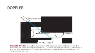
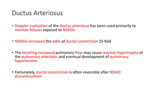
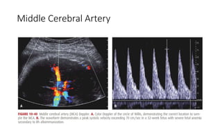
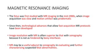
3D and 4D sonography provide static or real-time 3D views of the fetus, respectively, but are not routinely used in standard exams. Doppler ultrasound evaluates blood flow in various fetal vessels like the umbilical artery, ductus arteriosus, uterine artery, middle cerebral artery, and ductus venosus to monitor fetal well-being. Abnormal flow patterns in these vessels can indicate conditions like fetal growth restriction. Fetal MRI provides superior anatomical images compared to ultrasound and can help further characterize abnormalities, guide fetal therapy, or assess complications in high-risk pregnancies.
This document discusses Doppler ultrasound and its application in obstetrics. Doppler ultrasound uses the Doppler effect to analyze blood flow and detect movement. It is used to assess the placenta and fetal circulation through Doppler assessment of the uterine arteries, umbilical arteries, cerebral arteries, and descending aorta. Abnormal Doppler waveforms can indicate placental insufficiency or fetal distress. Precise technique and angle of insonation are important to obtain accurate readings. Doppler ultrasound provides valuable information for evaluating fetal wellbeing, growth, and detecting anomalies.
Doppler ultrasound provides important information about blood flow that cannot be determined through standard gray-scale ultrasound alone. It allows observation of flow velocity and direction, aiding in diagnosis of various vascular diseases. Doppler works by detecting the frequency shift of sound waves reflected from moving objects like blood cells. There are two main types - Pulsed Wave Doppler which measures flow at a selected sample volume, and Continuous Wave Doppler which evaluates flow over a range of depths. Doppler ultrasound offers advantages over standard gray-scale such as assessing blockages, plaque, abnormal veins, and fetal well-being during pregnancy without known health risks.
Ultrasound has become an essential tool in obstetrics, allowing visualization of the fetus and assessment of growth and well-being. Doppler ultrasound can evaluate blood flow in fetal and maternal vessels. While ultrasound is generally safe, concerns have been raised about potential neurological effects with prolonged or frequent use. Estimation of fetal weight and biophysical profiling helps monitor high-risk pregnancies. Overall, ultrasound has dramatically improved prenatal care and outcomes over the past decades.
Doppler ultrasonography is used to analyze blood flow velocity in fetal vessels. The umbilical artery, middle cerebral artery, uterine artery, ductus venosus, inferior vena cava, and umbilical vein are examined. Normal and abnormal waveforms in these vessels provide information about placental and fetal wellbeing. Abnormal waveforms can indicate issues like intrauterine growth restriction and increased risk of fetal demise. Doppler is a valuable tool for assessing fetal health through non-invasive analysis of blood flow velocities.
Emergency sonography in Pediatrics has evolved to become one of the most versatile
modalities for diagnosing and guiding
treatment of critically ill patients.
This document discusses central line placement, including:
1) It provides a brief history of central line development and discusses their increasing common use today.
2) Major uses of central lines include access for volume infusion, determination of cardiovascular function, and nutrition.
3) Potential complications include mechanical issues like arterial puncture or cannulation in 5-19% of cases, and infectious complications in 5-26% of cases.
Doppler ultrasound uses sound waves to evaluate blood flow and was first used in 1977 to study umbilical artery flow. It measures the Doppler shift in echoes from moving blood cells to determine flow velocity. Fetal vessels commonly assessed include the umbilical artery, which provides information on placental perfusion, and the middle cerebral artery, which indicates fetal oxygenation status. Doppler ultrasound can detect signs of fetal hypoxia like increased end-diastolic flow in the MCA, and meta-analyses found it reduces perinatal deaths and inductions when used for monitoring high-risk pregnancies.
Bedside Ultrasound in Neurosurgery Part 1/3Liew Boon Seng
This document provides an overview of ultrasound training in neurosurgery. It introduces various applications of ultrasound in neurosurgery such as fetal neurosonograms, cranial ultrasonography of newborns, assessing ventricular shunt patency, spinal ultrasound in infants, and transcranial insonation of blood vessels. It also summarizes techniques for different types of Doppler imaging and discusses pathologies that can be detected using ultrasound like intraventricular hemorrhage.
This document provides an overview of Doppler ultrasound in the assessment of fetal and placental blood flow during the second trimester of pregnancy. It discusses the history and development of Doppler ultrasound techniques. It then covers the basic principles of Doppler ultrasound and details the specific vessels that should be examined, including the umbilical artery, middle cerebral artery, uterine artery, thoracic aorta, inferior vena cava, ductus venosus, and umbilical vein. Normal and abnormal waveforms are presented for each vessel. The uses and clinical significance of Doppler assessments are described.
Doppler ultrasound is useful for detecting hepatic artery complications after liver transplantation. Common findings in the early postoperative period include transient high resistance in the hepatic artery Doppler waveform, which typically resolves without complications. Hepatic artery thrombosis appears as an absence of Doppler flow in the hepatic artery and branches. Hepatic artery stenosis may be suggested by a prolonged systolic acceleration time and low resistance index. Pseudoaneurysms present as cystic lesions that fill with color Doppler. Arterial steal syndromes can potentially cause hepatic hypoperfusion but findings are nonspecific. Contrast enhanced ultrasound can aid in evaluation when Doppler findings are unclear.
This document provides an overview of Doppler ultrasonography in obstetrics. It discusses the prerequisites for Doppler studies including the ultrasound machine capabilities and patient preparation. It describes the indications for Doppler including fetal growth abnormalities and maternal risk factors. It examines in detail the various fetal and maternal vessels that can be assessed by Doppler including the umbilical artery, middle cerebral artery, and uterine arteries. It outlines the normal and abnormal waveforms seen in each vessel and their clinical significance.
ultrasonography in obstetrics, usg in obstetrics, ultrasound in obstetrics, doppler in obstetrics, usg doppler in obstetrics, signs in ultrasound, anomaly scan, pregnancy scan, ultrasound in pregnancy,
This document discusses the role of color Doppler ultrasound in antepartum fetal surveillance. It begins by outlining the purposes of fetal surveillance, which include reducing fetal death and optimizing delivery timing. It then discusses various maternal and fetal conditions that require increased surveillance due to risks of chronic hypoxia. The document covers different methods of antepartum surveillance and provides detailed explanations of Doppler ultrasound principles, techniques like uterine and umbilical artery Doppler, and how abnormal Doppler readings can predict complications like fetal growth restriction.
1. Doppler ultrasound uses pulsed wave Doppler to measure blood flow velocity in a sample volume. It can calculate indices like the pulsatility index (PI) and resistive index (RI) to assess vascular resistance.
2. PI and RI provide information about resistance and elasticity in blood vessels. They are not affected by angle of insonation and help evaluate conditions like fetal growth restriction.
3. Uterine artery PI in particular measures uteroplacental perfusion, with higher values implying impaired placentation and increased risk of complications like preeclampsia.
Normal doppler spectral pattern of abdominal and limb vessels finalNipun Gupta
This document provides information on normal Doppler patterns of abdominal vessels. It begins by covering Doppler physics principles. It then discusses normal Doppler flow patterns seen in the abdominal aorta, ductus venosus, celiac artery, superior mesenteric artery, and mesenteric arteries. For each vessel, it describes anatomy, imaging recommendations, and typical Doppler waveform patterns. The document serves as an educational guide for residents to learn how to properly evaluate and interpret Doppler ultrasound of the abdominal vasculature.
Doppler ultrasound can be used in obstetrics and gynecology in several ways. It allows assessment of blood flow in various fetal and maternal vessels. In pregnancy, Doppler is commonly used to evaluate blood flow in the umbilical artery, middle cerebral artery, uterine arteries, ductus venosus and other vessels. Abnormal flow patterns in these vessels can indicate fetal growth restriction, hypoxia, or the risk of conditions like preeclampsia. Doppler provides important information about fetal well-being and helps manage high-risk pregnancies.
This document provides information about ultrasound imaging, including:
- Ultrasound uses high-frequency sound waves to produce images of the inside of the body without radiation.
- It is widely used due to its availability, low cost, speed, and ability to image in real-time.
- Common uses include imaging organs like the heart, liver, kidneys, and monitoring fetal development.
- The procedure involves a transducer sending sound waves into the body and receiving echoes to produce images.
This document discusses Doppler ultrasound principles and techniques. It begins by explaining the basic principles of how Doppler ultrasound measures blood flow velocity. It describes continuous wave and pulsed wave Doppler, and factors that affect Doppler measurements like ultrasound frequency, beam-to-flow angle, and aliasing. It also summarizes different ultrasound flow modes like color flow imaging, spectral Doppler, and power Doppler, when each is used, and how they complement each other.
• What is Ultrasound imaging?
• Why Ultrasound?
• Common Uses
• History
• Properties of Ultrasound
• Equipment types
• How does the procedure work?
• Physics
• Benefits and Risks etc.
This document provides information about Prof. Narendra Malhotra, including his academic and professional qualifications, positions held, awards received, publications, lectures given, and organizations founded. It then provides an objectives and agenda for a talk on using color doppler to assess fetal growth restriction (FGR). The talk covers doppler principles, applications in evaluating the uteroplacental and fetal circulations in FGR, implications of abnormal doppler waveforms, and new applications such as in assessing fetal anemia and 3D doppler. It proposes a staging system for FGR based on doppler findings to determine timely delivery.
Ultrasound uses high-frequency sound waves to produce images of the inside of the body in real-time without using radiation. It is widely used due to its availability, low cost, speed, and ability to show internal structures and blood flow. Common uses include examining organs like the heart, liver, and kidneys, as well as guiding procedures, imaging breasts and blood vessels, and assessing fetal development in pregnancies. The procedure works by a transducer sending sound waves into the body and receiving echoes to create images based on the return signal.
Dental radiology related to pedodonticsSunny Purohit
This document discusses the use of dental radiology for pediatric patients. It begins with a brief history of x-rays and their early use in dentistry. Various types of dental radiographs are described, including intraoral periapical, bitewing, occlusal, and panoramic images. Indications for different radiographs in children are provided. Techniques for improving pediatric patient cooperation and reducing radiation exposure are also outlined, such as desensitization and use of positioning devices. Potential radiation effects are discussed as well as safety measures like lead aprons. Guidelines for prescribing dental radiographs in pediatric patients from the AAPD are also mentioned.
This document provides information on ultrasound technology and its use in obstetrics. It describes how ultrasound works using piezoelectric crystals, discusses imaging modes including 2D, 3D and Doppler, and covers applications of ultrasound in evaluating early pregnancy, fetal anatomy and growth, and screening for fetal anomalies. Key examination parameters are outlined for assessing gestational age, fetal anatomy and abnormalities.
Cardiovascular CT is a valuable tool for evaluating congenital heart disease in children. It provides high spatial and temporal resolution to depict complex anatomy. Key applications include assessing pulmonary blood flow in pulmonary atresia, vascular rings prior to surgery, coronary artery anomalies, and postoperative complications. Careful patient preparation and protocols are needed given pediatric concerns. CT enables simultaneous evaluation of vascular structures, airways, and cardiac function to comprehensively evaluate complex congenital heart disease.
This document discusses various cardiac testing modalities including chest radiography, cardiac catheterization, nuclear cardiology, and intravascular ultrasound. Chest radiography uses X-rays to image the chest and can detect conditions like pneumonia and congestive heart failure. Cardiac catheterization inserts a catheter into the heart and uses dye and X-rays to evaluate heart function and detect issues like blockages. Nuclear cardiology uses radioactive tracers and imaging to evaluate blood flow and identify areas of damaged heart muscle. Intravascular ultrasound attaches an ultrasound probe to a catheter to image the inside of arteries and detect plaque buildup.
Ultra Sound Guided Regional Analgesia!(USG-RA) :What is Good or bad about it???Prof. Mridul Panditrao
Ultra Sound Guided Regional Analgesia (USG-RA) provides several benefits over traditional landmark-based techniques:
1) Nerves, blood vessels, and other structures can be visualized in real-time to accurately place the needle and avoid unintended punctures.
2) Spread of local anesthetic can be seen as it surrounds the targeted nerves.
3) Individual nerves can be identified and blocked more precisely.
4) Procedures like supraclavicular blocks that were previously risky can be performed safely.
While USG-RA requires an ultrasound machine and two providers, the advantages of improved accuracy, success rates, and safety outweigh the few disadvantages.
Ultrasound has many useful applications in critical care. It can reinvigorate the physical exam by providing valuable information about patients with limited mobility. Basic ultrasound skills should be part of critical care training, as brief training allows intensivists to perform limited transthoracic echocardiography and change patient management in many cases. Ultrasound is portable, avoids radiation, and can be repeated as needed at the bedside. It is useful for diagnosing problems like venous thrombosis, pulmonary diseases, and acute respiratory failure. The BLUE protocol allows rapid ultrasound evaluation of the lungs. Bedside echocardiography also has applications in critical care for assessing hemodynamics, infections, and postoperative complications.
CT Angiography is an important technique for diagnosing pulmonary embolism (PE). It allows direct visualization of blood clots in the lungs. A 16-slice CT scan can cover the entire chest in less than 10 seconds with 1mm resolution, evaluating vessels down to the 6th order branches. While CTPA is fast, non-invasive and highly sensitive and specific, limitations include potential allergic reactions to contrast dye or risks for patients with kidney problems or pregnancy. Proper technique including timing of contrast injection is important to avoid motion artifacts.
Fetal monitoring aims to assess fetal wellbeing during pregnancy and labor. This document provides guidelines for interpreting cardiotocography (CTG) traces and responding to patterns. CTGs should consider gestational age, fetal growth, movements, and any conditions affecting fetal wellbeing. Antenatally, reduced fetal movements or abnormal fundal height measurements may warrant further assessment. During labor, CTG is recommended for high-risk pregnancies and can identify non-reassuring patterns like late decelerations indicating possible hypoxia. Interpretation requires evaluating baseline rate, variability, decelerations, and accelerations in the context of the clinical situation.
This document provides guidance on antenatal care. It discusses the importance of preconception care, screening and risk assessment during pregnancy, and the essential components of antenatal visits. The goals of antenatal care are to ensure the best outcomes for women and babies by screening for problems, assessing risk, treating issues, providing medications and information. Key aspects covered include taking a medical history, conducting physical exams, estimating gestation, performing essential screening tests, discussing medications and vaccines, creating a management plan, and covering topics for subsequent routine prenatal visits.
This document provides guidelines for preventing mother-to-child transmission of HIV (PMTCT) in antenatal care settings. There are four key elements of PMTCT care: primary HIV prevention, preventing unintended pregnancies among HIV+ women, preventing transmission from mother to child, and treatment/support for HIV+ women and their families. The goals of PMTCT in antenatal care are to identify all HIV+ women, provide same-day ART to optimize health and prevent transmission, and ensure viral suppression through treatment. All pregnant women should be tested for HIV and receive counseling. HIV+ women initiate lifelong ART regardless of CD4 count or clinical stage, while HIV- women receive repeat testing during pregnancy and breastfeeding.
This document provides guidance on performing and managing caesarean deliveries. It discusses:
- The need for caesarean delivery capabilities 24/7 at district hospitals and ability to perform emergency c-sections within 1 hour.
- Testing fetal lung maturity before elective c-sections if gestational age is uncertain.
- Preparation steps like consent, blood availability, and ensuring an experienced surgeon.
- Precautions against hemorrhage like oxytocin administration and careful surgical technique.
- Managing hemorrhage through measures like massaging the uterus, giving uterotonics, exploring for bleeding sources, and considering compression sutures.
- Postoperative orders around analgesia, fluids, thrombosis
Induction of labour is the artificial initiation of labour to achieve a vaginal delivery. Common indications include post-term pregnancy, hypertension disorders, and pre-labour rupture of membranes. The document discusses assessing the need for induction and balancing risks to the mother and baby. It provides guidance on methods for induction including membranes sweeping, prostaglandins, misoprostol, and oxytocin administration. Risks like uterine hyperstimulation are addressed. Special considerations for fetal demise, ruptured membranes, and scarred uteruses are also covered.
This document provides guidance on diagnosing and treating infections during pregnancy and the postpartum period. It discusses abnormal vaginal discharge, sexually transmitted infections like candidiasis, gonorrhea, chlamydia and trichomoniasis. It also addresses genital warts, ulcers, syphilis, urinary tract infections, acute pyelonephritis, and malaria. For each condition, it describes signs and symptoms, recommended testing, and treatment guidelines. It emphasizes treating sexually transmitted infections syndromically and the importance of notifying partners for examination and treatment.
This document provides guidelines for managing medical disorders in pregnancy, including anemia, diabetes mellitus, and cardiac disease. For anemia, it outlines screening, prevention, and treatment protocols. It describes gestational and pregestational diabetes and their management. For cardiac disease, it discusses referral criteria and managing labor and delivery for high-risk patients. The overall aim is to provide optimal care for both mother and baby's health outcomes.
1) Tuberculosis (TB) is a major cause of maternal mortality in South Africa. All pregnant women, especially those with HIV, should be screened for TB at antenatal visits.
2) Symptom screening involves asking about cough, fever, night sweats, and weight loss. A TB test (GeneXpert) is also required for pregnant women with new HIV diagnoses or known HIV.
3) If TB is diagnosed, treatment should begin promptly according to national guidelines. For drug-resistant TB, consultation with infectious disease specialists is recommended due to high mortality risk.
1) Bleeding in early pregnancy, defined as before 22 weeks, can be caused by miscarriage, ectopic pregnancy, molar pregnancy, or other issues. A rapid assessment including vital signs and exam is needed.
2) Miscarriages are categorized as safe, unsafe, threatening, inevitable, incomplete, or septic and management depends on the category and gestational age. Manual vacuum aspiration is preferred for evacuating the uterus under 16 weeks.
3) Post-miscarriage care involves screening for physical and mental health issues, providing counseling and information, and discussing family planning options.
Pregnant and postpartum women with COVID-19 should receive supportive care. While pregnant women are not more likely to get infected, those who do contract COVID-19, especially in the third trimester, are at higher risk of severe outcomes. COVID-19 testing criteria are the same for pregnant women as non-pregnant adults. Preventative measures include vaccination, masks, distancing, and hygiene. COVID-19 vaccination is recommended in pregnancy to protect both mother and baby. Mild cases can be isolated at home but moderate or severe cases require hospital admission. Mode of delivery depends on obstetric needs and maternal stability.
This document provides guidance on the management of antepartum haemorrhage (APH), or bleeding during pregnancy prior to delivery. It discusses causes of APH including placental abnormalities, infections, trauma, and unknown causes. It provides recommendations for emergency management at clinics, community health centers, and hospitals. Specific guidance is given for managing placenta praevia, abruptio placentae, and APH of unknown origin. Recommendations include IV fluids, blood transfusions, ultrasound exams, monitoring vital signs, and determining need for transfer or delivery.
Hypertensive disorders in pregnancy (HDP) are a common cause of maternal and infant health problems and death. HDP include gestational hypertension, preeclampsia, and eclampsia. Risk factors include being young, older than 35, having previous HDP, obesity, diabetes, or kidney disease. Symptoms of severe preeclampsia include headaches, vision issues, low platelets, elevated liver enzymes, pain in the upper right abdomen, HELLP syndrome, or high creatinine. All pregnant people should take calcium and those at higher risk may benefit from low-dose aspirin. HDP requires frequent monitoring, control of blood pressure, delivery by 38 weeks for gestational hypertension or earlier for pre
This document discusses gender-based violence and provides guidance for health workers in responding to GBV. It begins by defining GBV and noting that 1 in 4 women in South Africa experience GBV during pregnancy. It then outlines the negative health impacts of untreated GBV for women and children. The document describes possible signs that a woman is experiencing violence and provides a screening tool for health workers. It provides guidance on first line support, safety planning, and self-care for health workers responding to disclosures of GBV.
The document summarizes various abnormalities that can occur during labour and their management. It discusses prolonged latent phase of labour, poor progress in the active phase, meconium staining of amniotic fluid, prolonged second stage of labour, vacuum extraction, fetal distress, cord prolapse, and shoulder dystocia. For each issue, it provides details on how to assess and manage the situation, including administering drugs, changing positioning, accelerating delivery, or transferring to a hospital if needed. The goal is to safely resolve any problems and deliver a healthy baby.
1. The document discusses fetal maturity and intrauterine growth restriction (IUGR), including definitions, clinical symptoms, signs, biochemical markers, and fetal maturity tests. Fetal maturity tests assess surfactant levels in amniotic fluid to predict risk of respiratory distress syndrome in newborns.
2. IUGR is defined as fetal weight below the 10th percentile and can be symmetric or asymmetric, early or late onset. It increases risks of complications. Management depends on gestational age and Doppler ultrasound results, with delivery generally between 34-37 weeks.
3. There is no worldwide consensus on specific management strategies for IUGR, and guidelines from organizations like RCOG and ACOG have some differences.
The document discusses how the fetus is able to survive as a semi-allograft within the mother's uterus despite having different genetic material. It was first proposed in 1953 that the fetus is able to evade maternal immune detection through lack of fetal antigen expression or maternal lymphocyte suppression. The document then explores various mechanisms by which the fetal-maternal interface avoids rejection, including lack of MHC class I expression on trophoblast cells, shifts in maternal immune cell profiles toward anti-inflammatory responses, and expression of inhibitory ligands on trophoblasts. Immune cells in both the peripheral maternal system and local decidua are adapted to tolerate the semi-allogeneic fetus through these various mechanisms.
Teratology is the study of birth defects and their causes. Some key points:
- Around 5% of newborns have a detectable birth defect, though the cause is unknown for 70% of cases. Less than 1% are due to medications.
- Teratogens are agents that cause permanent changes to embryonic or fetal development, and can cause malformations (teratogen), altered growth (trophogen), or interference with organ maturation (hadegen).
- Studying teratogenicity in humans is difficult due to ethical concerns, so animal studies are also used but not definitive. Counseling women exposed to potential teratogens is important to avoid anxiety.
- Amniotic fluid serves several important roles in fetal development including allowing movement, swallowing, breathing and protecting the fetus.
- The normal volume of amniotic fluid increases throughout pregnancy reaching around 800mL by the mid-third trimester. Abnormally low (oligohydramnios) or high (hydramnios) volumes can occur.
- Hydramnios, which complicates 1-2% of pregnancies, has many potential causes including fetal anomalies, diabetes or infections. It can lead to pregnancy complications like cesarean delivery. Oligohydramnios also has various causes like renal abnormalities and medications and is associated with adverse outcomes such as pulmonary hypoplasia
This document discusses various fetal disorders, focusing on fetal anemia, hydrops fetalis, and thrombocytopenia. It describes the main causes of fetal anemia as red blood cell alloimmunization and various infections. Doppler evaluation and fetal blood sampling are used to identify and monitor anemia. Left untreated, anemia can lead to heart failure, hydrops, and death. However, intrauterine transfusions have dramatically improved survival rates. Hydrops fetalis refers to fluid accumulation and can result from immune or nonimmune causes. For immune hydrops, the main cause is red blood cell alloimmunization, while aneuploidy and infections are common nonimmune causes. Thrombocytopenia can
Genetics is the study of genes, heredity, and inherited traits. Medical genetics deals with genetic causes of human diseases. The document discusses several types of chromosomal abnormalities including trisomies like Down syndrome, Edwards syndrome, and Patau syndrome. It also discusses sex chromosome abnormalities such as Turner syndrome, Klinefelter syndrome, and other conditions. Structural abnormalities of chromosomes including deletions, duplications, translocations, and microdeletions are also summarized. Chromosomal abnormalities are a major cause of genetic diseases and pregnancy complications.
Promoting Wellbeing - Applied Social Psychology - Psychology SuperNotesPsychoTech Services
A proprietary approach developed by bringing together the best of learning theories from Psychology, design principles from the world of visualization, and pedagogical methods from over a decade of training experience, that enables you to: Learn better, faster!
Does Over-Masturbation Contribute to Chronic Prostatitis.pptxwalterHu5
In some case, your chronic prostatitis may be related to over-masturbation. Generally, natural medicine Diuretic and Anti-inflammatory Pill can help mee get a cure.
Muktapishti is a traditional Ayurvedic preparation made from Shoditha Mukta (Purified Pearl), is believed to help regulate thyroid function and reduce symptoms of hyperthyroidism due to its cooling and balancing properties. Clinical evidence on its efficacy remains limited, necessitating further research to validate its therapeutic benefits.
Cell Therapy Expansion and Challenges in Autoimmune DiseaseHealth Advances
There is increasing confidence that cell therapies will soon play a role in the treatment of autoimmune disorders, but the extent of this impact remains to be seen. Early readouts on autologous CAR-Ts in lupus are encouraging, but manufacturing and cost limitations are likely to restrict access to highly refractory patients. Allogeneic CAR-Ts have the potential to broaden access to earlier lines of treatment due to their inherent cost benefits, however they will need to demonstrate comparable or improved efficacy to established modalities.
In addition to infrastructure and capacity constraints, CAR-Ts face a very different risk-benefit dynamic in autoimmune compared to oncology, highlighting the need for tolerable therapies with low adverse event risk. CAR-NK and Treg-based therapies are also being developed in certain autoimmune disorders and may demonstrate favorable safety profiles. Several novel non-cell therapies such as bispecific antibodies, nanobodies, and RNAi drugs, may also offer future alternative competitive solutions with variable value propositions.
Widespread adoption of cell therapies will not only require strong efficacy and safety data, but also adapted pricing and access strategies. At oncology-based price points, CAR-Ts are unlikely to achieve broad market access in autoimmune disorders, with eligible patient populations that are potentially orders of magnitude greater than the number of currently addressable cancer patients. Developers have made strides towards reducing cell therapy COGS while improving manufacturing efficiency, but payors will inevitably restrict access until more sustainable pricing is achieved.
Despite these headwinds, industry leaders and investors remain confident that cell therapies are poised to address significant unmet need in patients suffering from autoimmune disorders. However, the extent of this impact on the treatment landscape remains to be seen, as the industry rapidly approaches an inflection point.
TEST BANK For Basic and Clinical Pharmacology, 14th Edition by Bertram G. Kat...rightmanforbloodline
TEST BANK For Basic and Clinical Pharmacology, 14th Edition by Bertram G. Katzung, Verified Chapters 1 - 66, Complete Newest Version.
Here is the updated list of Top Best Ayurvedic medicine for Gas and Indigestion and those are Gas-O-Go Syp for Dyspepsia | Lavizyme Syrup for Acidity | Yumzyme Hepatoprotective Capsules etc
Adhd Medication Shortage Uk - trinexpharmacy.comreignlana06
The UK is currently facing a Adhd Medication Shortage Uk, which has left many patients and their families grappling with uncertainty and frustration. ADHD, or Attention Deficit Hyperactivity Disorder, is a chronic condition that requires consistent medication to manage effectively. This shortage has highlighted the critical role these medications play in the daily lives of those affected by ADHD. Contact : +1 (747) 209 – 3649 E-mail : sales@trinexpharmacy.com
Local Advanced Lung Cancer: Artificial Intelligence, Synergetics, Complex Sys...Oleg Kshivets
Overall life span (LS) was 1671.7±1721.6 days and cumulative 5YS reached 62.4%, 10 years – 50.4%, 20 years – 44.6%. 94 LCP lived more than 5 years without cancer (LS=2958.6±1723.6 days), 22 – more than 10 years (LS=5571±1841.8 days). 67 LCP died because of LC (LS=471.9±344 days). AT significantly improved 5YS (68% vs. 53.7%) (P=0.028 by log-rank test). Cox modeling displayed that 5YS of LCP significantly depended on: N0-N12, T3-4, blood cell circuit, cell ratio factors (ratio between cancer cells-CC and blood cells subpopulations), LC cell dynamics, recalcification time, heparin tolerance, prothrombin index, protein, AT, procedure type (P=0.000-0.031). Neural networks, genetic algorithm selection and bootstrap simulation revealed relationships between 5YS and N0-12 (rank=1), thrombocytes/CC (rank=2), segmented neutrophils/CC (3), eosinophils/CC (4), erythrocytes/CC (5), healthy cells/CC (6), lymphocytes/CC (7), stick neutrophils/CC (8), leucocytes/CC (9), monocytes/CC (10). Correct prediction of 5YS was 100% by neural networks computing (error=0.000; area under ROC curve=1.0).
TEST BANK For Community Health Nursing A Canadian Perspective, 5th Edition by...Donc Test
TEST BANK For Community Health Nursing A Canadian Perspective, 5th Edition by Stamler, Verified Chapters 1 - 33, Complete Newest Version Community Health Nursing A Canadian Perspective, 5th Edition by Stamler, Verified Chapters 1 - 33, Complete Newest Version Community Health Nursing A Canadian Perspective, 5th Edition by Stamler Community Health Nursing A Canadian Perspective, 5th Edition TEST BANK by Stamler Test Bank For Community Health Nursing A Canadian Perspective, 5th Edition Pdf Chapters Download Test Bank For Community Health Nursing A Canadian Perspective, 5th Edition Pdf Download Stuvia Test Bank For Community Health Nursing A Canadian Perspective, 5th Edition Study Guide Test Bank For Community Health Nursing A Canadian Perspective, 5th Edition Ebook Download Stuvia Test Bank For Community Health Nursing A Canadian Perspective, 5th Edition Questions and Answers Quizlet Test Bank For Community Health Nursing A Canadian Perspective, 5th Edition Studocu Test Bank For Community Health Nursing A Canadian Perspective, 5th Edition Quizlet Test Bank For Community Health Nursing A Canadian Perspective, 5th Edition Stuvia Community Health Nursing A Canadian Perspective, 5th Edition Pdf Chapters Download Community Health Nursing A Canadian Perspective, 5th Edition Pdf Download Course Hero Community Health Nursing A Canadian Perspective, 5th Edition Answers Quizlet Community Health Nursing A Canadian Perspective, 5th Edition Ebook Download Course hero Community Health Nursing A Canadian Perspective, 5th Edition Questions and Answers Community Health Nursing A Canadian Perspective, 5th Edition Studocu Community Health Nursing A Canadian Perspective, 5th Edition Quizlet Community Health Nursing A Canadian Perspective, 5th Edition Stuvia Community Health Nursing A Canadian Perspective, 5th Edition Test Bank Pdf Chapters Download Community Health Nursing A Canadian Perspective, 5th Edition Test Bank Pdf Download Stuvia Community Health Nursing A Canadian Perspective, 5th Edition Test Bank Study Guide Questions and Answers Community Health Nursing A Canadian Perspective, 5th Edition Test Bank Ebook Download Stuvia Community Health Nursing A Canadian Perspective, 5th Edition Test Bank Questions Quizlet Community Health Nursing A Canadian Perspective, 5th Edition Test Bank Studocu Community Health Nursing A Canadian Perspective, 5th Edition Test Bank Quizlet Community Health Nursing A Canadian Perspective, 5th Edition Test Bank Stuvia
Histololgy of Female Reproductive System.pptxAyeshaZaid1
Dive into an in-depth exploration of the histological structure of female reproductive system with this comprehensive lecture. Presented by Dr. Ayesha Irfan, Assistant Professor of Anatomy, this presentation covers the Gross anatomy and functional histology of the female reproductive organs. Ideal for students, educators, and anyone interested in medical science, this lecture provides clear explanations, detailed diagrams, and valuable insights into female reproductive system. Enhance your knowledge and understanding of this essential aspect of human biology.
1. 3D U/S, Dopplers and Fetal
MRI
Themba Hospital FCOG(SA) Part 1 Tutorials
By Dr N.E Manana
2. THREE- AND FOUR-DIMENSIONAL
SONOGRAPHY
• During the last two decades, three-dimensional (3-D) sonography has
gone from a novelty to a standard feature of most modern ultrasound
equipment
• 3-D sonography is not routinely used during a standard examination
nor considered a required modality.
• However, it may be a component of various specialized evaluations
• Unlike 2-D scanning, which appears to be in “real time,” 3-D imaging
is static.
• There is also four-dimensional (4-D) sonography, also known as real-
time 3-D sonography
5. Umbilical Artery
• Umbilical artery Doppler has been subjected to more rigorous assessment
than has any previous test of fetal health.
• The umbilical artery differs from other vessels in that it normally has
forward flow throughout the cardiac cycle
• The amount of flow during diastole increases as gestation advances
• The International Society of Ultrasound in Obstetrics and Gynecology
recommends that umbilical artery Doppler measurements be made in a
free loop of cord
7. Ductus Arteriosus
• Doppler evaluation of the ductus arteriosus has been used primarily to
monitor fetuses exposed to NSAIDs
• NSAIDs increased the odds of ductal constriction 15-fold
• The resulting increased pulmonary flow may cause reactive hypertrophy of
the pulmonary arterioles and eventual development of pulmonary
hypertension
• Fortunately, ductal constriction is often reversible after NSAID
discontinuation.
8. Uterine Artery
• Uterine blood flow is estimated to increase from 50 mL/min early in
gestation to 500 to 750 mL/min by term
• The uterine artery Doppler waveform is characterized by high diastolic
flow velocities and by highly turbulent flow
• Increased resistance to flow and development of a diastolic notch are
associated with later development of gestational hypertension,
preeclampsia, and fetal-growth restriction
10. Ductus Venosus
• The ductus venosus is imaged as it branches from the umbilical vein
at approximately the level of the diaphragm.
• The waveform is biphasic and normally has forward flow throughout
the cardiac cycle.
• The first peak reflects ventricular systole, and the second is diastolic
filling. These are followed by a nadir during atrial contraction—
termed the a-wave
• It is believed that there is a progression of Doppler findings in
preterm fetuses with growth restriction, such that umbilical artery
Doppler abnormalities occur first, followed by those in the middle
cerebral artery and then the ductus venosus
11. MAGNETIC RESONANCE IMAGING
• The fetus was first studied with MR imaging in the mid-1980s, when image
acquisition was slow and motion artifact was problematic
• Since then, technological advances that allow fast-acquisition MR protocols
have been developed
• Image resolution with MR is often superior to that with sonography
because it is not as hindered by bony interfaces
• MR may be a useful adjunct to sonography in evaluating and further
characterizing suspected fetal abnormalities.
15. Adjunct to Fetal Therapy
• As indications for fetal therapy have increased, MR imaging has been
become more routinely used to outline abnormalities preoperatively
• At some centers, before laser ablation of placental anastomoses for twin-
twin transfusion syndrome, MR imaging is performed to assess the brain
for IVH or periventricular leukomalacia
• If fetal surgery is considered for sacrococcygeal teratomas, MR imaging
may identify tumor extension into the fetal pelvis
• The clinical importance of identifying women with placenta accreta
Most 3-D scanning uses a special transducer developed for this purpose. After a region of interest is identified, a 3-D volume is acquired that may be rendered to display images of any plane— axial, sagittal, coronal, or even oblique—within that volume.
Sequential “slices” can be generated, similar to computed tomographic (CT) or MR images.
Technique applications include evaluation of intracranial anatomy in the sagittal plane, for example, the corpus callosum, and imaging of the palate and skeletal system (Benacerraf, 2006; Pilu, 2008; Timor-Tritsch, 2000).
One application of 4-D imaging has been to improve visualization of cardiac anatomy
Addition of an inversion-mode algorithm may aid imaging of blood flow within the heart and great vessels and may even permit measurement of ventricular blood volume (Goncalves, 2004
That said, comparisons of 3-D with conventional 2-D sonography for the diagnosis of most congenital anomalies have not demonstrated an improvement in overall detection (Goncalves, 2006; Reddy, 2008).
The American College of Obstetricians and Gynecologists (2011) currently recommends that 3-D ultrasound be used only as an adjunct to conventional sonography
When sound waves strike a moving target, the frequency of the waves reflected back is shifted proportionate to the velocity and direction of that moving target—a phenomenon known as the Doppler shift
Because the magnitude and direction of the frequency shift depend on the relative motion of the moving target, Doppler can be used to evaluate flow within blood vessels
An important component of the equation is the angle of insonation, abbreviated as theta (θ), which is the angle between the sound waves from the transducer and flow within the vessel
Measurement error becomes large when θ is not close to zero, in other words, when blood flow is not coming directly toward or away from the transducer.
For this reason, ratios are often used to compare different waveform components, allowing cosine θ to cancel out of the equation
Figure 10-38 is a schematic of the Doppler waveform and describes the three ratios commonly used.
The simplest is the systolic-diastolic ratio (S/D ratio), which compares the maximal (or peak) systolic flow with end-diastolic flow to evaluate downstream impedance to flow
Continuous wave Doppler equipment has two separate types of crystals—one transmits high-frequency sound waves, and another continuously captures signals.
In M-mode imaging, continuous wave Doppler is used to evaluate motion through time, however, it cannot image individual vessels.
Pulsed-wave Doppler uses only one crystal, which transmits the signal and then waits until the returning signal is received before transmitting another one.
It allows precise targeting and visualization of the vessel of interest.
Pulsed-wave Doppler can be configured to allow color-flow mapping—such that blood flowing toward the transducer is displayed in red and that flowing away from the transducer appears in blue
The S/D ratio normally decreases from approximately 4.0 at 20 weeks to 2.0 at term, and it is generally less than 3.0 after 30 weeks.
Because of downstream impedance to flow, more end-diastolic flow is observed at the placental cord insertion than at the fetal ventral wall.
Thus, abnormalities such as absent or reversed end-diastolic flow will appear first at the fetal cord insertion site.
However, the Society for Maternal Fetal Medicine has recommended that assessment be performed close to the ventral wall insertion to optimize reproducibility
The waveform is considered abnormal if the S/D ratio is above the 95th percentile for gestational age.
In extreme cases of growth restriction, end-diastolic flow may become absent or even reversed
Such reversal of end-diastolic flow has been associated with greater than 70-percent obliteration of the small muscular arteries in placental tertiary stem villi
Umbilical artery Doppler is a useful adjunct in the management of pregnancies complicated by fetal-growth restriction, and it has been associated with improved outcome in such cases (American College of Obstetricians and Gynecologists, 2013)
It is not recommended for complications other than growth restriction.
Similarly, it is not recommended as a screening tool for identifying pregnancies that will subsequently be complicated by growth restriction
Abnormal umbilical artery Doppler findings should prompt a complete fetal evaluation if not already done, as such findings are associated with major fetal anomalies and aneuploidy
The Society for Maternal Fetal Medicine has recommended that so long as fetal surveillance remains reassuring, pregnancies with fetal-growth restriction and absent end-diastolic flow in the umbilical artery may be managed expectantly until delivery at 34 weeks, and those with reversed end-diastolic flow managed expectantly until delivery at 32 weeks
Indomethacin, which is used by some for tocolysis, may cause ductal constriction or closure, particularly when used in the third trimester
Because ductal constriction is a potentially serious complication that should be avoided, the duration of NSAID administration is typically limited to less than 72 hours, and women taking NSAIDs are closely monitored so that these can be discontinued if ductal constriction is identified.
Women with chronic hypertension who had increased uterine artery impedance at 16 to 20 weeks were at increased risk to develop superimposed preeclampsia
Even so, the predictive value of uterine artery Doppler testing is low, and screening is not recommended in either high-risk or low-risk pregnancies
In a report from a workshop on prenatal imaging held by the NICHD, Reddy and associates (2008) concluded that perinatal benefits of uterine artery Doppler screening have not yet been demonstrated.
Doppler interrogation of the middle cerebral artery (MCA) has been investigated and applied clinically for fetal anemia detection and fetal-growth restriction evaluation
Anatomically, the path of the MCA is such that flow often approaches the transducer “head-on,” allowing for accurate determination of flow velocity
The MCA is imaged in an axial view of the head at the base of the skull, ideally within 2 mm of the internal carotid artery origin.
Velocity measurement is optimal when the insonating angle is close to zero, and no more than 30 degrees of angle correction should be used
When fetal anemia is present, the peak systolic velocity is increased due to increased cardiac output and decreased blood viscosity
This has permitted the reliable, noninvasive detection of fetal anemia in cases of blood-group alloimmunization.
More than a decade ago, Mari and colleagues (2000) demonstrated that an MCA peak systolic velocity threshold of 1.50 multiples of the median (MoM) could reliably identify fetuses with moderate or severe anemia.
In most referral centers, MCA peak systolic velocity has replaced invasive testing with amniocentesis for fetal anemia detection
MCA Doppler has also been studied as an adjunct in the evaluation of fetal-growth restriction.
Fetal hypoxemia is believed to result in increased blood flow to the brain, heart, and adrenal glands, leading to increased end-diastolic flow in the MCA.
This phenomenon, “brain-sparing,” is actually a misnomer, as it is not protective for the fetus but rather is associated with perinatal morbidity and mortality
The utility of MCA Doppler to aid the timing of delivery is uncertain.
It has not been evaluated in randomized trials nor adopted as standard practice in the management of growth restriction (American College of Obstetricians and Gynecologists, 2013; Berkley, 2012).
Fetal position poses more of a challenge in imaging the ductus venosus than it does with either the umbilical artery or the middle cerebral artery
However, there is wide variability in manifestation of these abnormalities (Berkley, 2012).
When severe fetal-growth restriction is present, cardiac dysfunction may lead to flow in the a-wave that is decreased, absent, and eventually reversed, along with pulsatile flow in the umbilical vein
Thus, ductus venosus abnormalities have potential to identify preterm growth-restricted fetuses that are at greatest risk for adverse outcome
The American College of Obstetricians and Gynecologists (2013) recently concluded that Doppler assessment of vessels other than the umbilical artery has not been shown to improve perinatal outcome and that its role in clinical practice remains uncertain
These newer protocols permit image acquisition in 1 second or less, which significantly reduces motion artifact and eliminates the need for sedation
MR imaging, however, is not portable, it is time-consuming, and its use is generally limited to referral centers with expertise in fetal imaging.
It may be helpful in the evaluation of complex abnormalities of the fetal CNS, thorax, gastrointestinal system, genitourinary system, and musculoskeletal system
MR has also been used in the evaluation of maternal pelvic masses, placental invasion, and abnormalities of the pelvic floor and cervix
The American College of Radiology and Society for Pediatric Radiology (2010) have developed a practice guideline for fetal MR imaging.
This guideline acknowledges that sonography is the screening modality of choice.
Moreover, it recommends that fetal MR imaging be used for problem solving to ideally contribute to prenatal diagnosis, counseling, treatment, and delivery planning
Human studies and tissue studies support the safety of fetal MR imaging.
Repetitive exposure of human lung fibroblasts to a static 1.5-T magnetic field has not been found to affect cellular proliferation
Fetal heart rate patterns have been evaluated before and during MR imaging, with no significant differences observed (Vadeyar, 2000).
Children exposed to MR as fetuses have not been found to have an increased incidence of disease or disability when tested at age 9 months or 3 years (Baker, 1994; Clements, 2000).
Health-care providers who are pregnant may work in and around an MR unit, but it is recommended that they not remain in the MR scanner magnet room—known as Zone IV—while an examination is in progress (American College of Radiology, 2013).
Gadolinium-based MR contrast agents should be avoided during pregnancy because of the potential for dissociation of the chelate molecule in the amnionic fluid (American College of Radiology, 2013).
These readily enter the fetal circulation and are excreted into the amnionic fluid via fetal urine. Here, they may remain for an indeterminate period before being reabsorbed. The longer the gadolinium-chelate molecule remains in a protected space such as the amnionic sac, the greater the potential for dissociation of the toxic gadolinium ion (American College of Radiology, 2013). In adults with renal disease, this contrast agent has been associated with development of nephrogenic systemic fibrosis, a potentially severe complication.