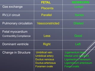
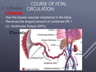
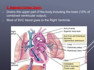
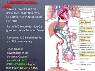
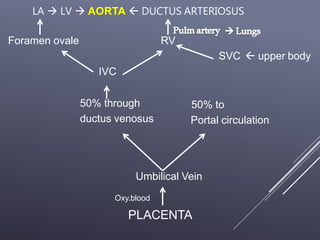
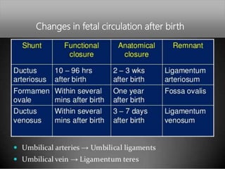
The fetal circulatory system differs significantly from the postnatal circulatory system in several ways. During fetal development, gas exchange occurs via the placenta rather than the lungs. As a result, the fetal circulatory system has four unique shunts that allow blood to bypass the lungs, including the foramen ovale, ductus arteriosus, ductus venosus, and umbilical vessels. This allows oxygenated blood from the placenta to reach the upper body while less oxygenated blood is shunted to the lower body and placenta for reoxygenation. At birth, closure of the shunts and expansion of the lungs causes the circulatory system to transition to adult configuration with
The conotruncus comprises collectively two myocardial subsegments, the conus and the truncus.
Conus is the myocardial segment between ventricle and semi lunar valves which gives rise to sub arterial coni.
Truncus is the fibrous segment between semi lunar valves and aortic sac which gives rise to great arteries.
A transesophageal echocardiogram (TEE) uses echocardiography to assess the structure and function of the heart. During the procedure, a transducer (like a microphone) sends out ultrasonic sound waves. When the transducer is placed at certain locations and angles, the ultrasonic sound waves move through the skin and other body tissues to the heart tissues, where the waves bounce or "echo" off of the heart structures. The transducer picks up the reflected waves and sends them to a computer. The computer displays the echoes as images of the heart walls and valves.
A traditional echocardiogram is done by putting the transducer on the surface of the chest. This is called a transthoracic echocardiogram. A transesophageal echocardiogram is done by inserting a probe with a transducer down the esophagus. This provides a clearer image of the heart because the sound waves do not have to pass through skin, muscle, or bone tissue. The TEE probe is much closer to the heart since the esophagus and heart are right next to each other.
Pulmonary atresia with intact interventricular septum Ramachandra Barik
PA/IVS is a rare congenital cardiac defect that consists of atresia of the pulmonary valve resulting in an absent connection between the right ventricular outflow tract (RVOT) and pulmonary arteries, and an intact ventricular septum that allows no connection between the right and left ventricles
venous drainage of the upper limb, median vein of forearm, deep veins, basilic vein, cephalic vein, median cubital vein, superficial vein, dorsal venous arch,
Blood from the placenta is carried to the fetus by the umbilical vein. In humans, less than a third of this enters the fetal ductus venosus and is carried to the inferior vena cava, while the rest enters the liver proper from the inferior border of the liver. The branch of the umbilical vein that supplies the right lobe of the liver first joins with the portal vein. The blood then moves to the right atrium of the heart. In the fetus, there is an opening between the right and left atrium (the foramen ovale), and most of the blood flows through this hole directly into the left atrium from the right atrium, thus bypassing pulmonary circulation. The continuation of this blood flow is into the left ventricle, and from there it is pumped through the aorta into the body. Some of the blood moves from the aorta through the internal iliac arteries to the umbilical arteries, and re-enters the placenta, where carbon dioxide and other waste products from the fetus are taken up and enter the maternal circulation.
This pdf is about the Schizophrenia.
For more details visit on YouTube; @SELF-EXPLANATORY;
https://www.youtube.com/channel/UCAiarMZDNhe1A3Rnpr_WkzA/videos
Thanks...!
Earliest Galaxies in the JADES Origins Field: Luminosity Function and Cosmic ...Sérgio Sacani
We characterize the earliest galaxy population in the JADES Origins Field (JOF), the deepest
imaging field observed with JWST. We make use of the ancillary Hubble optical images (5 filters
spanning 0.4−0.9µm) and novel JWST images with 14 filters spanning 0.8−5µm, including 7 mediumband filters, and reaching total exposure times of up to 46 hours per filter. We combine all our data
at > 2.3µm to construct an ultradeep image, reaching as deep as ≈ 31.4 AB mag in the stack and
30.3-31.0 AB mag (5σ, r = 0.1” circular aperture) in individual filters. We measure photometric
redshifts and use robust selection criteria to identify a sample of eight galaxy candidates at redshifts
z = 11.5 − 15. These objects show compact half-light radii of R1/2 ∼ 50 − 200pc, stellar masses of
M⋆ ∼ 107−108M⊙, and star-formation rates of SFR ∼ 0.1−1 M⊙ yr−1
. Our search finds no candidates
at 15 < z < 20, placing upper limits at these redshifts. We develop a forward modeling approach to
infer the properties of the evolving luminosity function without binning in redshift or luminosity that
marginalizes over the photometric redshift uncertainty of our candidate galaxies and incorporates the
impact of non-detections. We find a z = 12 luminosity function in good agreement with prior results,
and that the luminosity function normalization and UV luminosity density decline by a factor of ∼ 2.5
from z = 12 to z = 14. We discuss the possible implications of our results in the context of theoretical
models for evolution of the dark matter halo mass function.
Nutraceutical market, scope and growth: Herbal drug technologyLokesh Patil
As consumer awareness of health and wellness rises, the nutraceutical market—which includes goods like functional meals, drinks, and dietary supplements that provide health advantages beyond basic nutrition—is growing significantly. As healthcare expenses rise, the population ages, and people want natural and preventative health solutions more and more, this industry is increasing quickly. Further driving market expansion are product formulation innovations and the use of cutting-edge technology for customized nutrition. With its worldwide reach, the nutraceutical industry is expected to keep growing and provide significant chances for research and investment in a number of categories, including vitamins, minerals, probiotics, and herbal supplements.
Professional air quality monitoring systems provide immediate, on-site data for analysis, compliance, and decision-making.
Monitor common gases, weather parameters, particulates.
Multi-source connectivity as the driver of solar wind variability in the heli...Sérgio Sacani
The ambient solar wind that flls the heliosphere originates from multiple
sources in the solar corona and is highly structured. It is often described
as high-speed, relatively homogeneous, plasma streams from coronal
holes and slow-speed, highly variable, streams whose source regions are
under debate. A key goal of ESA/NASA’s Solar Orbiter mission is to identify
solar wind sources and understand what drives the complexity seen in the
heliosphere. By combining magnetic feld modelling and spectroscopic
techniques with high-resolution observations and measurements, we show
that the solar wind variability detected in situ by Solar Orbiter in March
2022 is driven by spatio-temporal changes in the magnetic connectivity to
multiple sources in the solar atmosphere. The magnetic feld footpoints
connected to the spacecraft moved from the boundaries of a coronal hole
to one active region (12961) and then across to another region (12957). This
is refected in the in situ measurements, which show the transition from fast
to highly Alfvénic then to slow solar wind that is disrupted by the arrival of
a coronal mass ejection. Our results describe solar wind variability at 0.5 au
but are applicable to near-Earth observatories.
3. Introduction:
"Nature is neither lazy nor devoid of foresight.
Having given the matter thought, she knows in
advance that the lung of the fetus does not require
the same arrangements of a perfected lung. She
has therefore anastomosed the pulmonary artery
with the aorta, and the left and right atria. . . .“
-Galen, 2nd Century
4. FETAL NEWBORN
Gas exchange Placenta Lungs
RV,LV circuit Parallel Series
Pulmonary circulation Vasoconstricted Dilated
Fetal myocardium
Contractility,Compliance Less Good
Dominant ventricle Right Left
Change in Structure Umbilical vein
Umbilical artery
Ductus venosus
Ductus arteriosus
Foramen ovale
Ligamentum teres
Medial umb ligament
Ligamentum venosum
Ligamentum arteriosum
Fossa ovalis
7. 2. Superior Vena Cava:
Drains the upper part of the body,including the brain (15% of
combined ventricular output).
Most of SVC blood goes to the Right Ventricle.
8. 3. INFERIOR VENA
CAVA:
DRAINS LOWER PART OF
BODY AND PLACENTA (70%
OF COMBINED VENTRICULAR
OUTPUT)
Part of IVC blood with high O2
goes into LA via Foramen Ovale.
Remaining IVC blood enter RV
and Pulmonary artery.
Since blood is
oxygenated in the
placenta, Oxygen
saturation in IVC
(PO2 =26-28%) is higher
than that in SVC (12-14%).
9. COURSE OF FETAL CIRCULATION:
Most of SVC blood (less oxygenated blood) goes intoRV.
Most of IVC blood (high O2 concentration) is directed by theCrista
Dividens to the LA through Foramen ovale.
Rest of IVC blood enters RV & pulmonaryartery.
Less oxygenated blood in Pulmonary artery flows through Ductus
Arteriosus to descending aorta and then to placenta for
oxygenation.
10. COURSE OF FETAL CIRCULATION:
The Result is:
Brain and coronary circulation receive blood with higher
concentration (PO2 = 28 mm Hg) than the lower part of the
body (PO2 = 24 mm Hg)
11. FETAL CIRCULATION: THE PATHWAY:
PLACENTA OXYGENATED BLOOD UMBILICAL VEIN
Hepatic
circulation
Bypasses liver & joins
IVC via ductus
venosus
Partially mixes with poorly oxygenated
IVC blood derived from lower part of
fetal body
12. FETAL CIRCULATION:
Combined lower body blood plus umbilical venous
blood flow (PO2 of ≈26–28 mm Hg) passes through
IVC to the Right atrium and is preferentially
directed across the foramen ovale to the left atrium.
The blood then flows into the left ventricle and is
ejected into the ascending aorta.
Fetal SVC blood, which is considerably less
oxygenated (PO2 of 12–14 mm Hg), enters the Right
atrium and preferentially traverses the tricuspid
valve, rather thanthe foramen ovale, and flows
primarily to the right ventricle.
13. FETAL CIRCULATION:
From the right ventricle Pulmonary
artery.
Because the pulmonary arterial circulation is
vasoconstricted, only about 10% of right ventricular
outflow enters the lungs.
The rest 90% blood (which has a PO2 of ≈18–22 mm
Hg) bypasses the lungs and flows through the
ductus arteriosus into the descending aorta to
perfuse the lower part of the fetal body.
It the returns to the placenta via the two umbilical
arteries.
14. Thus, upper part of fetal body (including coronary & cerebral
arteries and those to upper extremities) is perfused exclusively
from the Left ventricle with blood that has a slightly higher PO2 ,
than the blood perfusing the lower part of the fetal body, which
is derived mostly from the Right ventricle.
Only a small volume of blood from the ascending aorta (10% of
fetal cardiac output) flows across the aortic isthmus to the
descending aorta.
15. Thus, upper part of fetal body (including coronary & cerebral
arteries and those to upper extremities) is perfused exclusively
from the Left ventricle with blood that has a slightly higher PO2 ,
than the blood perfusing the lower part of the fetal body, which
is derived mostly from the Right ventricle.
Only a small volume of blood from the ascending aorta (10% of
fetal cardiac output) flows across the aortic isthmus to the
descending aorta.
16. LA LV AORTA DUCTUS ARTERIOSUS
Foramen ovale RV
SVC upper body
IVC
50% through
ductus venosus
50% to
Portal circulation
Umbilical Vein
Oxy.blood
PLACENTA
18. FETAL CIRCULATION:
The total fetal cardiac output—the combined output of both
the left and right ventricles—is ≈450mL/kg/min.
Descending aortic blood flow :
-65% returns to placenta;
-Remaining 35% perfuses the fetal organs & tissues.
Right ventricular output is about 1.3 times the left
ventricular flow.
Thus, during fetal life the rightventricle
-is pumping against systemic blood pressure
-is performing greater volume of work than LV.