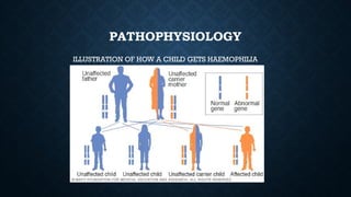
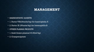
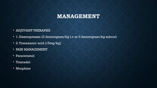
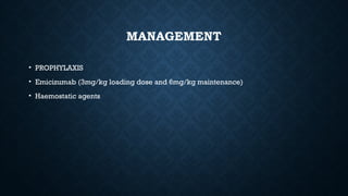
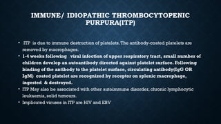
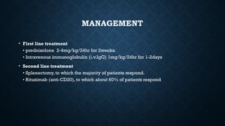
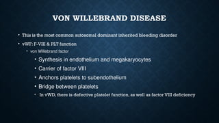
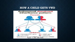
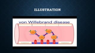
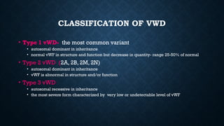
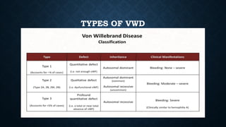
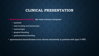
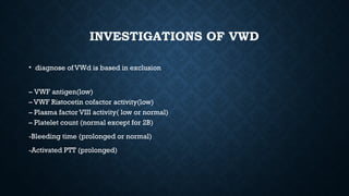
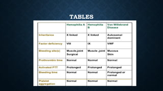
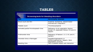
BLEEDING
DISORDERS
BY SAEED MAZIMBA
PRESENTATION CONTENTS
• HAEMOSTASIS
• HAEMOPHILIA (A & B)
• IMMUNE/ IDIOPATHIC THROMBOCYTOPENIC PURPURA(ITP)
• VON WILLEBRAND DISEASE
• REFERENCES
INTRODUCTION BLEEDING DISORDERS
• These are qualitative and quantitative disorders of the various components of the
coagulation cascade and fibrinolytic pathways that results into bleeding
• These disorders can either be congenital or acquired
HEMOSTASIS
• Is the mechanism of stoppage of bleeding after injury of a blood vessel via blood clot
formation and control
• It involve five different components
1. Vascular response
2. Platelet plug formation
3. Coagulation cascade
4. Fibrinolytic system
5. Anticoagulants
HEMOSTASIS
• Hemostasis is divide into two
• 1. primary hemostasis
• 2. secondary hemostasis
PRIMARY HEMOSTASIS
• Is the formation of a weak platelet plug which is achieved in four phases
• 1.Vasoconstriction
• 2. Platelet adhesion
• 3. Platelet activation
• 4. Platelet aggregation
PRIMARY HEMOSTASIS
SECONDARY HEMOSTASIS
• Is the cascade of enzymatic reactions that ultimately results in the conversion of
fibrinogen to fibrin.
• Secondary hemostasis has three pathways each with specific factors;
• 1. Extrinsic pathway (factor III and VII)
• 2. Intrinsic pathway (factor XII, XI, IX and VIII)
• 3. Common pathway ( X,V, II, I and XIII)
CLOTTING FACTORS
CLOTTING CASCADE
CLOTTING
• Intact endothelial lining protects from stimulation of coagulation
• Subendothelium is highly thrombogenic
• Vascular injury exposes subendothelium collagen
• Exposure of endothelial derived vWF
• Stimulation of platelet response ensues
• Also leads to stimulation of anti clotting regulatory mechanisms
BLEEDING DISORDER
• Disorders involve
• Coagulation deficiency (haemophilia A&B)
• Blood vessel disorder (von willebrand disease)
• Platelet dysfunction(ITP)
HAEMOPHILIA
• Haemophilia is a group of congenital bleeding disorders caused by deficiency of
coagulation factor VIII (haemophilia A) or factor IX (haemophilia B) or rarely factor
XI (haemophilia C). Haemophilia A and B are X-linked, whereas haemophilia C is
autosomal recessive.
• Haemophilia A is the classical disease, haemophilia B is also called Christmas
disease
• The deficiency is due to a genetic mutation in the specific clotting factor gene.
However, 30% of cases are non-hereditary resulting from spontaneous mutations
without any family history of haemophilia.
HAEMOPHILIA
• EPIDERMIOLOGY
• About 1 125 000 people wide are haemophilic
• Globally haemophilia A affects 1 in 5000 male births
• Haemophilia A is more common than haemophilia B, representing 80 to 85% of the total
haemophilia population
• About 1700-2000 individuals are estimated to have haemophilia in Zambia
PATHOPHYSIOLOGY
The haemophilia gene sits on the X-chromosome hence the mode of inheritance is X-linked.This
entails that only males are affected; they inherit the abnormal gene from their mothers. Affected
fathers pass the gene to their daughters who become carriers
Bleeding from injuries resulting from activities of daily living is a common occurrence.When this
occurs, the body has inherent mechanisms to stop the bleeding.This is done with the help of
clotting factors of which Factors VIII and IX participate in conjunction with others. In patients with
haemophilia, due to deficiency of factor VIII and/or IX, the clotting mechanism is impaired resulting
in spontaneous, easy and prolonged bleeding. Factor VIII as a co-factor to Factor IX is responsible
for the activation of factor X which has a pivotal role in the coagulation cascade, in intrinsic pathways
for formation of a stable fibrin clot to arrest haemorrhage.
PATHOPHYSIOLOGY
ILLUSTRATION OF HOW A CHILD GETS HAEMOPHILIA
CLINICAL PRESENTATION
• The degree of clinical manifestation of haemophilia depends on severity
CLINICAL PRESENTATION
• Clinical manifestations (hemophilia A & B are indistinguishable). These include;
• Excessive bleeding into various parts of the body
• hemarthroses
• hematomas
• hematuria
• hemorrhage into the central nervous system
• mucous membrane hemorrhage
• dental and surgical bleeding
• Ecchymosis
• Joint pain
ECCHYMOSIS
HEMARTHROSES
• Bleeding into joints accounts for about 75% of bleeding episodes in severely
affected patients
• The joints most frequently involved: knees, elbows, ankles, shoulders , wrists and hips
• Repeated hemarthroses results in destruction of articular cartilage, synovial
hypertrophy and inflammation
• The major complication of repeated bleeding is joint deformity complicated by
muscle atrophy and soft tissue contractures
HEMARTHROSES
NEUROLOGICAL COMPLICATIONS
• Hemorrhage into the central nervous system is the most dangerous event in hemophilic patients
• Intracranial bleeding may be spontaneous or follows trauma, which may be trivial.
• SUBDURAL OR EPIDURAL HEMATOMA
• Hemorrhage into the spinal canal can result in paraplegia
• Peripheral nerve compression is a frequent complication of muscle hematomas, particularly in
the extremities
• SDW
• SSD
COMPLICATIONS
• Acute severe bleed in the cavities
• Psoas bleed
• Infections
• Anthropathy
• Inhibitors develop in 25% of Hemophilia A patients
INVESTIGATIONS
• Bleeding time (normal 7 minutes)
• Prothrombin time (normal <15 seconds)
• Activated partial thromboplastin time (prolonged >45 seconds)
• Serum factor VIII and IX assay (low)
• Mixing study
MANAGEMENT
• HAEMOSTATIC AGENTS
• 1. Factor VIII(30units/kg) for haemophilia A
• 2. Factor IX (50units/kg) for haemophilia B
• OTHER PLASMA PRODUTS
• 1. fresh frozen plasma(15-20ml/kg)
• 2. Cryoprecipitate
MANAGEMENT
• ADJUVANT THERAPIES
• 1. Desmopressin (0.3microgram/kg i.v or 0.4microgram/kg subcut)
• 2.Tranexamic acid (15mg/kg)
• PAIN MANAGEMENT
• Paracetamol
• Tramadol
• Morphine
MANAGEMENT
• PROPHYLAXIS
• Emicizumab (3mg/kg loading dose and 6mg/kg maintenance)
• Haemostatic agents
IMMUNE/ IDIOPATHIC THROMBOCYTOPENIC
PURPURA(ITP)
• ITP is due to immune destruction of platelets.The antibody-coated platelets are
removed by macrophages.
• 1-4 weeks following viral infection of upper respiratory tract, small number of
children develop an autoantibody directed against platelet surface. Following
binding of the antibody to the platelet surface, circulating antibody(IgG OR
IgM) coated platelet are recognized by receptor on splenic macrophage,
ingested & destroyed.
• ITP May also be associated with other autoimmune disorder, chronic lymphocytic
leukaemia, solid tumours.
• Implicated viruses in ITP are HIV and EBV
CLINICAL PRESENTATIONS
1-the classic presentation of ITP from 1-4 years old with sudden onset is generalized petechiae &
purpura.
2-often there is bleeding from gums & mucous membrane.
3- splenomegaly are rare, also lymphadenopathy or hepatosplenomegaly.
4-70 to 80% of children who present with acute ITP will have spontaneous resolution of their ITP
within 6 months, if not the disease become chronic
5-less than 1% of cases develop intracranial hemorrhage.
6-epistaxis
7-menorrhagia
CLINICAL PRESENTATIONS
INVESTIGATIONS
Diagnosis of ITP usually is based on clinical presentation and the platelet count
LAB:
Bleeding Time (high>7minutes)
Full blood count ( low platelet count < 30,000)
Clotting times (normal)
Morphology
Peripheral Blood
thrombocytopenia, abnormally large platelets ( Giant platelets)
Marrow
Normal or Increased megakaryocyte
MANAGEMENT
• Patients with platelet counts >30 × 109/L generally require no treatment unless they
are about to undergo a surgical procedure or have spontaneous bruising or
bleeding.
• Platelet transfusions are reserved for intracranial or other extreme haemorrhage,
where emergency splenectomy may be justified
MANAGEMENT
• First line treatment
• prednisolone 2-4mg/kg/24hr for 2weeks.
• Intravenous immunoglobulin (i.v.IgG) 1mg/kg/24hr for 1-2days
• Second line treatment
• Splenectomy, to which the majority of patients respond.
• Rituximab (anti-CD20), to which about 60% of patients respond
VON WILLEBRAND DISEASE
• This is the most common autosomal dominant inherited bleeding disorder
• vWF: F-VIII & PLT function
• von Willebrand factor
• Synthesis in endothelium and megakaryocytes
• Carrier of factor VIII
• Anchors platelets to subendothelium
• Bridge between platelets
• In vWD, there is defective platelet function, as well as factor VIII deficiency
HOW A CHILD GETS VWD
ILLUSTRATION
CLASSIFICATION OF VWD
• Type 1 vWD- the most common variant
• autosomal dominant in inheritance
• normal vWF in structure and function but decrease in quantity- range 25-50% of normal
• Type 2 vWD (2A, 2B, 2M, 2N)
• autosomal dominant in inheritance
• vWF is abnormal in structure and/or function
• Type 3 vWD
• autosomal recessive in inheritance
• the most severe form characterized by very low or undetectable level of vWF
TYPES OF VWD
CLINICAL PRESENTATION
• Mucocutaneous bleeding- the most common symptom
• epistaxis
• easy bruising and hematomas
• menorrhagia
• gingival bleeding
• gastrointestinal bleeding
• spontaneous hemarthroses occur almost exclusively in patients with type 3 vWD
INVESTIGATIONS OF VWD
• diagnose of VWd is based in exclusion
– VWF antigen(low)
– VWF Ristocetin cofactor activity(low)
– Plasma factor VIII activity( low or normal)
– Platelet count (normal except for 2B)
-Bleeding time (prolonged or normal)
-Activated PTT (prolonged)
MANAGEMENT
•
-Plasma-derived factor VIII concentrates containing intact vWF and are the current
mainstay of replacement therapy.
-Desmopressin is a treatment option where possible
MANAGEMENT
• Factor viii dosage for vWD
Type 1
• Loading dose: 40 to 60 international units vWF:RCo/kg IV
• Maintenance dose: 40 to 50 international units vWF:RCo/kg IV every 8 to 12 hours for 3 days
to keep vWF:RCo trough greater than 50%
Type 2
• Loading dose: 60 to 80 international units vWF:RCo/kg IV
• Maintenance dose: 40 to 60 international units vWF:RCo/kg IV every 8 to 12 hours for 3 days
to keep VWF:RCo trough greater than 50%
Type 3
• Loading dose: 60 to 80 international units vWF:RCo/kg IV
• Maintenance dose: 40 to 60 international units vWF:RCo/kg IV every 12 to 24 hours for 5 to 7
days, keeping vWF:RCo and FVIII troughs greater than 50%
MANAGEMENT
• Desmopressin Dosage in vWD
• 0.3 mcg/kg i.v once slowly over 15-30 minutes
• Intranasal: 1 spray (1.5 mg/mL) in each nostril one time
TABLES
TABLES
REFERENCES
• ZAMBIA NATIONAL GUIDELINE FOR THE MANAGEMENT OF HAEMOPHILIA
• NELSONS ESSENTIAL OF PAEDIATRICS
• PAEDIATRIC BOARD STUDY GUIDE
• APPROACH TO BLEEDING DISORDER ( BY DR MWANDAMA)
• BLEEDING DISORDER (BY DR CHUNDA)

COMMON BLEEDING DISORDERS IN CHILDREN_TWO.pptx

  • 1.
  • 2.
    PRESENTATION CONTENTS • HAEMOSTASIS •HAEMOPHILIA (A & B) • IMMUNE/ IDIOPATHIC THROMBOCYTOPENIC PURPURA(ITP) • VON WILLEBRAND DISEASE • REFERENCES
  • 3.
    INTRODUCTION BLEEDING DISORDERS •These are qualitative and quantitative disorders of the various components of the coagulation cascade and fibrinolytic pathways that results into bleeding • These disorders can either be congenital or acquired
  • 4.
    HEMOSTASIS • Is themechanism of stoppage of bleeding after injury of a blood vessel via blood clot formation and control • It involve five different components 1. Vascular response 2. Platelet plug formation 3. Coagulation cascade 4. Fibrinolytic system 5. Anticoagulants
  • 5.
    HEMOSTASIS • Hemostasis isdivide into two • 1. primary hemostasis • 2. secondary hemostasis
  • 6.
    PRIMARY HEMOSTASIS • Isthe formation of a weak platelet plug which is achieved in four phases • 1.Vasoconstriction • 2. Platelet adhesion • 3. Platelet activation • 4. Platelet aggregation
  • 7.
  • 8.
    SECONDARY HEMOSTASIS • Isthe cascade of enzymatic reactions that ultimately results in the conversion of fibrinogen to fibrin. • Secondary hemostasis has three pathways each with specific factors; • 1. Extrinsic pathway (factor III and VII) • 2. Intrinsic pathway (factor XII, XI, IX and VIII) • 3. Common pathway ( X,V, II, I and XIII)
  • 9.
  • 10.
  • 11.
    CLOTTING • Intact endotheliallining protects from stimulation of coagulation • Subendothelium is highly thrombogenic • Vascular injury exposes subendothelium collagen • Exposure of endothelial derived vWF • Stimulation of platelet response ensues • Also leads to stimulation of anti clotting regulatory mechanisms
  • 12.
    BLEEDING DISORDER • Disordersinvolve • Coagulation deficiency (haemophilia A&B) • Blood vessel disorder (von willebrand disease) • Platelet dysfunction(ITP)
  • 13.
    HAEMOPHILIA • Haemophilia isa group of congenital bleeding disorders caused by deficiency of coagulation factor VIII (haemophilia A) or factor IX (haemophilia B) or rarely factor XI (haemophilia C). Haemophilia A and B are X-linked, whereas haemophilia C is autosomal recessive. • Haemophilia A is the classical disease, haemophilia B is also called Christmas disease • The deficiency is due to a genetic mutation in the specific clotting factor gene. However, 30% of cases are non-hereditary resulting from spontaneous mutations without any family history of haemophilia.
  • 14.
    HAEMOPHILIA • EPIDERMIOLOGY • About1 125 000 people wide are haemophilic • Globally haemophilia A affects 1 in 5000 male births • Haemophilia A is more common than haemophilia B, representing 80 to 85% of the total haemophilia population • About 1700-2000 individuals are estimated to have haemophilia in Zambia
  • 15.
    PATHOPHYSIOLOGY The haemophilia genesits on the X-chromosome hence the mode of inheritance is X-linked.This entails that only males are affected; they inherit the abnormal gene from their mothers. Affected fathers pass the gene to their daughters who become carriers Bleeding from injuries resulting from activities of daily living is a common occurrence.When this occurs, the body has inherent mechanisms to stop the bleeding.This is done with the help of clotting factors of which Factors VIII and IX participate in conjunction with others. In patients with haemophilia, due to deficiency of factor VIII and/or IX, the clotting mechanism is impaired resulting in spontaneous, easy and prolonged bleeding. Factor VIII as a co-factor to Factor IX is responsible for the activation of factor X which has a pivotal role in the coagulation cascade, in intrinsic pathways for formation of a stable fibrin clot to arrest haemorrhage.
  • 16.
    PATHOPHYSIOLOGY ILLUSTRATION OF HOWA CHILD GETS HAEMOPHILIA
  • 17.
    CLINICAL PRESENTATION • Thedegree of clinical manifestation of haemophilia depends on severity
  • 18.
    CLINICAL PRESENTATION • Clinicalmanifestations (hemophilia A & B are indistinguishable). These include; • Excessive bleeding into various parts of the body • hemarthroses • hematomas • hematuria • hemorrhage into the central nervous system • mucous membrane hemorrhage • dental and surgical bleeding • Ecchymosis • Joint pain
  • 19.
  • 20.
    HEMARTHROSES • Bleeding intojoints accounts for about 75% of bleeding episodes in severely affected patients • The joints most frequently involved: knees, elbows, ankles, shoulders , wrists and hips • Repeated hemarthroses results in destruction of articular cartilage, synovial hypertrophy and inflammation • The major complication of repeated bleeding is joint deformity complicated by muscle atrophy and soft tissue contractures
  • 21.
  • 22.
    NEUROLOGICAL COMPLICATIONS • Hemorrhageinto the central nervous system is the most dangerous event in hemophilic patients • Intracranial bleeding may be spontaneous or follows trauma, which may be trivial. • SUBDURAL OR EPIDURAL HEMATOMA • Hemorrhage into the spinal canal can result in paraplegia • Peripheral nerve compression is a frequent complication of muscle hematomas, particularly in the extremities • SDW • SSD
  • 23.
    COMPLICATIONS • Acute severebleed in the cavities • Psoas bleed • Infections • Anthropathy • Inhibitors develop in 25% of Hemophilia A patients
  • 24.
    INVESTIGATIONS • Bleeding time(normal 7 minutes) • Prothrombin time (normal <15 seconds) • Activated partial thromboplastin time (prolonged >45 seconds) • Serum factor VIII and IX assay (low) • Mixing study
  • 25.
    MANAGEMENT • HAEMOSTATIC AGENTS •1. Factor VIII(30units/kg) for haemophilia A • 2. Factor IX (50units/kg) for haemophilia B • OTHER PLASMA PRODUTS • 1. fresh frozen plasma(15-20ml/kg) • 2. Cryoprecipitate
  • 26.
    MANAGEMENT • ADJUVANT THERAPIES •1. Desmopressin (0.3microgram/kg i.v or 0.4microgram/kg subcut) • 2.Tranexamic acid (15mg/kg) • PAIN MANAGEMENT • Paracetamol • Tramadol • Morphine
  • 27.
    MANAGEMENT • PROPHYLAXIS • Emicizumab(3mg/kg loading dose and 6mg/kg maintenance) • Haemostatic agents
  • 28.
    IMMUNE/ IDIOPATHIC THROMBOCYTOPENIC PURPURA(ITP) •ITP is due to immune destruction of platelets.The antibody-coated platelets are removed by macrophages. • 1-4 weeks following viral infection of upper respiratory tract, small number of children develop an autoantibody directed against platelet surface. Following binding of the antibody to the platelet surface, circulating antibody(IgG OR IgM) coated platelet are recognized by receptor on splenic macrophage, ingested & destroyed. • ITP May also be associated with other autoimmune disorder, chronic lymphocytic leukaemia, solid tumours. • Implicated viruses in ITP are HIV and EBV
  • 29.
    CLINICAL PRESENTATIONS 1-the classicpresentation of ITP from 1-4 years old with sudden onset is generalized petechiae & purpura. 2-often there is bleeding from gums & mucous membrane. 3- splenomegaly are rare, also lymphadenopathy or hepatosplenomegaly. 4-70 to 80% of children who present with acute ITP will have spontaneous resolution of their ITP within 6 months, if not the disease become chronic 5-less than 1% of cases develop intracranial hemorrhage. 6-epistaxis 7-menorrhagia
  • 30.
  • 31.
    INVESTIGATIONS Diagnosis of ITPusually is based on clinical presentation and the platelet count LAB: Bleeding Time (high>7minutes) Full blood count ( low platelet count < 30,000) Clotting times (normal) Morphology Peripheral Blood thrombocytopenia, abnormally large platelets ( Giant platelets) Marrow Normal or Increased megakaryocyte
  • 32.
    MANAGEMENT • Patients withplatelet counts >30 × 109/L generally require no treatment unless they are about to undergo a surgical procedure or have spontaneous bruising or bleeding. • Platelet transfusions are reserved for intracranial or other extreme haemorrhage, where emergency splenectomy may be justified
  • 33.
    MANAGEMENT • First linetreatment • prednisolone 2-4mg/kg/24hr for 2weeks. • Intravenous immunoglobulin (i.v.IgG) 1mg/kg/24hr for 1-2days • Second line treatment • Splenectomy, to which the majority of patients respond. • Rituximab (anti-CD20), to which about 60% of patients respond
  • 34.
    VON WILLEBRAND DISEASE •This is the most common autosomal dominant inherited bleeding disorder • vWF: F-VIII & PLT function • von Willebrand factor • Synthesis in endothelium and megakaryocytes • Carrier of factor VIII • Anchors platelets to subendothelium • Bridge between platelets • In vWD, there is defective platelet function, as well as factor VIII deficiency
  • 35.
    HOW A CHILDGETS VWD
  • 36.
  • 37.
    CLASSIFICATION OF VWD •Type 1 vWD- the most common variant • autosomal dominant in inheritance • normal vWF in structure and function but decrease in quantity- range 25-50% of normal • Type 2 vWD (2A, 2B, 2M, 2N) • autosomal dominant in inheritance • vWF is abnormal in structure and/or function • Type 3 vWD • autosomal recessive in inheritance • the most severe form characterized by very low or undetectable level of vWF
  • 38.
  • 39.
    CLINICAL PRESENTATION • Mucocutaneousbleeding- the most common symptom • epistaxis • easy bruising and hematomas • menorrhagia • gingival bleeding • gastrointestinal bleeding • spontaneous hemarthroses occur almost exclusively in patients with type 3 vWD
  • 40.
    INVESTIGATIONS OF VWD •diagnose of VWd is based in exclusion – VWF antigen(low) – VWF Ristocetin cofactor activity(low) – Plasma factor VIII activity( low or normal) – Platelet count (normal except for 2B) -Bleeding time (prolonged or normal) -Activated PTT (prolonged)
  • 41.
    MANAGEMENT • -Plasma-derived factor VIIIconcentrates containing intact vWF and are the current mainstay of replacement therapy. -Desmopressin is a treatment option where possible
  • 42.
    MANAGEMENT • Factor viiidosage for vWD Type 1 • Loading dose: 40 to 60 international units vWF:RCo/kg IV • Maintenance dose: 40 to 50 international units vWF:RCo/kg IV every 8 to 12 hours for 3 days to keep vWF:RCo trough greater than 50% Type 2 • Loading dose: 60 to 80 international units vWF:RCo/kg IV • Maintenance dose: 40 to 60 international units vWF:RCo/kg IV every 8 to 12 hours for 3 days to keep VWF:RCo trough greater than 50% Type 3 • Loading dose: 60 to 80 international units vWF:RCo/kg IV • Maintenance dose: 40 to 60 international units vWF:RCo/kg IV every 12 to 24 hours for 5 to 7 days, keeping vWF:RCo and FVIII troughs greater than 50%
  • 43.
    MANAGEMENT • Desmopressin Dosagein vWD • 0.3 mcg/kg i.v once slowly over 15-30 minutes • Intranasal: 1 spray (1.5 mg/mL) in each nostril one time
  • 44.
  • 45.
  • 46.
    REFERENCES • ZAMBIA NATIONALGUIDELINE FOR THE MANAGEMENT OF HAEMOPHILIA • NELSONS ESSENTIAL OF PAEDIATRICS • PAEDIATRIC BOARD STUDY GUIDE • APPROACH TO BLEEDING DISORDER ( BY DR MWANDAMA) • BLEEDING DISORDER (BY DR CHUNDA)