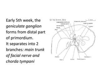
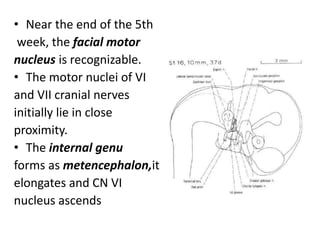
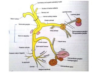
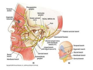
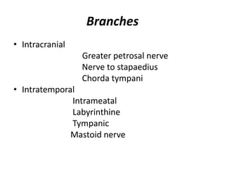
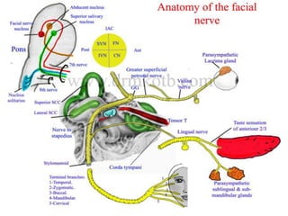
surgical anatomy of nose is a humble attempt to make the anatomy of nose simpler and easy for medical students and fellow physicians. at the end of the presentation the students will be able to identify all the structures.
FACIAL NERVE AND IT'S APPLIED ANATOMY AND IT'S SIGNIFICANCE FOR A DENTIST ALONG WITH THE CAUTIONS TO AVOID AN IATROGENIC INJURY TO FACIAL NERVE AND THE MANAGEMENT OF A PATIENT OF FACIAL NERVE DISORDER DURING ENDODONTIC PROCEDURES
The Larynx: Anatomy, Function, and Disorders
Introduction
The larynx, commonly known as the voice box, is a vital structure in the human body responsible for a multitude of functions, the most prominent of which is voice production. This complex organ plays a crucial role in breathing, swallowing, and protecting the airway. Understanding the anatomy, function, and common disorders of the larynx is essential for grasping its significance in our daily lives. In this comprehensive 2000-word essay, we will explore the larynx in detail, delving into its anatomy, function, the mechanics of voice production, common laryngeal disorders, and their treatment.
I. Anatomy of the Larynx
The larynx is a complex structure located in the neck, connecting the lower part of the pharynx to the trachea. It comprises several cartilages, muscles, ligaments, and other anatomical components that work together to facilitate various functions. To understand the larynx better, it is crucial to break down its anatomy into its constituent parts.
Cartilages
A. Thyroid Cartilage: The thyroid cartilage, often referred to as the Adam's apple, is the most prominent and easily palpable cartilage of the larynx. It is made up of two fused plates and provides structural support to the front of the larynx.
B. Cricoid Cartilage: The cricoid cartilage is a ring-like structure that sits just below the thyroid cartilage. It plays a crucial role in connecting the larynx to the trachea and provides structural support.
C. Epiglottis: The epiglottis is a leaf-shaped cartilage located behind the tongue root. It acts as a lid to cover the entrance of the trachea during swallowing, preventing food and liquids from entering the airway.
D. Arytenoid Cartilages: These paired cartilages are located on top of the cricoid cartilage. They play a pivotal role in controlling vocal cord tension and movement.
E. Corniculate and Cuneiform Cartilages: These smaller cartilages are positioned within the aryepiglottic folds and aid in maintaining the laryngeal structure.
Muscles
A. Intrinsic Laryngeal Muscles: These muscles are responsible for controlling the position and tension of the vocal cords. Key intrinsic muscles include the cricothyroid, thyroarytenoid, lateral cricoarytenoid, posterior cricoarytenoid, and interarytenoid muscles.
B. Extrinsic Laryngeal Muscles: Extrinsic muscles are responsible for moving the larynx as a whole, helping with functions such as swallowing and speech. The sternothyroid, thyrohyoid, and omohyoid muscles are examples of extrinsic laryngeal muscles.
Vocal Cords
The vocal cords, or vocal folds, are a pair of muscular structures located within the larynx. They are composed of layers of mucous membrane, muscle, and connective tissue. The true vocal cords, also known as the vocal ligaments, are the structures primarily responsible for sound production. They are capable of opening and closing rapidly to produce sound when air flows through them.
facial nerve is the seventh cranial nerve supplies the submandibular, sublingual, lacrimal glands, the mucosal glands of the nose, palate, pharynx and taste fibres, and on being injured it leads to loss of lacrimation, loss of salivation, loss of taste sensation and paralysis of the muscles of facial expression.
facial nerve is the 7th cranial nerve. it supplies the parts of the face and also the muscles of mastication. it helps in the expression of the face too.
Facial nerve and its extracranial and intracranial rotssonambohra2
facial nerve its origin and insertion and its extracranial and intracranial roots and its branches and clinical significance and its related syndromes explained well along with treatment plan
NVBDCP.pptx Nation vector borne disease control programSapna Thakur
NVBDCP was launched in 2003-2004 . Vector-Borne Disease: Disease that results from an infection transmitted to humans and other animals by blood-feeding arthropods, such as mosquitoes, ticks, and fleas. Examples of vector-borne diseases include Dengue fever, West Nile Virus, Lyme disease, and malaria.
These simplified slides by Dr. Sidra Arshad present an overview of the non-respiratory functions of the respiratory tract.
Learning objectives:
1. Enlist the non-respiratory functions of the respiratory tract
2. Briefly explain how these functions are carried out
3. Discuss the significance of dead space
4. Differentiate between minute ventilation and alveolar ventilation
5. Describe the cough and sneeze reflexes
Study Resources:
1. Chapter 39, Guyton and Hall Textbook of Medical Physiology, 14th edition
2. Chapter 34, Ganong’s Review of Medical Physiology, 26th edition
3. Chapter 17, Human Physiology by Lauralee Sherwood, 9th edition
4. Non-respiratory functions of the lungs https://academic.oup.com/bjaed/article/13/3/98/278874
Title: Sense of Taste
Presenter: Dr. Faiza, Assistant Professor of Physiology
Qualifications:
MBBS (Best Graduate, AIMC Lahore)
FCPS Physiology
ICMT, CHPE, DHPE (STMU)
MPH (GC University, Faisalabad)
MBA (Virtual University of Pakistan)
Learning Objectives:
Describe the structure and function of taste buds.
Describe the relationship between the taste threshold and taste index of common substances.
Explain the chemical basis and signal transduction of taste perception for each type of primary taste sensation.
Recognize different abnormalities of taste perception and their causes.
Key Topics:
Significance of Taste Sensation:
Differentiation between pleasant and harmful food
Influence on behavior
Selection of food based on metabolic needs
Receptors of Taste:
Taste buds on the tongue
Influence of sense of smell, texture of food, and pain stimulation (e.g., by pepper)
Primary and Secondary Taste Sensations:
Primary taste sensations: Sweet, Sour, Salty, Bitter, Umami
Chemical basis and signal transduction mechanisms for each taste
Taste Threshold and Index:
Taste threshold values for Sweet (sucrose), Salty (NaCl), Sour (HCl), and Bitter (Quinine)
Taste index relationship: Inversely proportional to taste threshold
Taste Blindness:
Inability to taste certain substances, particularly thiourea compounds
Example: Phenylthiocarbamide
Structure and Function of Taste Buds:
Composition: Epithelial cells, Sustentacular/Supporting cells, Taste cells, Basal cells
Features: Taste pores, Taste hairs/microvilli, and Taste nerve fibers
Location of Taste Buds:
Found in papillae of the tongue (Fungiform, Circumvallate, Foliate)
Also present on the palate, tonsillar pillars, epiglottis, and proximal esophagus
Mechanism of Taste Stimulation:
Interaction of taste substances with receptors on microvilli
Signal transduction pathways for Umami, Sweet, Bitter, Sour, and Salty tastes
Taste Sensitivity and Adaptation:
Decrease in sensitivity with age
Rapid adaptation of taste sensation
Role of Saliva in Taste:
Dissolution of tastants to reach receptors
Washing away the stimulus
Taste Preferences and Aversions:
Mechanisms behind taste preference and aversion
Influence of receptors and neural pathways
Impact of Sensory Nerve Damage:
Degeneration of taste buds if the sensory nerve fiber is cut
Abnormalities of Taste Detection:
Conditions: Ageusia, Hypogeusia, Dysgeusia (parageusia)
Causes: Nerve damage, neurological disorders, infections, poor oral hygiene, adverse drug effects, deficiencies, aging, tobacco use, altered neurotransmitter levels
Neurotransmitters and Taste Threshold:
Effects of serotonin (5-HT) and norepinephrine (NE) on taste sensitivity
Supertasters:
25% of the population with heightened sensitivity to taste, especially bitterness
Increased number of fungiform papillae
Ethanol (CH3CH2OH), or beverage alcohol, is a two-carbon alcohol
that is rapidly distributed in the body and brain. Ethanol alters many
neurochemical systems and has rewarding and addictive properties. It
is the oldest recreational drug and likely contributes to more morbidity,
mortality, and public health costs than all illicit drugs combined. The
5th edition of the Diagnostic and Statistical Manual of Mental Disorders
(DSM-5) integrates alcohol abuse and alcohol dependence into a single
disorder called alcohol use disorder (AUD), with mild, moderate,
and severe subclassifications (American Psychiatric Association, 2013).
In the DSM-5, all types of substance abuse and dependence have been
combined into a single substance use disorder (SUD) on a continuum
from mild to severe. A diagnosis of AUD requires that at least two of
the 11 DSM-5 behaviors be present within a 12-month period (mild
AUD: 2–3 criteria; moderate AUD: 4–5 criteria; severe AUD: 6–11 criteria).
The four main behavioral effects of AUD are impaired control over
drinking, negative social consequences, risky use, and altered physiological
effects (tolerance, withdrawal). This chapter presents an overview
of the prevalence and harmful consequences of AUD in the U.S.,
the systemic nature of the disease, neurocircuitry and stages of AUD,
comorbidities, fetal alcohol spectrum disorders, genetic risk factors, and
pharmacotherapies for AUD.
Prix Galien International 2024 Forum ProgramLevi Shapiro
June 20, 2024, Prix Galien International and Jerusalem Ethics Forum in ROME. Detailed agenda including panels:
- ADVANCES IN CARDIOLOGY: A NEW PARADIGM IS COMING
- WOMEN’S HEALTH: FERTILITY PRESERVATION
- WHAT’S NEW IN THE TREATMENT OF INFECTIOUS,
ONCOLOGICAL AND INFLAMMATORY SKIN DISEASES?
- ARTIFICIAL INTELLIGENCE AND ETHICS
- GENE THERAPY
- BEYOND BORDERS: GLOBAL INITIATIVES FOR DEMOCRATIZING LIFE SCIENCE TECHNOLOGIES AND PROMOTING ACCESS TO HEALTHCARE
- ETHICAL CHALLENGES IN LIFE SCIENCES
- Prix Galien International Awards Ceremony
Couples presenting to the infertility clinic- Do they really have infertility...Sujoy Dasgupta
Dr Sujoy Dasgupta presented the study on "Couples presenting to the infertility clinic- Do they really have infertility? – The unexplored stories of non-consummation" in the 13th Congress of the Asia Pacific Initiative on Reproduction (ASPIRE 2024) at Manila on 24 May, 2024.
micro teaching on communication m.sc nursing.pdfAnurag Sharma
Microteaching is a unique model of practice teaching. It is a viable instrument for the. desired change in the teaching behavior or the behavior potential which, in specified types of real. classroom situations, tends to facilitate the achievement of specified types of objectives.
Flu Vaccine Alert in Bangalore Karnatakaaddon Scans
As flu season approaches, health officials in Bangalore, Karnataka, are urging residents to get their flu vaccinations. The seasonal flu, while common, can lead to severe health complications, particularly for vulnerable populations such as young children, the elderly, and those with underlying health conditions.
Dr. Vidisha Kumari, a leading epidemiologist in Bangalore, emphasizes the importance of getting vaccinated. "The flu vaccine is our best defense against the influenza virus. It not only protects individuals but also helps prevent the spread of the virus in our communities," he says.
This year, the flu season is expected to coincide with a potential increase in other respiratory illnesses. The Karnataka Health Department has launched an awareness campaign highlighting the significance of flu vaccinations. They have set up multiple vaccination centers across Bangalore, making it convenient for residents to receive their shots.
To encourage widespread vaccination, the government is also collaborating with local schools, workplaces, and community centers to facilitate vaccination drives. Special attention is being given to ensuring that the vaccine is accessible to all, including marginalized communities who may have limited access to healthcare.
Residents are reminded that the flu vaccine is safe and effective. Common side effects are mild and may include soreness at the injection site, mild fever, or muscle aches. These side effects are generally short-lived and far less severe than the flu itself.
Healthcare providers are also stressing the importance of continuing COVID-19 precautions. Wearing masks, practicing good hand hygiene, and maintaining social distancing are still crucial, especially in crowded places.
Protect yourself and your loved ones by getting vaccinated. Together, we can help keep Bangalore healthy and safe this flu season. For more information on vaccination centers and schedules, residents can visit the Karnataka Health Department’s official website or follow their social media pages.
Stay informed, stay safe, and get your flu shot today!
Title: Sense of Smell
Presenter: Dr. Faiza, Assistant Professor of Physiology
Qualifications:
MBBS (Best Graduate, AIMC Lahore)
FCPS Physiology
ICMT, CHPE, DHPE (STMU)
MPH (GC University, Faisalabad)
MBA (Virtual University of Pakistan)
Learning Objectives:
Describe the primary categories of smells and the concept of odor blindness.
Explain the structure and location of the olfactory membrane and mucosa, including the types and roles of cells involved in olfaction.
Describe the pathway and mechanisms of olfactory signal transmission from the olfactory receptors to the brain.
Illustrate the biochemical cascade triggered by odorant binding to olfactory receptors, including the role of G-proteins and second messengers in generating an action potential.
Identify different types of olfactory disorders such as anosmia, hyposmia, hyperosmia, and dysosmia, including their potential causes.
Key Topics:
Olfactory Genes:
3% of the human genome accounts for olfactory genes.
400 genes for odorant receptors.
Olfactory Membrane:
Located in the superior part of the nasal cavity.
Medially: Folds downward along the superior septum.
Laterally: Folds over the superior turbinate and upper surface of the middle turbinate.
Total surface area: 5-10 square centimeters.
Olfactory Mucosa:
Olfactory Cells: Bipolar nerve cells derived from the CNS (100 million), with 4-25 olfactory cilia per cell.
Sustentacular Cells: Produce mucus and maintain ionic and molecular environment.
Basal Cells: Replace worn-out olfactory cells with an average lifespan of 1-2 months.
Bowman’s Gland: Secretes mucus.
Stimulation of Olfactory Cells:
Odorant dissolves in mucus and attaches to receptors on olfactory cilia.
Involves a cascade effect through G-proteins and second messengers, leading to depolarization and action potential generation in the olfactory nerve.
Quality of a Good Odorant:
Small (3-20 Carbon atoms), volatile, water-soluble, and lipid-soluble.
Facilitated by odorant-binding proteins in mucus.
Membrane Potential and Action Potential:
Resting membrane potential: -55mV.
Action potential frequency in the olfactory nerve increases with odorant strength.
Adaptation Towards the Sense of Smell:
Rapid adaptation within the first second, with further slow adaptation.
Psychological adaptation greater than receptor adaptation, involving feedback inhibition from the central nervous system.
Primary Sensations of Smell:
Camphoraceous, Musky, Floral, Pepperminty, Ethereal, Pungent, Putrid.
Odor Detection Threshold:
Examples: Hydrogen sulfide (0.0005 ppm), Methyl-mercaptan (0.002 ppm).
Some toxic substances are odorless at lethal concentrations.
Characteristics of Smell:
Odor blindness for single substances due to lack of appropriate receptor protein.
Behavioral and emotional influences of smell.
Transmission of Olfactory Signals:
From olfactory cells to glomeruli in the olfactory bulb, involving lateral inhibition.
Primitive, less old, and new olfactory systems with different path
New Directions in Targeted Therapeutic Approaches for Older Adults With Mantl...i3 Health
i3 Health is pleased to make the speaker slides from this activity available for use as a non-accredited self-study or teaching resource.
This slide deck presented by Dr. Kami Maddocks, Professor-Clinical in the Division of Hematology and
Associate Division Director for Ambulatory Operations
The Ohio State University Comprehensive Cancer Center, will provide insight into new directions in targeted therapeutic approaches for older adults with mantle cell lymphoma.
STATEMENT OF NEED
Mantle cell lymphoma (MCL) is a rare, aggressive B-cell non-Hodgkin lymphoma (NHL) accounting for 5% to 7% of all lymphomas. Its prognosis ranges from indolent disease that does not require treatment for years to very aggressive disease, which is associated with poor survival (Silkenstedt et al, 2021). Typically, MCL is diagnosed at advanced stage and in older patients who cannot tolerate intensive therapy (NCCN, 2022). Although recent advances have slightly increased remission rates, recurrence and relapse remain very common, leading to a median overall survival between 3 and 6 years (LLS, 2021). Though there are several effective options, progress is still needed towards establishing an accepted frontline approach for MCL (Castellino et al, 2022). Treatment selection and management of MCL are complicated by the heterogeneity of prognosis, advanced age and comorbidities of patients, and lack of an established standard approach for treatment, making it vital that clinicians be familiar with the latest research and advances in this area. In this activity chaired by Michael Wang, MD, Professor in the Department of Lymphoma & Myeloma at MD Anderson Cancer Center, expert faculty will discuss prognostic factors informing treatment, the promising results of recent trials in new therapeutic approaches, and the implications of treatment resistance in therapeutic selection for MCL.
Target Audience
Hematology/oncology fellows, attending faculty, and other health care professionals involved in the treatment of patients with mantle cell lymphoma (MCL).
Learning Objectives
1.) Identify clinical and biological prognostic factors that can guide treatment decision making for older adults with MCL
2.) Evaluate emerging data on targeted therapeutic approaches for treatment-naive and relapsed/refractory MCL and their applicability to older adults
3.) Assess mechanisms of resistance to targeted therapies for MCL and their implications for treatment selection
HOT NEW PRODUCT! BIG SALES FAST SHIPPING NOW FROM CHINA!! EU KU DB BK substit...GL Anaacs
Contact us if you are interested:
Email / Skype : kefaya1771@gmail.com
Threema: PXHY5PDH
New BATCH Ku !!! MUCH IN DEMAND FAST SALE EVERY BATCH HAPPY GOOD EFFECT BIG BATCH !
Contact me on Threema or skype to start big business!!
Hot-sale products:
NEW HOT EUTYLONE WHITE CRYSTAL!!
5cl-adba precursor (semi finished )
5cl-adba raw materials
ADBB precursor (semi finished )
ADBB raw materials
APVP powder
5fadb/4f-adb
Jwh018 / Jwh210
Eutylone crystal
Protonitazene (hydrochloride) CAS: 119276-01-6
Flubrotizolam CAS: 57801-95-3
Metonitazene CAS: 14680-51-4
Payment terms: Western Union,MoneyGram,Bitcoin or USDT.
Deliver Time: Usually 7-15days
Shipping method: FedEx, TNT, DHL,UPS etc.Our deliveries are 100% safe, fast, reliable and discreet.
Samples will be sent for your evaluation!If you are interested in, please contact me, let's talk details.
We specializes in exporting high quality Research chemical, medical intermediate, Pharmaceutical chemicals and so on. Products are exported to USA, Canada, France, Korea, Japan,Russia, Southeast Asia and other countries.
Pulmonary Thromboembolism - etilogy, types, medical- Surgical and nursing man...VarunMahajani
Disruption of blood supply to lung alveoli due to blockage of one or more pulmonary blood vessels is called as Pulmonary thromboembolism. In this presentation we will discuss its causes, types and its management in depth.
2. 1. Introduction
2. Embryology
3. Nuclei of origin
4. Course & Relations
5. Branches of facial nerve
6. Functional components
7. Ganglia associated with facial nerve
8. Blood supply
10.Variations of nerve
11.Identification of facial nerve
12.Applied aspect
13.Conclusion
3. • The Facial nerve is the seventh of twelve
paired cranial nerves, it is a mixed nerve with
motor and sensory roots.
• It emerges from the brain stem between the
pons and the medulla, controls the muscles of
facial expression except levator palpebrae
superioris which is supplied by occulomotor
nerve
4. • It functions in the conveyance of taste
sensations from the anterior two thirds of the
tongue and oral cavity
• It also supplies preganglionic parasympathetic
fibres to several head and neck ganglia
5. Embryology
• The facial nerve is developmentally derived
from the hyoid arch, which is the second
branchial arch
• The motor division of facial nerve is derived
from the basal plate of the embryonic pons
• The sensory division originates from the
cranial neural crest
6. • Facial nerve course, branching pattern, and
anatomical relationships are established
during the first 3 months of prenatal life.
• The nerve is not fully developed until about 4
years of age.
• The first identifiable Facial Nerve tissue is seen
at the third week of gestation- facioacoustic
primordium or crest
7. • By the end of the 4th week, the facial and
acoustic portions are more distinct
• The facial portion extends to placode
• The acoustic portion terminates on otocyst
8.
9. Early 5th week, the
geniculate ganglion
forms from distal part
of primordium.
It separates into 2
branches: main trunk
of facial nerve and
chorda tympani
10. • Near the end of the 5th
week, the facial motor
nucleus is recognizable.
• The motor nuclei of VI
and VII cranial nerves
initially lie in close
proximity.
• The internal genu
forms as metencephalon,it
elongates and CN VI
nucleus ascends
11. Proximal branches form in the 6th week,
posterior auricular branch, branch of digastric.
Early 7th week, geniculate ganglion is well-
defined and facial nerve roots are recognizable.
The nervus intermedius arises from the ganglion
and passes to brainstem. Motor root fibers pass
mainly caudal to ganglion.
12. • Early 8th week, temporofacial and
cervicofacial divisions present.
• Late 8th week,5 major peripheral subdivisions
present
13. Nucleui of Origin
1. Motor nucleus of facial nerve :
• It lies in the lower part of the pons.
2. Superior salivatory nucleus :
• It lies in the pons lateral to the main motor
nucleus of VII and gives rise to secretomotor
parasympathetic fibers that pass in greater
superficial petrosal nerve and chorda
tympani.
14. • 3. Nucleus solitarus :
It lies in the medulla, receives the taste sensation
from the anterior 2/3 of the tongue via the central
processes of the cells of the geniculate ganglion of
the facial nerve.
• 4. Lacrimatory nucleus :
Through these fibers to acoustic meatus & back
of auricle through communication from auricular
branch of vagus. These fibers terminate in main
sensory nucleus & spinal nucleus of 5 th nerve.
15.
16. COURSE OF FACIAL NERVE
• Internal course: the motor fibres passes
dorsally and medially forming a loop around
the abducent nucleus in the floor of the 4th
ventricle forming facial colliculus
• Superficial origin: at the pontomedullary
angle above the inferior cerebellar peduncle.
17. The facial nerve is formed mainly of
two parts:
• 1- Facial nerve proper (motor): arising from facial
motor nucleus in pons.
• 2- Nervus intermedius: it is the sensory root of
facial lies position between the facial proper and
vestibulcochlear nerve in the pontocerebellar
angle. Carrying para-sympathetic fibers (from
superior salivary nucleus) and taste fibers ( to the
solitarynucleus).
19. I- The intrapetrous course:
The nerve passes laterally with the vestibulocochlear
nerve (CN VIII) to the internal auditary meatus. At the
bottom of the meatus the nerve enters the facial bony
canal where it runs laterally above the vestibule of inner
ear.
Reaching the medial wall of the middle ear, it bends
sharply backwards above the promontory (forming its
genu) where the genicular ganglion is found.
It then arches downwards in the medial wall of the
middle ear to reach the stylomastoid foramen
20.
21.
22. II- Extracranial course:
• As it emerges from the stylomastoid foramen,
it runs forwards in the substance of the
parotid gland crosses the styloid process, the
retromandibular vein and the external carotid
artery.
• It divides behind the neck of the mandible into
its terminal branches which come out of the
anteromedial surface of the gland.
27. • Within the facial canal:
• 1- Nerve to stapedius: supplies the stapedius
muscle.
• 2- Greater superfacial petrosal nerve (GSPN) :
arises from the genicular ganglion
28. • The greater superficial petrosal nerve joins the
deep petrosal nerve from the sympathetic
plexus on the internal carotid artery in carotid
canal to form the nerve of the pterygoid canal
(vidian nerve) which passes through the
pterygoid canal to the pterygopalatine fossa
and ends in the pterygopalatine ganglion.
29. 3- Chorda tympani nerve:
• It arises from the facial nerve 6 mm above the
stylomastoid foramen and runs upwards to
perforate the posterior bony wall of the
tympanic cavity.
• It then passes forwards on the medial surface
of the tympanic membrane between its
fibrous and mucous layers crossing the
handle of the malleus.
30. • It comes out of the tympanic cavity through
the petrotympanic fissure to the
infratemporal fossa where it joins the lingual
nerve.
• Through the lingual nerve, it supplies both the
submandibular and sublingual salivary
glands by secretomotor fibres and taste fibers
from the anterior 2/3 of the tongue
31. II- At the exit from the stylomastoid foramen
1- Posterior auricular nerve:
to the auricularis posterior and the occipital
belly of the occipitofrontalis muscle.
2- Digastric branch:
to the posterior belly of digastric muscle
3- Stylohyoid branch:
to the stylohyoid muscle
32. TERMINAL BRANCHES
• The temporal branches of the facial nerve (frontal
branch of the facial nerve) crosses the zygomatic
arch to the temporal region, supplying the auricularis
anterior and superior, and joining with the
zygomaticotemporal branch of the maxillary nerve,
and with the auriculotemporal branch of the
mandibular nerve.
33. The more anterior branches supply the frontalis,
the orbicularis oculi, and corrugator supercilii,
and join the supraorbital and lacrimal branches
of the ophthalmic.
The temporal branch acts as the efferent limb of
the corneal reflex.
34. • The zygomatic branches of the facial nerve
(malar branches) run across the zygomatic
bone to the lateral angle of the orbit.
• Here they supply the Orbicularis oculi, and
join with filaments from the lacrimal nerve
and the zygomaticofacial branch of the
maxillary nerve.
35. • The Buccal Branches of the facial nerve
(infraorbital branches), of larger size than the
rest of the branches, pass horizontally forward
to be distributed below the orbit and around
the mouth.
36. • The marginal mandibular branch of the facial
nerve passes forward beneath the platysma
and depressor anguli oris.
• It supplies the muscles of the lower lip and
chin, and communicating with the mental
branch of the inferior alveolar nerve.
37. • The cervical branch of the facial nerve runs
forward
• It forms a series of arches across the side of
the neck over the suprahyoid region.
• One branch descends to join the cervical
cutaneous nerve from the cervical plexus;
others supply the Platysma. Also supplies the
depressor anguli oris.
38.
39. Facial Nerve: Functional Components
1. Special Visceral Efferent/Branchial Motor
2. General Visceral Efferent/Parasympathetic
3. General Sensory Afferent/Sensory
4. Special Visceral Afferent/Taste
40.
41. GANGLIA ASSOCIATED
WITH THE FACIAL NERVE
• Geniculate ganglion
• Submandibular ganglion
• Pterygopalatine ganglion
42. • Geniculate Ganglion
The geniculate ganglion (from Latin genu, for
"knee") is an L-shaped collection of fibers and
sensory neurons of the facial nerve located in
the facial canal of the head.
43.
44. Submandibular Ganglion
The submandibular ganglion is small and
fusiform in shape. It is situated above the deep
portion of the submandibular gland, on the
hyoglossus muscle, near the posterior border of
the mylohyoid muscle.
45. Pterygopalatine Ganglion
• The pterygopalatine ganglion (meckel's
ganglion,nasal ganglion or sphenopalatine
ganglion) is a parasympathetic ganglion found
in the pterygopalatine fossa.
46. Facial Nerve blood supply
The facial nerve gets it’s blood supply from 4
vessels:
Anterior inferior cerebellar artery – at the
cerebellopontine angle
Labyrinthine artery (branch of anterior inferior
cerebellar artery) – within internal acoustic meatus
Superficial petrosal artery (branch of
middlemeningeal artery) – geniculate ganglion and
nearby parts
47. • Stylomastoid artery
(branch of posterior auricular artery) – mastoid
segment
• Posterior auricular artery supplies the facial
nerve at & distal to stylomastoid foramen
• Venous drainage parallels the arterial blood
supply
48. Variations of Facial Nerve
• 1. Buccal branch usually single, two branches
in 15% cases
• 2. Marginal mandibular branch – pass below
the lower border of mandible, incidence
varying between 20-50%
• 3. Cervical branch – 20% cases, two branches
49. 4. Katz and Catalano reported cases (3%)
presenting two main trunks, known as the major
and minor trunks of facial nerve.
5. Baker and Conley reported trifurcation,
quadrifurcation, or even a plexiform branching
pattern of the trunk of the facial nerve
50.
51. Testing of Facial Nerve Branches
Testing the temporal branches of the facial nerve
• To test the function of the temporal branches of
the facial nerve, a patient is asked to frown and
wrinkle his or her forehead.
• Testing the Zygomatic branches of the facial
nerve
The patient is asked to close their eyes tightly.
52. • Testing the buccal branches of the facial
nerve
• Puff up cheeks (buccinator).
• Smile and show teeth (orbicularis oris).
• Tap with finger over each cheek to detect
ease of air expulsion on the affected side.
53. • The marginal mandibular nerve may be
injured during surgery in the neck region,
especially during excision of the
submandibular salivary gland or during neck
dissections.
55. • Damage to facial nerve is possible in severe
maxillofacial surgeries with basilar skull
fractures anywhere in the area of course of
the nerve and would result in ipsilateral
paralysis of the muscles of facial expression
• Of concern to the surgeon is the close
proximity of the main trunk of facial nerve
where it exits the stylomastoid foramen and
mandibular condyle
56.
57. After exiting the stylomastoid foramen, which is
situated posterolateral to stylomastoid process,
the nerve enters the substance of parotid gland
where it divides into its upper and lower
divisions just posterior to the mandible.
The approximate distance from the lowest
pointof the external bony auditory meatus to
the bifurcation of the facial nerve is 2.3 cm
58. • Posterior to the parotid gland,the nerve is
atleast 2cm deep into the skin surface,from
this point the two branches curve around the
posterior mandible,where they form plexus
between the parotid gland and the masseter
muscle
59. The terminal branches of facial nerve then
spread in a fan like fashion as five separate
nerves
Temporal branch :
It exits the parotid gland anterior to superficial
temporal artery,During an open approach to
the TMJ, violation of this branch is possible
60. Zygomatic Branch :
• Its course is antero superior crossing the
zygomatic bone
• Inadvertent damage may occur to this nerve
during open reduction of zygomatic arch or
with the use of a byrd screw or zygomatic
hook during closed approaches
61. Buccal Branch:
• It runs almost horizontally and will often
divide into separate branch above and below
parotid duct as it runs anteriorly.
• Injury is possible in association with soft tissue
trauma to the cheek region
62. Marginal mandibular branch:
• It extends anteriorly and inferiorly within the
substance of parotid gland, there may be two or
three branches of this nerve.
• These branches run anteriorly parallel to inferior
border of mandible and in some cases the course
of the nerve is above the inferior border.
• In essentially all cases the nerve is located above
the inferior border of mandible beyond the facial
artery.
63. • The marginal mandibular branch is an
important structure encountered at the
inferior border of the mandible just beneath
the platysma muscle fibres.
• During an open approach to the mandibular
angle and body area.
64. • For this reason, an initial incision made
approximately 1 to 1.5cm below the inferior
border which prevents direct exposure or
trauma to the nerve
65. • Cervical Branch:
The cervical branch exits the parotid gland
above its inferior pole and runs downwards
underneath the platysma muscle.
66. • The surgeon must be mindful of the facial
nerves intimate involvement with the TMJ,
specially when performing surgical
approaches to the joint.
• The temporal and zygomatic branches are at
increased risk during pre auricular approach
and the marginal mandibular branch during
submandibular approach
• The intra oral approach to the TMJ has
minimal risk to the branches of facial nerve
which is its major advantage
67. Identification of Facial Nerve
The four commonly used landmarks in
identification of the facial nerve trunk during
surgical procedures are:
The tragal pointer
The posterior belly of digastric muscle
The junction of the bony cartilaginous ear canal.
The tympanomastoid suture
68.
69. Pre-auricular incision with its
variations:
The pre-auricular incision is sited just anterior
to pinna or alternatively around the tragus and
at the junction of the ear and the scalp
superiorly. It is then directed obliquely forwards
and upwards at an angle of 45°. Usually
posterior branch of superficial temporal artery
requires ligation while its anterior branch and
auriculotemporal nerve are retracted anteriorly.
70.
71. Al Kayat and Bramley (1979) modification -
used for a wider exposure.
a question mark shaped skin incision which avoids main
vessels and nerves
About 2 cm above the malar arch, the temporalis fascia
splits into 2 parts, which can be easily identified by fat
globules between 2 layers which form an important
landmark.
In this, temporal facia and superficial temporal artery are
reflected with skin flap. Later helps in better healing of the
flap.
Under no circumstances should the inferior end of the skin
incision be extended below the lobe of the ear as it
increases the risk of damage to main trunk of facial nerve. It
is particularly important in children where it may be quite
superficial.
72. The length of the facial nerve which is visible to
the surgeon is about 1.3 cm. It divides into
temporofacial and cervicofacial divisions at a
point vertically below the lowest part of bony
external auditory meatus at a distance of 2.3 +
0.28cm; shortest distance being 1.5 cm.
The distance between lowest point of posterior
glenoid tubercle to bifurcation of facial nerve is
3.0 + 0.3 cm; shortest distance being 2.4 cm
(Alkayat and Bramley, 1979).
75. 1. Supra nuclear type:
• Features:
a) Paralysis of lower part of face (opposite side)
b) Partial paralysis of upper part of face
c) Normal taste and saliva secretion
d) Stapedius not paralysed
76.
77. 2. Nuclear type:
Millard cobbler syndrome
• Features:
a) Paralysis of facial muscle (same side)
b) Paralysis of lateral rectus
c) Internal strabismus
78. 3. Infranuclear lesion
a) At internal acoustic meatus
Features:
i. Paralysis of secretomotor fibers
ii. Hyper acusis
iii. Loss of corneal reflex
iv. Taste fibers unaffected
v. Facial expression and movements paralysed
79.
80. b) Injury distal to geniculate ganglion
Features:
i. Complete motor paralysis (same side)
ii. No hyperacusis
iii. Loss of corneal reflex
iv. Taste fibers affected
v. Facial expression and movements paralysed
vi. Pronounced reaction of degeneration
81. c) Injury at stylomastoid foramen
• Condition known as Bell’s Palsy
Idiopathic and sudden onset of ipsilateral lower
motor neuron facial palsy.
83. • First described more than a century ago by Sir
Charles Bell
• Main cause of Bell's palsy is latent herpes
viruses (herpes simplex virus type 1 and
herpes zoster virus), which are reactivated
from cranial nerve ganglia
84. • Bell's phenomenon is the upward diversion of
the eye ball on attempted closure of the lid is
seen when eye closure is incomplete.
85. I.Unilateral involvement
II. Inability to smile, close eye or raise eyebrow
III. Whistling impossible
IV. Drooping of corner of the mouth
V. Inability to close eyelid (Bell’s sign)
VI. Inability to wrinkle forehead
VII. Loss of blinking reflex
VIII.Slurred speech
IX. Mask like appearance of face
X. Loss/ alteration of taste
86. Medical treatment
Corticosteroids :
Prednisolone 1 mg/kg/day 7-10 days
Corticosteroids combine with antiviral drug is
better
Acyclovir 400 mg 5 times/day
Famciclovir and valacyclovir 500 mg bid
87. Surgical treatment
Facial nerve decompression
Indication:
Completely paralysis
Appropriate time for surgery is 2-3 weeks after
paralysis
89. Acute and chronic otitis media
• Otitis media is an infection in the middle ear,
which can spread to the facial nerve and
inflame it, causing compression of the nerve in
its canal.
90. • Neurosarcoidosis
• Facial nerve paralysis, sometimes bilateral, is
a common manifestation of neurosarcoidosis
(sarcoidosis of the nervous system).
• Itself a rare condition.
92. Cardiofacial Syndrome
• Unilateral facial paralysis involving only the
lower lip and congenital heart disease
• The facial paralysis in these patients involves
only those muscles concerned with pulling the
lower lip downwards and outwards
• These are the mentalis, depressor labii
inferioris and depressor anguli oris muscles
93. • All are supplied by the mandibular marginal
branch of the facial nerve.
• Lesions of this nerve have been recognized in
adults and children for many years.
• The paralysis is only recognizable when the
patient talks, smiles or cries.
94. Treacher collins syndrome
(mandibulo facial dysostosis)
There is a set of typical symptoms within Treacher Collins
Syndrome
The OMENS classification was developed as a
comprehensive and stage-based approach to differentiate
the diseases.
O; orbital asymmetry
M; mandibular hypoplasia
E; auricular deformity
N; nerve development
S; soft-tissue disease
95. • Facial Nerve involvement in Treacher collins
syndrome
N0: No facial nerve involvement.
N1: Upper facial nerve involvement (temporal or
zygomatic branches).
N2: Lower facial nerve involvement (buccal,
mandibular or cervical).
N3: All branches affected.
96. Goldenhars syndrome
(oculoauriculo vertebral dysplasia)
• It is a wide spectrum of congenital anomalies
that involves structures arising from the first
and second branchial arches.
• Features of hemi facial microsomia, anotia,
vertebral anomalies, congenital facial nerve
palsy.
97.
98. Conclusion
Surgeons have to pay attention to minimize the risk of
complication during parotidectomy.
The best means of reducing iatrogenic facial nerve injury, in
parotid surgery, still remains a clear understanding of the
anatomy, good surgical technique with the use of multiple
anatomic landmarks.
Pre-operative discussion and consent for surgery, tailored
according to the age and health of the patient as well as the
behavior of the tumor, are mandatory.
Furthermore, the patient has to be informed about the
cosmetic sequelae of the incision and all patients have to be
told that facial nerve paralysis or paresis is possible and can
be partial or total, temporary or permanent.