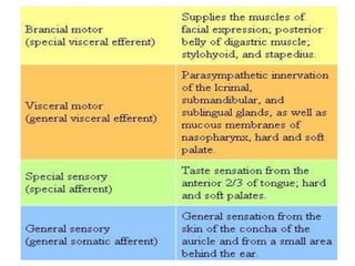
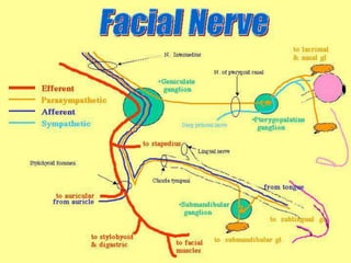
FACIAL NERVE AND IT’S
BRANCHES
DR. MANOJ KUMAR SINGH
1st YR PG
PHYSICAL MEDICINE AND REHABILITATION
PMCH PATNA
INTRODUCTION
*Develops from 2nd pharyngeal arch.
*MIXED Nerve.
*7TH CRANIAL NERVE.
SURFACE MARKING
Marked by a short horizontal line which joins the
following two points
• A point at the middle of the anterior border of
the mastoid process. The stylomastoid
foramen lies 2 cm deep to this point.
• A second point behind the neck of the
mandible. Here the nerve divides into its 5
branches for the facial muscles.
Functional Components
• Special visceral or branchial efferent, to
muscles responsible for facial expression and
for elevation of the hyoid bone.
• General visceral efferent or parasympathetic.
These fibers are secretomotor to the
submandibular and sublingual salivary gland,
lacrimal gland, and glands of the nose, the
palate and the pharynx.
 General visceral afferent component carries afferent
impulses from the previously mentioned glands.
 Special visceral afferent fibers carry taste sensations
from the anterior two-thirds of the tongue except
from valate papillae and from palate.
 General somatic afferent fibers probably innervate a
part of the skin of the ear. The nerve
does not give any direct branches to the ear,
but some fibers may reach it through
communications with the vagus nerve.
Proprioceptive impulses from muscles of face
travel through branches of trigeminal nerve to
reach the mesencephalic nucleus of the nerve.
Nuclei
The fibers of the nerve arise from 4 nuclei
situated in the lower pons.
1. Motor nucleus of branchiomotor
2. Superior salivatory nucleus or
parasympathetic
3. Lacrimatory nucleus is also parasympathetic
4. Nucleus of tractus solitarius which is
gustatory and also receives afferent fibers
from the glands
• The motor nucleus lies deep in the reticular
formation of the lower pons.
• The part of the nucleus that supplies muscles
of the upper part of the face receives
corticonuclear fibers from the motor cortex of
both the right and left sides.
• The part of the nucleus that supplies muscles
of the lower part of the face receive
corticonuclear fibers only from the opposite
cerebral hemisphere.
Course and Relations
• The facial nerve is attached to the brainstem
by 2 roots, motor and sensory (nervus
intermedius).
• The 2 roots of the facial nerve are attached to
the lower border of the pons just medial to
the 8th cranial nerve.
• The 2 roots run laterally and forwards, with
the 8th nerve to reach the internal acoustic
meatus.
• Inside the meatus, the motor root lies in a
groove on the 8th nerve, with the sensory root
intervening.
• Here the 7th and 8th nerves are accompanied
by labyrinthine vessels.
• At the bottom or fundus of the meatus, the 2
roots. Sensory and motor use to form a single
trunk, which lies in the petrous part of
temporal bone.
• Within the canal, the course of the nerve can
be divided into 3 parts by 2 bends.
• The first part is directed laterally above the
vestibule
• The second part runs backward in relation to
the medial wall of the middle ear, above the
promontry.
• The third part is directed vertically
downwards behind the promontry.
• The 1st bend at the junction of the 1st and 2nd
parts is sharp. It lies over the anterosuperior
part of the promontry called the genu.
• The 2nd bend is gradual, and lies b/w
promontry and aditus to mastoid antrum
• The facial nerve leaves the skull by passing
through the stylomastoid foramen.
• In its extracranial course, the facial nerve
crosses the lateral side of the base of styloid
process.
• It enters the posteromedial surface of parotid
gland, runs forwards though the gland
crossing the retromandibular vein and ECA.
• Behind the neck of the mandible it divides
into its 5 terminal branches which emerge
along the anterior border of the parotid gland.
Branches and Distribution
Within the facial canal
• Greater petrosal nerve
• Nerve to stapedius : arises opposite the
pyramid of middle ear and supplies the
stapedius muscle.
• Chords tympani : arises in the vertical part of
facial canal about 6 mm above the
stylomastoid foramen. It carries preganglionic
secretmotor fibers to the submandibular and
sublingual salivary glands and taste fibers
from the anterior two-thirds of the tongue.
At its exit from stylomastoid foramen
• Posterior auricular : arises just below the
stylomastoid foamen and supplies auricularis
posterior, occipitalis and intrinsic muscles on
the back of auricle.
• Digastric : arises close to the posterior
auricular. It is short and supplies the posterior
belly of the digastric.
• Stylohyoid : arise with the digastric branch, is
long and supplies stylohyoid muscle.
Terminal branches within parotid gland
• Temporal : cross zygomatic arch and supply
auricularis anterior, auricularis superior,
intrinsic muscles on the lateral side of ear,
frontalis, orbicularis oculi, corrugator supercili.
• Zygomatic : run across the zygomatic bone
and supply the orbicularis oculi.
• Buccal : 2 in number. Upper branch runs
above the parotid gland and lower below it
and supplies the muscle in that vicinity.
• Marginal mandibular : runs below the angle of
mandible deep to platysma. It crosses the
body of mandible and supplies muscles of the
lower lip and chin.
• Cervical : emerges from apex of parotid gland,
and runs downwards and forwards in the neck
to supply the platysma.
Ganglia
Geniculate ganglion
• Located on the 1st bend of facial nerve, in
relation to the medial wall of the middle ear.
• It is a sensory ganglion.
• The taste fibers present in the nerve are
peripheral processes of pseudounipolar
neurons present in the geniculate ganglion.
Submandibular ganglion
• Parasympathetic ganglion.
• For relay of secretomotor fibres to the
submandibular and sublingual glands.
Pterygopalatine ganglion
• Parasympathetic ganglion.
• Secretomotor fibers meant for the lacrimal
gland relay in this ganglion.
Clinical anatomy
• Supranulear and infranuclear lesions.
• In supranulear lesions; usually a part of
hemiplegia, only lower part of the opposite
side of the face is paralyzed. The upper part
with the frontalis and orbicularis oculi escapes
due to bilateral representation in the cerebra
cortex.
• In infranuclear lesions, known as Bell’s palsy,
the whole of the face of the same side gets
paralyzed. The face becomes asymmetrical
and is drawn up to the normal side. The
affected side is motionless. Wrinkles
disappear from the forehead. Eye cannot be
closed. Food accumulates b/w cheek and
teeth during mastigation.
• The symptoms according to the level of injury
of facial nerve.
• At internal auditory meatus; loss of
lacrimation, stapedial reflex, taste from most
of anterior two-third of tongue, lack of
salivation and paralysis of muscles of facial
expression.
• Below geniculate ganglion; loss of stapedial
reflex, taste from anterior two-
third of tongue, lack of salivation and paralayis
of facial expression muscles.
• Region b/w nerve to stapedius and chorda
tympani : loss of taste from anterior two-third
of tongue, lack of salivation and paralysis of
facial expression muscles.
• Region below stylomastoid foramen : paralysis
of facial expression muscles.
THANKS

Facial nerve and it’s branches

  • 1.
    FACIAL NERVE ANDIT’S BRANCHES DR. MANOJ KUMAR SINGH 1st YR PG PHYSICAL MEDICINE AND REHABILITATION PMCH PATNA
  • 2.
    INTRODUCTION *Develops from 2ndpharyngeal arch. *MIXED Nerve. *7TH CRANIAL NERVE.
  • 3.
    SURFACE MARKING Marked bya short horizontal line which joins the following two points • A point at the middle of the anterior border of the mastoid process. The stylomastoid foramen lies 2 cm deep to this point. • A second point behind the neck of the mandible. Here the nerve divides into its 5 branches for the facial muscles.
  • 4.
    Functional Components • Specialvisceral or branchial efferent, to muscles responsible for facial expression and for elevation of the hyoid bone. • General visceral efferent or parasympathetic. These fibers are secretomotor to the submandibular and sublingual salivary gland, lacrimal gland, and glands of the nose, the palate and the pharynx.
  • 5.
     General visceralafferent component carries afferent impulses from the previously mentioned glands.  Special visceral afferent fibers carry taste sensations from the anterior two-thirds of the tongue except from valate papillae and from palate.  General somatic afferent fibers probably innervate a part of the skin of the ear. The nerve
  • 6.
    does not giveany direct branches to the ear, but some fibers may reach it through communications with the vagus nerve. Proprioceptive impulses from muscles of face travel through branches of trigeminal nerve to reach the mesencephalic nucleus of the nerve.
  • 8.
    Nuclei The fibers ofthe nerve arise from 4 nuclei situated in the lower pons. 1. Motor nucleus of branchiomotor 2. Superior salivatory nucleus or parasympathetic 3. Lacrimatory nucleus is also parasympathetic 4. Nucleus of tractus solitarius which is gustatory and also receives afferent fibers from the glands
  • 9.
    • The motornucleus lies deep in the reticular formation of the lower pons. • The part of the nucleus that supplies muscles of the upper part of the face receives corticonuclear fibers from the motor cortex of both the right and left sides. • The part of the nucleus that supplies muscles of the lower part of the face receive corticonuclear fibers only from the opposite cerebral hemisphere.
  • 10.
    Course and Relations •The facial nerve is attached to the brainstem by 2 roots, motor and sensory (nervus intermedius). • The 2 roots of the facial nerve are attached to the lower border of the pons just medial to the 8th cranial nerve. • The 2 roots run laterally and forwards, with the 8th nerve to reach the internal acoustic meatus.
  • 11.
    • Inside themeatus, the motor root lies in a groove on the 8th nerve, with the sensory root intervening. • Here the 7th and 8th nerves are accompanied by labyrinthine vessels. • At the bottom or fundus of the meatus, the 2 roots. Sensory and motor use to form a single trunk, which lies in the petrous part of temporal bone.
  • 12.
    • Within thecanal, the course of the nerve can be divided into 3 parts by 2 bends. • The first part is directed laterally above the vestibule • The second part runs backward in relation to the medial wall of the middle ear, above the promontry. • The third part is directed vertically downwards behind the promontry. • The 1st bend at the junction of the 1st and 2nd parts is sharp. It lies over the anterosuperior part of the promontry called the genu.
  • 13.
    • The 2ndbend is gradual, and lies b/w promontry and aditus to mastoid antrum • The facial nerve leaves the skull by passing through the stylomastoid foramen. • In its extracranial course, the facial nerve crosses the lateral side of the base of styloid process. • It enters the posteromedial surface of parotid gland, runs forwards though the gland crossing the retromandibular vein and ECA.
  • 14.
    • Behind theneck of the mandible it divides into its 5 terminal branches which emerge along the anterior border of the parotid gland.
  • 15.
  • 16.
    Within the facialcanal • Greater petrosal nerve • Nerve to stapedius : arises opposite the pyramid of middle ear and supplies the stapedius muscle.
  • 17.
    • Chords tympani: arises in the vertical part of facial canal about 6 mm above the stylomastoid foramen. It carries preganglionic secretmotor fibers to the submandibular and sublingual salivary glands and taste fibers from the anterior two-thirds of the tongue.
  • 18.
    At its exitfrom stylomastoid foramen • Posterior auricular : arises just below the stylomastoid foamen and supplies auricularis posterior, occipitalis and intrinsic muscles on the back of auricle. • Digastric : arises close to the posterior auricular. It is short and supplies the posterior belly of the digastric. • Stylohyoid : arise with the digastric branch, is long and supplies stylohyoid muscle.
  • 19.
    Terminal branches withinparotid gland • Temporal : cross zygomatic arch and supply auricularis anterior, auricularis superior, intrinsic muscles on the lateral side of ear, frontalis, orbicularis oculi, corrugator supercili. • Zygomatic : run across the zygomatic bone and supply the orbicularis oculi. • Buccal : 2 in number. Upper branch runs above the parotid gland and lower below it
  • 20.
    and supplies themuscle in that vicinity. • Marginal mandibular : runs below the angle of mandible deep to platysma. It crosses the body of mandible and supplies muscles of the lower lip and chin. • Cervical : emerges from apex of parotid gland, and runs downwards and forwards in the neck to supply the platysma.
  • 22.
  • 23.
    Geniculate ganglion • Locatedon the 1st bend of facial nerve, in relation to the medial wall of the middle ear. • It is a sensory ganglion. • The taste fibers present in the nerve are peripheral processes of pseudounipolar neurons present in the geniculate ganglion.
  • 24.
    Submandibular ganglion • Parasympatheticganglion. • For relay of secretomotor fibres to the submandibular and sublingual glands.
  • 25.
    Pterygopalatine ganglion • Parasympatheticganglion. • Secretomotor fibers meant for the lacrimal gland relay in this ganglion.
  • 26.
    Clinical anatomy • Supranulearand infranuclear lesions. • In supranulear lesions; usually a part of hemiplegia, only lower part of the opposite side of the face is paralyzed. The upper part with the frontalis and orbicularis oculi escapes due to bilateral representation in the cerebra cortex.
  • 27.
    • In infranuclearlesions, known as Bell’s palsy, the whole of the face of the same side gets paralyzed. The face becomes asymmetrical and is drawn up to the normal side. The affected side is motionless. Wrinkles disappear from the forehead. Eye cannot be closed. Food accumulates b/w cheek and teeth during mastigation.
  • 28.
    • The symptomsaccording to the level of injury of facial nerve. • At internal auditory meatus; loss of lacrimation, stapedial reflex, taste from most of anterior two-third of tongue, lack of salivation and paralysis of muscles of facial expression. • Below geniculate ganglion; loss of stapedial reflex, taste from anterior two-
  • 29.
    third of tongue,lack of salivation and paralayis of facial expression muscles. • Region b/w nerve to stapedius and chorda tympani : loss of taste from anterior two-third of tongue, lack of salivation and paralysis of facial expression muscles. • Region below stylomastoid foramen : paralysis of facial expression muscles.
  • 30.