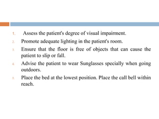
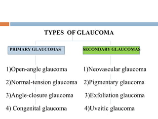
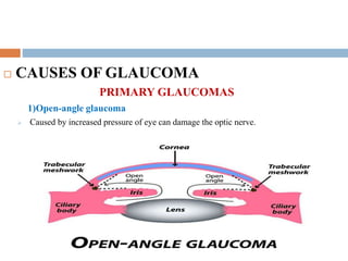
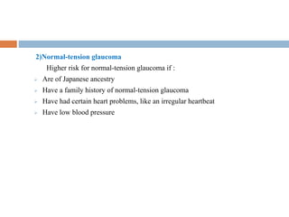
DISORDERS OF EYE
CATARACT
 INTRODUCTION
A cataract is a cloudy lens.
The lens is positioned behind the coloured part of eye (iris). The lens
focuses light that passes into eye, producing clear, sharp images on the
retina
 DEFINITION
A condition in which the lens of the eye becomes
cloudy symptoms include blurred, cloudy or double
vision , sensitivity to light and difficulty seeing at
night.
 TYPES OF CATARACT
 Age related cataract
 Traumatic cataract
 Radiation cataract
 Pediatric cataract
 Secondary cataract
 ETIOLOGY
 Smoking
 Ultraviolet radiation
 Long term use of steroid medication
 Diabetes
 Trauma
 Radiation therapy
 Over production of antioxidant
 RISK FACTORS
 Older age
 Heavy alcohol use
 Obesity
 High blood pressure
 Previous eye injuries
 A family history of cataract
 Too much sun exposure
 Diabetes
 Exposure to radiation from X-ray and the cancer treatment.
PATHOPHYSIOLOGY
The lens is made of water and protein fibers
Age-related and Due to etiological factors
Cause proteins and fibers within the lenses to break down and clump together
lenses in eyes become less flexible, less transparent and thicker
Resulting cloudy lens
Cataract scatters and blocks the light passes through the lens
Preventing a producing clear, sharp images on the retina
As a result, vision becomes blurred.
 SIGN AND SYMPTOMS
 Clouded, blurred or dim vision
 Increasing difficulty with vision at night
 Sensitivity to light and glare
 Need for brighter light for reading and other activities
 Seeing "halos" around lights
 Frequent changes in eyeglass or prescription
 Fading or yellowing of colours
 Double vision in a single eye
 DIGNOSTIC EVALUATION
 Visual acuity test.
 Slit-lamp examination.
 Retinal exam.
 Applanation tonometry.
 MANAGEMENT
1)Small incision surgery
CATARACT SURGERY 2)Large incision surgery
3) Femtosecond (FS)
laser surgery
 NURSING DIGNOSIS
1. Disturbed Sensory Perception(Visual) related to cataract as evidenced
by verbal complaints of vision problems such as cloudy and hallow
vision and inability to see properly.
2. Risk for Injury related to loss of vision and reduced visual acuity.
3. Deficient knowledge related to new diagnosis of cataracts as evidenced
by patient's verbalization of "I want to know more about my new
diagnosis and care“.
4. Risk for ineffective therapeutic regimen secondary to cataract related to
un familiarity with the treatment plan.
 INTERVENTION
1. Assess the vision ability of the patient using an eye chart, slit-lamp
exam, and retinal exam.
2. Encourage the patient to have regular checkups with an ophthalmologist
at least once a year.
3. Encourage the patient to promote sufficient lighting at home.
4. Encourage the patient to always wear the prescribed glasses.
5. Prepare the patient for cataract surgery if indicated.
6. Encourage the patient to add foods containing vitamins C, E, beta-
carotene, zinc, and copper in daily diet.
1. Assess the patient's degree of visual impairment.
2. Promote adequate lighting in the patient's room.
3. Ensure that the floor is free of objects that can cause the
patient to slip or fall.
4. Advise the patient to wear Sunglasses specially when going
outdoors.
5. Place the bed at the lowest position. Place the call bell within
reach.
GLAUCOMA
 INTRODUCTION
 Glaucoma develops when the optic nerve becomes
damaged. As this nerve gradually deteriorates, blind
spots develop in vision.
 DEFINITION
 Glaucoma is a group of eye conditions that damage
the optic nerve . The optic nerve send the visual
information from eye to brain and is vital for good
vision.
TYPES OF GLAUCOMA
PRIMARY GLAUCOMAS SECONDARY GLAUCOMAS
1)Open-angle glaucoma 1)Neovascular glaucoma
2)Normal-tension glaucoma 2)Pigmentary glaucoma
3)Angle-closure glaucoma 3)Exfoliation glaucoma
4) Congenital glaucoma 4)Uveitic glaucoma
 CAUSES OF GLAUCOMA
PRIMARY GLAUCOMAS
1)Open-angle glaucoma
 Caused by increased pressure of eye can damage the optic nerve.
2)Normal-tension glaucoma
Higher risk for normal-tension glaucoma if :
 Are of Japanese ancestry
 Have a family history of normal-tension glaucoma
 Have had certain heart problems, like an irregular heartbeat
 Have low blood pressure
3) Angle-closure glaucoma
 The outer edge of the iris blocks fluid from draining out of the front of the eye
causing a sudden increase in eye pressure.
Conti………
4) Congenital glaucoma
 In congenital glaucoma, babies are born with a problem in their eye that
keeps fluid from draining normally.
SECONDARY GLAUCOMAS
1)Neovascular glaucoma
 Neovascular glaucoma happens when the eye makes extra blood vessels that
cover the part of your eye where fluid would normally drain. It’s usually caused
by another medical condition, like diabetes or high blood pressure
2)Pigmentary glaucoma
 Pigmentary glaucoma happens when the pigment (colour) from iris (the
coloured part of eye) flakes off and blocks fluid from draining out of your
eye.
3)Exfoliation glaucoma
 Exfoliation glaucoma happens when extra material to deposit on parts of the
eye and block fluid from draining.
4)Uveitic glaucoma
 Uveitic glaucoma can happen in people who have uveitis , a condition
that causes swelling and inflammation in the eye.
 OTHER CAUSES
 Cataracts
 Eye tumours
 Eye injuries
 Optic nerve damage
 RISK FACTORS
o High intraocular pressure
o Age over 55
o Black, Asian
o Family history of glaucoma
o Certain medical conditions, such as diabetes, migraines, high
blood pressure and sickle cell anaemia
o Corneas that are thin in the centre
o Extreme nearsightedness or farsightedness
o Eye injury
o eye surgery
o Use of corticosteroid medicines, especially eye drops, for a long
time
 PATHOPHYSIOLOGY
 SIGN AND SYMPTOMS
1. Eye pain
2. Nausea and vomiting
3. Redness of eye
4. Headache
5. Tenderness around eye
6. Seeing ring around lights
7. Blurred vision
8. Low vision
9. Narrowed vision (Blind spots)s
 DIGNOSIS AND TEST
 Dilated eye exam
 Gonioscopy
 Optical Cohrance Tomography (OCT)
 Occular pressure test (Tomography)
 Patchymetri
 Slit lamp examination
 Visual acuity test
 MEDICAL MANAGEMENT
 Eye drops
Prescription eye drop medicines include:
1. Prostaglandins.
2. Rho kinase inhibitor.
3. Nitric oxide.
4. Miotic or cholinergic agents.
5. Beta blockers.
6. Carbonic anhydrase inhibitors.
 SURGICAL MANAGEMENT
 Laser therapy.
 Electrocautery
 Drainage Implant Surgery
 EX-PRESS Mini-shunt
 Ahmed Glaucoma Valve
 Contact Transcleral Laser Cycloablation (CTLC)
 NURSING DIGNOSIS
 Anxiety related to possible vision loss.
 Disturbed Sensory Perception related to visual impairment.
 Ineffective health maintenance related to knowledge
deficit.
 Risk for injury related to vision loss.
 Self care deficit related to vision loss.
CONJUNCTIVITIS
 INTRODUCTION
COMMON CONDITION OF CONJUNCTIVA
CONJUNCTIVITIS (PINK EYE)
SUBCONJUNCTIVAL
HEMORRHAGE
 CONJUNCTIVITIS (PINK EYE)
 Pink eye is a common eye infection .Its caused by allergens ,
irritants ,bacteria and viruses.
 SUBCONJUNCTIVAL HEMORRHAGE
 A subconjunctival hemorrhage occurs when a tiny blood
vessel breaks just underneath the conjunctiva.
The conjunctiva can't absorb blood very quickly, so the blood
gets trapped.
 ETIOLOGY
 Viral infection
 Bacteria
 Allergies such as shampoos, dirt, smoke, and pool chlorine
 A chemical splash in eye
 A foreign object in eye
 In newborn, a blocked tear duct.
 RISK FACTORS
 Exposure to someone infected with conjunctivitis
 Exposure to allergens
 Using contact lenses
 TYPES
 Viral conjunctivitis
 Adenovirus
 Herpes simplex virus
 Varicella zoster virus
 Bacterial conjunctivitis
 Allergic conjunctivitis
 SIGN AND SYMOTOMS
 Redness
 Itching
 Discharge
 Pain that doesn’t go in few days
 Light sensitivity
 Blurred vision
 Double vision
 Vision is getting worse.
 COMPLICATIONS
 Eye pain
 Feeling that something is stuck in eyes.
 Blurred vision
 Light sensitivity.

cataract.pptx

  • 1.
  • 3.
    CATARACT  INTRODUCTION A cataractis a cloudy lens. The lens is positioned behind the coloured part of eye (iris). The lens focuses light that passes into eye, producing clear, sharp images on the retina
  • 4.
     DEFINITION A conditionin which the lens of the eye becomes cloudy symptoms include blurred, cloudy or double vision , sensitivity to light and difficulty seeing at night.
  • 5.
     TYPES OFCATARACT  Age related cataract  Traumatic cataract  Radiation cataract  Pediatric cataract  Secondary cataract
  • 6.
     ETIOLOGY  Smoking Ultraviolet radiation  Long term use of steroid medication  Diabetes  Trauma  Radiation therapy  Over production of antioxidant
  • 7.
     RISK FACTORS Older age  Heavy alcohol use  Obesity  High blood pressure  Previous eye injuries  A family history of cataract  Too much sun exposure  Diabetes  Exposure to radiation from X-ray and the cancer treatment.
  • 8.
    PATHOPHYSIOLOGY The lens ismade of water and protein fibers Age-related and Due to etiological factors Cause proteins and fibers within the lenses to break down and clump together lenses in eyes become less flexible, less transparent and thicker Resulting cloudy lens Cataract scatters and blocks the light passes through the lens Preventing a producing clear, sharp images on the retina As a result, vision becomes blurred.
  • 9.
     SIGN ANDSYMPTOMS  Clouded, blurred or dim vision  Increasing difficulty with vision at night  Sensitivity to light and glare  Need for brighter light for reading and other activities  Seeing "halos" around lights  Frequent changes in eyeglass or prescription  Fading or yellowing of colours  Double vision in a single eye
  • 10.
     DIGNOSTIC EVALUATION Visual acuity test.
  • 11.
  • 12.
  • 13.
  • 14.
     MANAGEMENT 1)Small incisionsurgery CATARACT SURGERY 2)Large incision surgery 3) Femtosecond (FS) laser surgery
  • 15.
     NURSING DIGNOSIS 1.Disturbed Sensory Perception(Visual) related to cataract as evidenced by verbal complaints of vision problems such as cloudy and hallow vision and inability to see properly. 2. Risk for Injury related to loss of vision and reduced visual acuity. 3. Deficient knowledge related to new diagnosis of cataracts as evidenced by patient's verbalization of "I want to know more about my new diagnosis and care“. 4. Risk for ineffective therapeutic regimen secondary to cataract related to un familiarity with the treatment plan.
  • 16.
     INTERVENTION 1. Assessthe vision ability of the patient using an eye chart, slit-lamp exam, and retinal exam. 2. Encourage the patient to have regular checkups with an ophthalmologist at least once a year. 3. Encourage the patient to promote sufficient lighting at home. 4. Encourage the patient to always wear the prescribed glasses. 5. Prepare the patient for cataract surgery if indicated. 6. Encourage the patient to add foods containing vitamins C, E, beta- carotene, zinc, and copper in daily diet.
  • 17.
    1. Assess thepatient's degree of visual impairment. 2. Promote adequate lighting in the patient's room. 3. Ensure that the floor is free of objects that can cause the patient to slip or fall. 4. Advise the patient to wear Sunglasses specially when going outdoors. 5. Place the bed at the lowest position. Place the call bell within reach.
  • 18.
    GLAUCOMA  INTRODUCTION  Glaucomadevelops when the optic nerve becomes damaged. As this nerve gradually deteriorates, blind spots develop in vision.  DEFINITION  Glaucoma is a group of eye conditions that damage the optic nerve . The optic nerve send the visual information from eye to brain and is vital for good vision.
  • 19.
    TYPES OF GLAUCOMA PRIMARYGLAUCOMAS SECONDARY GLAUCOMAS 1)Open-angle glaucoma 1)Neovascular glaucoma 2)Normal-tension glaucoma 2)Pigmentary glaucoma 3)Angle-closure glaucoma 3)Exfoliation glaucoma 4) Congenital glaucoma 4)Uveitic glaucoma
  • 20.
     CAUSES OFGLAUCOMA PRIMARY GLAUCOMAS 1)Open-angle glaucoma  Caused by increased pressure of eye can damage the optic nerve.
  • 21.
    2)Normal-tension glaucoma Higher riskfor normal-tension glaucoma if :  Are of Japanese ancestry  Have a family history of normal-tension glaucoma  Have had certain heart problems, like an irregular heartbeat  Have low blood pressure
  • 22.
    3) Angle-closure glaucoma The outer edge of the iris blocks fluid from draining out of the front of the eye causing a sudden increase in eye pressure.
  • 23.
    Conti……… 4) Congenital glaucoma In congenital glaucoma, babies are born with a problem in their eye that keeps fluid from draining normally.
  • 24.
    SECONDARY GLAUCOMAS 1)Neovascular glaucoma Neovascular glaucoma happens when the eye makes extra blood vessels that cover the part of your eye where fluid would normally drain. It’s usually caused by another medical condition, like diabetes or high blood pressure 2)Pigmentary glaucoma  Pigmentary glaucoma happens when the pigment (colour) from iris (the coloured part of eye) flakes off and blocks fluid from draining out of your eye.
  • 25.
    3)Exfoliation glaucoma  Exfoliationglaucoma happens when extra material to deposit on parts of the eye and block fluid from draining. 4)Uveitic glaucoma  Uveitic glaucoma can happen in people who have uveitis , a condition that causes swelling and inflammation in the eye.
  • 26.
     OTHER CAUSES Cataracts  Eye tumours  Eye injuries  Optic nerve damage
  • 27.
     RISK FACTORS oHigh intraocular pressure o Age over 55 o Black, Asian o Family history of glaucoma o Certain medical conditions, such as diabetes, migraines, high blood pressure and sickle cell anaemia o Corneas that are thin in the centre o Extreme nearsightedness or farsightedness o Eye injury o eye surgery o Use of corticosteroid medicines, especially eye drops, for a long time
  • 28.
  • 29.
     SIGN ANDSYMPTOMS 1. Eye pain 2. Nausea and vomiting 3. Redness of eye 4. Headache 5. Tenderness around eye 6. Seeing ring around lights 7. Blurred vision 8. Low vision 9. Narrowed vision (Blind spots)s
  • 30.
     DIGNOSIS ANDTEST  Dilated eye exam  Gonioscopy  Optical Cohrance Tomography (OCT)  Occular pressure test (Tomography)  Patchymetri  Slit lamp examination  Visual acuity test
  • 31.
     MEDICAL MANAGEMENT Eye drops Prescription eye drop medicines include: 1. Prostaglandins. 2. Rho kinase inhibitor. 3. Nitric oxide. 4. Miotic or cholinergic agents. 5. Beta blockers. 6. Carbonic anhydrase inhibitors.
  • 32.
     SURGICAL MANAGEMENT Laser therapy.  Electrocautery  Drainage Implant Surgery  EX-PRESS Mini-shunt  Ahmed Glaucoma Valve  Contact Transcleral Laser Cycloablation (CTLC)
  • 33.
     NURSING DIGNOSIS Anxiety related to possible vision loss.  Disturbed Sensory Perception related to visual impairment.  Ineffective health maintenance related to knowledge deficit.  Risk for injury related to vision loss.  Self care deficit related to vision loss.
  • 34.
  • 35.
    COMMON CONDITION OFCONJUNCTIVA CONJUNCTIVITIS (PINK EYE) SUBCONJUNCTIVAL HEMORRHAGE
  • 36.
     CONJUNCTIVITIS (PINKEYE)  Pink eye is a common eye infection .Its caused by allergens , irritants ,bacteria and viruses.  SUBCONJUNCTIVAL HEMORRHAGE  A subconjunctival hemorrhage occurs when a tiny blood vessel breaks just underneath the conjunctiva. The conjunctiva can't absorb blood very quickly, so the blood gets trapped.
  • 37.
     ETIOLOGY  Viralinfection  Bacteria  Allergies such as shampoos, dirt, smoke, and pool chlorine  A chemical splash in eye  A foreign object in eye  In newborn, a blocked tear duct.  RISK FACTORS  Exposure to someone infected with conjunctivitis  Exposure to allergens  Using contact lenses
  • 38.
     TYPES  Viralconjunctivitis  Adenovirus  Herpes simplex virus  Varicella zoster virus  Bacterial conjunctivitis  Allergic conjunctivitis
  • 39.
     SIGN ANDSYMOTOMS  Redness  Itching  Discharge  Pain that doesn’t go in few days  Light sensitivity  Blurred vision  Double vision  Vision is getting worse.
  • 40.
     COMPLICATIONS  Eyepain  Feeling that something is stuck in eyes.  Blurred vision  Light sensitivity.