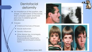
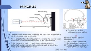
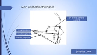
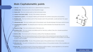
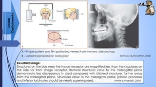
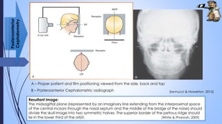
3. INTRODUCTION
๏ด Cephalometric radiography = standardized and reproducible form of skull
radiography used extensively in orthodontic to assess the relationships of the
teeth to the jaws and the jaws to the rest of the facial skeleton. (Whaites & Drage,
2013)
๏ด Orthodontic = Greek word (orthos means โstraight, perfect or properโ ; dontos
means โteethโ)
๏ด a branch of dentistry that specializes in treating patients with improper
positioning of teeth when the mouth is closed (malocclusion), which results
in an improper bite
๏ด Includes treating and controlling various aspects of facial growth
(dentofacial orthopaedics) and the shape and development of the jaw.
(Medical News Today, 2014)
๏ด Mainly used to study of growth patterns in the craniofacial complex.
๏ด Concern on malocclusion and dentofacial deformity (Proffit, Fields & Sarver, 2007)
4. Malocclusion
๏ด Abnormal alignment of the teeth and the
way in which the upper and lower teeth fit
together.
๏ด The upper teeth overlap slightly outside the
lower teeth in normal person in which allows
the projections (cusps) on each tooth to fit
in the depressions of the opposite tooth.
๏ด Causes:
๏ด Habitual thumb-sucking and/ or tongue
thrusting (push the tongue up against
the front teeth)
๏ด Size mismatch between jaw and teeth
๏ด Lost teeth
๏ด Certain birth defects of the jaw or
fracture
(Murchison, 2014)
(Smile Simplicity, 2015)
5. Dentofacial
deformity
๏ด An imbalance of the position, size,
shape, or orientation of the bones
that comprise upper and lower
jaws due to skeletal growth
disturbance
๏ด Causes:
๏ด Childhood fractures of the jaw
๏ด Muscle dysfunction
๏ด Genetic influences
๏ด Other disease (eg. Acromegaly,
Rheumatoid Arthritis,
Hemimandibular Hypertrophy,
Malformed and Supernumerary
Teeth)
(Proffit, Fields & Sarver, 2007)
6. INDICATIONS
๏ด Orthodontics
๏ด Initial diagnosis to confirm of underlying skeletal and tissue deformities
๏ด Treatment planning
๏ด Monitoring treatment planning
๏ด Appraisal of treatment progress
๏ด Research purposes
๏ด Orthognathic surgery
๏ด Pre-operative evaluation of skeletal and soft tissue patterns
๏ด Assist in treatment planning
๏ด Post-operative appraisal of the result of surgery and long term follow-up
study
๏ด Research purposes
(Whaites & Drage, 2013; Mitchell, 2013)
7. PRINCIPLES
๏ด Cephalostat is a machine that holds the head in a set position in
relation to x-ray tube and the film
๏ด Place at patientโs external auditory meati and the central beam
is directed towards the ear rods and to the center of film.
๏ด Patientโs head in vertical axis is standardized by ensuring
patientโs Frankfort plane is horizontal, manually or placing a
mirror at the patientโs head level
๏ Frankfort plane : line
joining porion and orbitale
!!!!!
Be ensure that the patientโs
midsagittal =
(1) Vertical and parallel to the
film (lateral cephalometric)
(2) Vertical and perpendicular to
the film (PA Cephalometry)
(Gill & Naini, 2011)
8. EQUIPMENT
Film size : 18 x 24 cm (commonly
used)
Intensifying screen (usually 18 x 24 cm)
Extraoral machine with
cephalostat
Head stabilizing and
ear rods
- Ensure
standardized patient
position
- To stabilize
patientโs head To attenuate the x-ray
beam selectively in the
facial soft tissue region
(Iannucci & Howerton, 2012)
10. Main Cephalometric points
1) Sella (S) - The centre of the sella turcica, (determined by inspection).
2) Orbitale (Or) - The lowest point on the infraorbital margin.
3) Nasion (N) - The most anterior point on the frontonasal suture.
4) Anterior nasal spine (ANS) - The tip of the anterior nasal spine.
5) Subspinale or point A - The deepest midline point between the anterior nasal spine and prosthion.
6) Prosthion (Pr) - The most anterior point of the alveolar crest in the premaxilla, usually between the upper
central incisors.
7) Infradentale (Id) - The most anterior point of the alveolar crest, situated between the lower central incisors.
8) Supramentale or point B - The deepest point in the bony outline between the infradentale and the
pogonion.
9) Pogonion (Pog) - The most anterior point of the bony chin.
10)Gnathion (Gn) - The most anterior and inferior point on the bony outline of the chin, situated equidistant
from pogonion and menton.
11)Menton (Me) - The lowest point on the bony outline of the mandibular symphysis.
12)Gonion (Go) - The most lateral external point at the junction of the horizontal and ascending rami of the
mandible. Note: The gonion is found by bisecting the angle formed by tangents to the posterior and inferior
borders of the mandible.
13)Posterior nasal spine (PNS) - The tip of the posterior spine of the palatine bone in the hard palate.
14)Articulare (Ar) - The point of intersection of the dorsal contours of the posterior border of the mandible and
temporal bone.
15)Porion (Po) - The uppermost point of the bony external auditory meatus, usually regarded as coincidental
with the uppermost point of the ear rods of the cephalostat.
(Whaites, 2003)
11. PROJECTIONS
The patient is positioned within the cephalostat, with the
sagittal plane of the head vertical and parallel to the
film and with the Frankford plane horizontal. The teeth
should generally be in maximum intercuspation.
The head is immobilized carefully with the apparatus
with the plastic ear rods being inserted gradually into the
external auditory meati.
The Aluminium wedge is positioned to cover the
anterior part of the film.
The equipment is designed to ensure that when the
patient is positioned correctly, the x-ray beam is
horizontal and centred on the ear rods
- -
The head-stabilizing apparatus of the
cephalostat is rotated through 90ยฐ
The patient is positioned in the
apparatus with the head tipped
forwards and with radiographic baseline
horizontal and perpendicular to the film
The head is immobilized within the
apparatus by inserting the plastic ear
rods into the external auditory meati
The fixed x-ray beam is horizontal with
the central ray centred through the
cervical spine at the level of the rami of
the mandible
Lateral
Cephalometry
Posteroanterior
Cephalometry
(Whaites & Drage, 2013;Iannucci & Howerton, 2012)
12. A โ Proper patient and film positioning viewed from the front, side and top
B โ Lateral Cephalometric radiograph
Resultant image:
Structures on the side near the image receptor are magnified less than the structures on
the side far from image receptor. Bilateral structures close to the midsagittal plane
demonstrate less discrepancy in sized compared with bilateral structures farther away
from the midsagittal plane. Structures close to the midsagittal plane (clinoid processes
and inferior turbinates should be nearly superimposed. (White & Pharoah, 2009)
Lateral
Cephalometry
(Iannucci & Howerton, 2012)
13. Cranial collimator
shadow
Head support
Ear rod
Aluminium wedge
filter
Cervical spine
collimator shadow
(Whaites, 2003)
Note the images of the ear rods should appear superimposed on each other. Other
shadows of cephalostat equipment and collimator are indicated.
14. A โ Proper patient and film positioning viewed from the side, back and top
B โ Posteroanterior Cephalometric radiograph
Resultant image:
The midsagittal plane (represented by an imaginary line extending from the interproximal space
of the central incisors through the nasal septum and the middle of the bridge of the noise) should
divide the skull image into two symmetric halves. The superior border of the petrous ridge should
lie in the lower third of the orbit. (White & Pharoah, 2009)
Posteroanterior
Cephalometry
(Iannucci & Howerton, 2012)
17. References
๏ด Gill, D. S. & Naini, F. B. (2011). Orthodontics โ Principles and Practice (eds). United Kingdom: John Wiley
and Sons - Blackwell
๏ด Iannucci, J. M. & Howerton (2012). Dental Radiography โ Principles and Technique (4th ed.). United States
of America, Elsevier Saunders
๏ด Medical News Today (2014). What is Orthodontics? Retrieved Apr 05, 2015 from
http://www.medicalnewstoday.com/articles/249482.php
๏ด Mitchell, L. (2013). Introduction to Orthodontics (4th ed.). United Kingdom: Oxford University Press
๏ด Murchison, D. F. (n.d.). Mulocclusion โ misaligned teeth. Retrieved Apr 05, 2015 from
http://www.merckmanuals.com/home/mouth-and-dental-disorders/symptoms-of-oral-and-dental-
disorders/malocclusion
๏ด Proffit, W. R. , Fields, H. W. & Sarver, D. M. (2007). Contemporary Orthodontics (4th ed.). Canada: Mosby
Elsevier
๏ด SmileSimplicity (2015). What is Mulocclusion? Retrieved Apr 05, 2015 from
http://www.smilesimplicity.com/malocclusion/
๏ด Whaites, E. & Drage, N. (2013). Essentials of Dental Radiography and Radiology (4th ed.). Retrieved from
https://books.google.com.my/books?id=lSSwAAAAQBAJ&pg=PR4&dq=Essentials+of+Dental+Radiograph
y+and+Radiology+4th+ed&hl=en&sa=X&ei=ya9hVafRM5STuASi5YDACg&ved=0CBwQ6AEwAA#v=onepa
ge&q=Essentials%20of%20Dental%20Radiography%20and%20Radiology%204th%20ed&f=false
๏ด White, S. C. & Pharoah, M. J. (2009). Oral Radiography โ Principles and Techniques (6th ed). United States
of America: Mosby Elsevier
Editor's Notes
Morphological analysis โ sagittal and vertical relationship of dentition, facial skeleton and soft tissue profile. This aid diagnosis particularly where there is a skeletal discrepancy or where anteroposterior movement of the incisors is planned.
Growth analysis โ superimposition of cephalograms taken over a period of time. This may help in deciding the timing of treatement especially those with skeletal III malocclusions
There are a number of different types of malocclusion. Some of the more common forms are:
1. Uppers overbite (buck teeth) โ can result from pacifier use or thumb sucking2. Underbite โ bottom teeth jut forward of the upper teeth3. Open bite โ occurs when upper and lower teeth do not overlap4. Overbite โ upper teeth overlap too far down over the lower teeth5. Spacing or crowding โ common condition which can often cause teeth not to come in properly resulting in impaction6. Cross bite โ this is a condition when any of the upper teeth fit on the wrong side of the lower teeth7. Rotation โ when a tooth turns out of its normal position8. Transposition โ when a tooth erupts in the wrong place
Was developed by B. Holly Broadbent, in the period after the First World War
Consists of an X-ray machine which is at a fixed distance from a set of ear posts (to fit into ptโs external auditory meatus and to stabilize ptโs head)
Central beam directed towards the ear posts
Patientโs head in vertical axis is standardized by ensuring the patientโs Frankfort plane is horizontal
Frankfort plane = line joining the porion (upper and outermost point on bony external auditory meatus) and orbitale (most inferior point on orbital margin)
If difficult to found the porion, can be refer to the uppermost surface of condylar head)