Downloaded 259 times


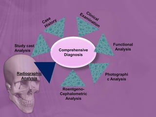
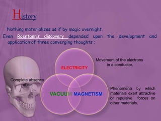
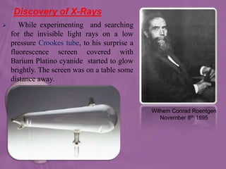
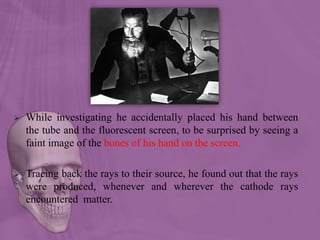
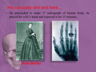
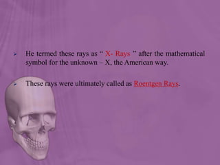
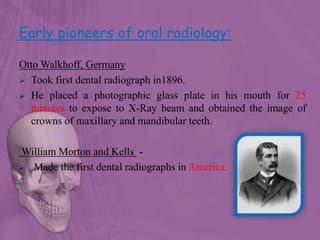
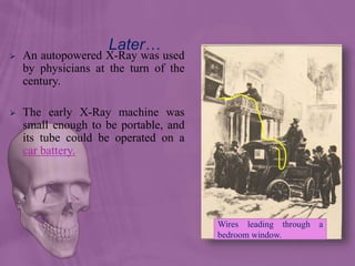

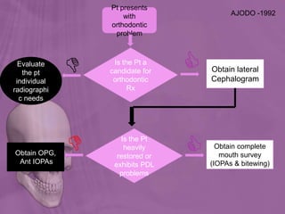
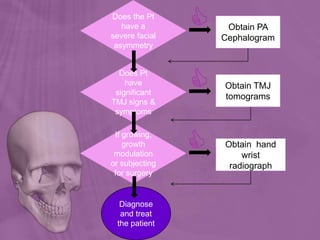
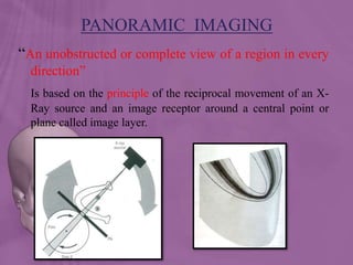
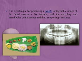


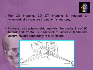


The document discusses various radiographic techniques used in orthodontic diagnosis. It begins with a brief history of x-rays and their discovery by Roentgen. It then summarizes several intraoral and extraoral radiographs used in orthodontics including panoramic radiographs, lateral cephalograms, posterior anterior views, and temporomandibular joint tomograms. It highlights the structures visualized and diagnostic information provided by each technique. The document also discusses digital radiography and its advantages over conventional radiography.


![ONFH[AVN HIP] -TRIPLE REGIME -A NOVAL SURGICAL CONCEPT .pptx](https://cdn.slidesharecdn.com/ss_thumbnails/onfhavnhip2026koaconcalicutdrgokuldevdrmashraf-260210064517-213ec005-thumbnail.jpg?width=640&height=640&fit=bounds)


![PERI-PROSTHETIC FRACTURE NAIL-PLATE CONSTRUCT [NPC].pptx](https://cdn.slidesharecdn.com/ss_thumbnails/drarunkumardrmohamedashrafperiprostheticfrasturenail-plateconstructnpc-260209164459-7e9d15a1-thumbnail.jpg?width=640&height=640&fit=bounds)


