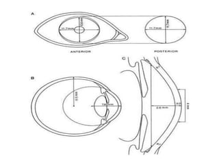
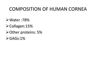
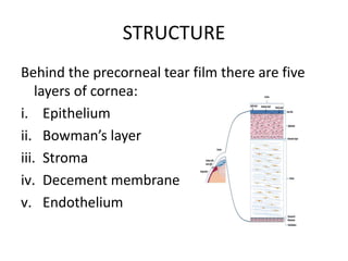
CORNEA
Shahana
Nethradhama school of optometry
The cornea is a transparent avascular tissue with
a smooth convex outer surface and concave
inner surface,which resembles a small watch
glass.
To meet the diverse functional demands the
cornea must be:
Transparent
Refract light
Contain the intraocular pressure
Provide a protective interface
DIMENSIONS
Anterior surface:
• Ellipitical,about 11.7mm horizontal and 10.6
mm vertically
Posterior surface:
• Circular , about 11.7 mm in diameter
Thickness:
• Centrally about 0.52mm
• Peripherally about 0.67mm
Surface area:
• About 1.3cm²
Optical zone:
• Cornea is almost a sphere,the central 1/3rd is called
optical zone about 5.4mm
Radius of curvature:
• Anterior surface – about 7.8mm
• Posterior surface – about 6.5mm
Refractive power:+43.1 D
Refractive index:1.376
COMPOSITION OF HUMAN CORNEA
Water :78%
Collagen:15%
Other proteins: 5%
GAGs:1%
STRUCTURE
Behind the precorneal tear film there are five
layers of cornea:
i. Epithelium
ii. Bowman’s layer
iii. Stroma
iv. Decement membrane
v. Endothelium
Epithelium:
The corneal epithelium is :
• Stratified,squamous and non keratinised
• Continuous with conjuctival epithelium at limbus
but having no goblet cells
• 50micron thick
• Consists of 5 or 6 layers of nucleated cells
resting on a basal lamina , namely
a) Basal cells
b) Wing cells
c) Surface cells
Bowman’s layer
• Modified region of anterior stroma
• Acellular homogeneous zone
• 8-14 micrometer thick
• Anterior surface is smooth and parallel with
corneal surface
Stroma: (substantia propria)
• About 500 micrometer thick
• Consists of regularly arranged lamellae of collagen
bundles,lie in proteoglycan ground substance with-
 200-300 bundles-centrally
 500 bundles-peripherally
 Width about 9 – 260 micrometer
 Height about 1.15-2micrometer
• small population of cells- keratocytes present.
• Keratocytes occupy 2.5 – 5 % of total stromal volume
and is responsible for synthesis and maintaining of
collagen and proteoglycan substance of stroma.
Arrangement of lamellae-
• Lamellae are arranged in layers and are
parallel with each other and with corneal
surface
• In deeper the stroma the lamellae form strap
like ribbons which run approximately at right
angles to those in consecutive layers
• At the limbus the bundles appeared to take a
circular course
Descemet’s membrane:
• It is the basal lamina of corneal endothelium
• First appears at 2nd month of gestation and
synthesis continue through out adult life
• About 10 micrometer thickness
Endothelium:
• It is a single layer of hexagonal,cuboidal cells attached
posterior ascept of decement membrane
• It is nuroectoderm in origin
• The endothelium can be viewed by specular reflection
using a slit-lamp biomicroscope at high magnification (25X -
40X)
• Corneal endothelium cells production is relatively fixed and
it is about 500,000
• At birth cells are 10 micrometer in height with age it
becomes flattened to 3-5 micrometer and 18-20
micrometer width
• Endothelium gets its nutrition and oxygen from aqueous
Blood supply of cornea
• The cornea is avascular structure
• Small loop derived from the anterior ciliary
vessels invade its periphery for about 1mm
and this loops are in the subconjunctival tissue
which overlaps the cornea.
Nerve supply of cornea
• Cornea is rich in sensory nerve supply derived
from ophthalmic division of trigeminal nerve
via anterior ciliary nerves and nerves to the
surrounding conjunctiva
• The nerve fibres become more visible when
the cornea is oedematous

Cornea

  • 1.
  • 2.
    The cornea isa transparent avascular tissue with a smooth convex outer surface and concave inner surface,which resembles a small watch glass. To meet the diverse functional demands the cornea must be: Transparent Refract light Contain the intraocular pressure Provide a protective interface
  • 3.
    DIMENSIONS Anterior surface: • Ellipitical,about11.7mm horizontal and 10.6 mm vertically Posterior surface: • Circular , about 11.7 mm in diameter
  • 4.
    Thickness: • Centrally about0.52mm • Peripherally about 0.67mm Surface area: • About 1.3cm² Optical zone: • Cornea is almost a sphere,the central 1/3rd is called optical zone about 5.4mm Radius of curvature: • Anterior surface – about 7.8mm • Posterior surface – about 6.5mm Refractive power:+43.1 D Refractive index:1.376
  • 6.
    COMPOSITION OF HUMANCORNEA Water :78% Collagen:15% Other proteins: 5% GAGs:1%
  • 7.
    STRUCTURE Behind the precornealtear film there are five layers of cornea: i. Epithelium ii. Bowman’s layer iii. Stroma iv. Decement membrane v. Endothelium
  • 8.
    Epithelium: The corneal epitheliumis : • Stratified,squamous and non keratinised • Continuous with conjuctival epithelium at limbus but having no goblet cells • 50micron thick • Consists of 5 or 6 layers of nucleated cells resting on a basal lamina , namely a) Basal cells b) Wing cells c) Surface cells
  • 9.
    Bowman’s layer • Modifiedregion of anterior stroma • Acellular homogeneous zone • 8-14 micrometer thick • Anterior surface is smooth and parallel with corneal surface
  • 10.
    Stroma: (substantia propria) •About 500 micrometer thick • Consists of regularly arranged lamellae of collagen bundles,lie in proteoglycan ground substance with-  200-300 bundles-centrally  500 bundles-peripherally  Width about 9 – 260 micrometer  Height about 1.15-2micrometer • small population of cells- keratocytes present. • Keratocytes occupy 2.5 – 5 % of total stromal volume and is responsible for synthesis and maintaining of collagen and proteoglycan substance of stroma.
  • 11.
    Arrangement of lamellae- •Lamellae are arranged in layers and are parallel with each other and with corneal surface • In deeper the stroma the lamellae form strap like ribbons which run approximately at right angles to those in consecutive layers • At the limbus the bundles appeared to take a circular course
  • 12.
    Descemet’s membrane: • Itis the basal lamina of corneal endothelium • First appears at 2nd month of gestation and synthesis continue through out adult life • About 10 micrometer thickness
  • 13.
    Endothelium: • It isa single layer of hexagonal,cuboidal cells attached posterior ascept of decement membrane • It is nuroectoderm in origin • The endothelium can be viewed by specular reflection using a slit-lamp biomicroscope at high magnification (25X - 40X) • Corneal endothelium cells production is relatively fixed and it is about 500,000 • At birth cells are 10 micrometer in height with age it becomes flattened to 3-5 micrometer and 18-20 micrometer width • Endothelium gets its nutrition and oxygen from aqueous
  • 14.
    Blood supply ofcornea • The cornea is avascular structure • Small loop derived from the anterior ciliary vessels invade its periphery for about 1mm and this loops are in the subconjunctival tissue which overlaps the cornea.
  • 15.
    Nerve supply ofcornea • Cornea is rich in sensory nerve supply derived from ophthalmic division of trigeminal nerve via anterior ciliary nerves and nerves to the surrounding conjunctiva • The nerve fibres become more visible when the cornea is oedematous