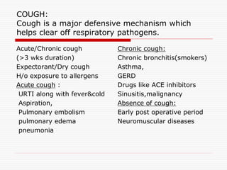
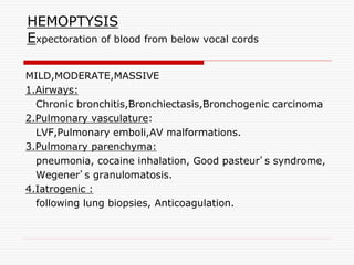
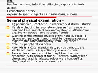
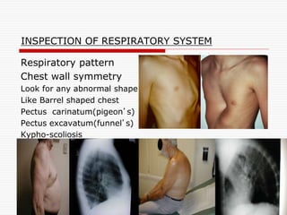
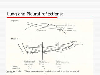
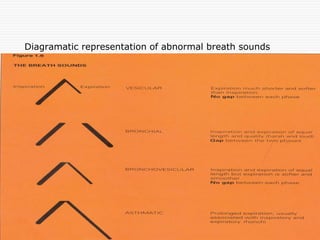
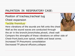
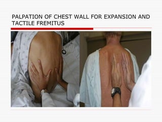
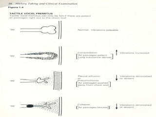
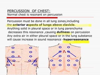
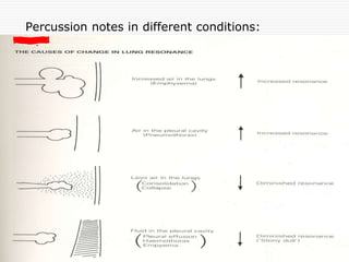
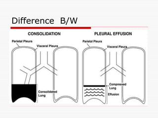
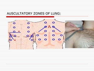
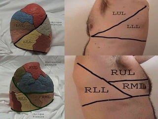
EXAMINATION OF RESPIRATORY SYSTEM
Objectives
 Common symptoms of the respiratory
system
 Signs of respiratory diseases
 Differential diagnoses of respiratory
diseases
HISTORY TAKING IN RESPIRATORY CASE
 COUGH
 DYSPNEA
 HEMOPTYSIS
 CHEST PAIN
SPUTUM
COUGH:
Cough is a major defensive mechanism which
helps clear off respiratory pathogens.
Acute/Chronic cough
(>3 wks duration)
Expectorant/Dry cough
H/o exposure to allergens
Acute cough :
URTI along with fever&cold
Aspiration,
Pulmonary embolism
pulmonary edema
pneumonia
Chronic cough:
Chronic bronchitis(smokers)
Asthma,
GERD
Drugs like ACE inhibitors
Sinusitis,malignancy
Absence of cough:
Early post operative period
Neuromuscular diseases
Complications of cough:
Cough syncope, Bronchospasm,
Urinary incontinence,Conjunctival haemorrhage
Dry cough
Drug induced ACE inhibitors
allergens
Expectoration
Black-Smokers
Brown-
puemonia,pneumococci
Green/yellowish-infection
Pink frothy-P. edema
Bloody –TB,bronchiectasis
malignancy,
Types of cough:
Bovine cough-RLN palsy
Barking cough-epiglotitis
Paroxysmal cough-pertussis
Stridor- laryngotracheo -
bronchitis
DYSPNEA:SHORTNESS OF BREATH
Acute/chronic
Position:PND,orthopnea
Exertion
Acute dyspnea:
Asthma,pneumothorax,
Pulmonary edema,ARDS
Pulmonary emboli
Pulmonary infections
Chronic dyspnea:
COPD,asthma,CCF.
Grades of Dyspnea:
1-No dyspnea on normal
activity
2-Dyspnea on more than
normal activity but
comfortable at rest
3-Dyspnea on minimal
activity,comfortable only
at rest
4-Dyspnea on less than
minimal activity
HEMOPTYSIS
Expectoration of blood from below vocal cords
MILD,MODERATE,MASSIVE
1.Airways:
Chronic bronchitis,Bronchiectasis,Bronchogenic carcinoma
2.Pulmonary vasculature:
LVF,Pulmonary emboli,AV malformations.
3.Pulmonary parenchyma:
pneumonia, cocaine inhalation, Good pasteur’s syndrome,
Wegener’s granulomatosis.
4.Iatrogenic :
following lung biopsies, Anticoagulation.
PMH:
H/o frequent lung infections, Allergies, exposure to toxic
agents
Occupational history:
exposur to specific agents as in asbestosis, silicosis
General physical examination –
ill ( pneumonia), cachectic, in respiratory distress, stridor
 Hands – clubbing in respiratory disease from cancer –
non small cell bronchial carcinoma, chronic inflammation
e.g. bronchiectasis, lung abscess, fibrosis
 Wasting of the intrinsic muscle of the hand suggest T1
lesions e.g. pancoast tumor, wrist tenderness suggests
hypertrophic osteoarthropathy from lung cancer
 Colour – peripheral cyanosis,
 Asterixis is a CO2 retention flap, pulsus paradoxus is
weakened pulse in inspiration.eg severe asthma
 Face – ptosis and constricted pupil from horners
syndrome, with pancoast tumor – cervical sympathetic
plexus and brachial plexus. colour – are tongue/lips
blue/purplish from central cyanosis
INSPECTION OF RESPIRATORY SYSTEM
Respiratory pattern
Chest wall symmetry
Look for any abnormal shape
Like Barrel shaped chest
Pectus carinatum(pigeon’s)
Pectus excavatum(funnel’s)
Kypho-scoliosis
Lung and Pleural reflections:
Diagramatic representation of abnormal breath sounds
PALPATION IN RESPIRATORY CASE:
Position of trachea,tracheal tug
Chest expansion
Tactile fremitus
Here vibrations of the sounds are felt onto the chest
Wall as the sound waves are conducted through
the air in the bronchi,bronchioles,alveoli, chest wall
Compare the strengths of these vibrations on either side of
Chest-front,back,over apical, middle and basal zones.
Increased TF:Pneumonic consolidation
Decreased TF:pleural effusion,collapse
PALPATION OF CHEST WALL FOR EXPANSION AND
TACTILE FREMITUS
PERCUSSION OF CHEST:
Normal chest is resonant on percussion
Percussion must be done in all lung zones,including
the anterior aspects of lungs above clavicle.
Anything solid in pleural space or in lung parenchyma
decreases this resonance ,causing dullness on percussion
Any extra air in either pleural space or in the lung substance
will cause increase in sound resonance -hyperresonance
Percussion notes in different conditions:
Difference B/W
AUSCULTATION OF CHEST
Normal sounds of breathing consists of an inspiratory phase
followed by a shorter,softer,expiratory phase with no gap.
This is called the normal vesicular breathing
TYPES OF BREATHING
Vesicular
Bronchial
Bronchovesicular
Added sounds
Wheeze-high pitched, muscial,
air through narrowed tubes.
Asthma,COPD
Crackles- fine, medium,
coarse. Not cleared by
coughing as in
Pulmonary edema
Crepitations-consolidation
Pleural rub- crackly,grating,dry
low pitched, machine-like in
Plueritis
AUSCULTATORY ZONES OF LUNG:
RESPIRATORY SOUNDS
TAKE A GUESS!
 m

Examination-of-respiratory-system-ppt

  • 1.
  • 2.
    Objectives  Common symptomsof the respiratory system  Signs of respiratory diseases  Differential diagnoses of respiratory diseases
  • 3.
    HISTORY TAKING INRESPIRATORY CASE  COUGH  DYSPNEA  HEMOPTYSIS  CHEST PAIN SPUTUM
  • 4.
    COUGH: Cough is amajor defensive mechanism which helps clear off respiratory pathogens. Acute/Chronic cough (>3 wks duration) Expectorant/Dry cough H/o exposure to allergens Acute cough : URTI along with fever&cold Aspiration, Pulmonary embolism pulmonary edema pneumonia Chronic cough: Chronic bronchitis(smokers) Asthma, GERD Drugs like ACE inhibitors Sinusitis,malignancy Absence of cough: Early post operative period Neuromuscular diseases
  • 5.
    Complications of cough: Coughsyncope, Bronchospasm, Urinary incontinence,Conjunctival haemorrhage Dry cough Drug induced ACE inhibitors allergens Expectoration Black-Smokers Brown- puemonia,pneumococci Green/yellowish-infection Pink frothy-P. edema Bloody –TB,bronchiectasis malignancy, Types of cough: Bovine cough-RLN palsy Barking cough-epiglotitis Paroxysmal cough-pertussis Stridor- laryngotracheo - bronchitis
  • 6.
    DYSPNEA:SHORTNESS OF BREATH Acute/chronic Position:PND,orthopnea Exertion Acutedyspnea: Asthma,pneumothorax, Pulmonary edema,ARDS Pulmonary emboli Pulmonary infections Chronic dyspnea: COPD,asthma,CCF. Grades of Dyspnea: 1-No dyspnea on normal activity 2-Dyspnea on more than normal activity but comfortable at rest 3-Dyspnea on minimal activity,comfortable only at rest 4-Dyspnea on less than minimal activity
  • 7.
    HEMOPTYSIS Expectoration of bloodfrom below vocal cords MILD,MODERATE,MASSIVE 1.Airways: Chronic bronchitis,Bronchiectasis,Bronchogenic carcinoma 2.Pulmonary vasculature: LVF,Pulmonary emboli,AV malformations. 3.Pulmonary parenchyma: pneumonia, cocaine inhalation, Good pasteur’s syndrome, Wegener’s granulomatosis. 4.Iatrogenic : following lung biopsies, Anticoagulation.
  • 8.
    PMH: H/o frequent lunginfections, Allergies, exposure to toxic agents Occupational history: exposur to specific agents as in asbestosis, silicosis General physical examination – ill ( pneumonia), cachectic, in respiratory distress, stridor  Hands – clubbing in respiratory disease from cancer – non small cell bronchial carcinoma, chronic inflammation e.g. bronchiectasis, lung abscess, fibrosis  Wasting of the intrinsic muscle of the hand suggest T1 lesions e.g. pancoast tumor, wrist tenderness suggests hypertrophic osteoarthropathy from lung cancer  Colour – peripheral cyanosis,  Asterixis is a CO2 retention flap, pulsus paradoxus is weakened pulse in inspiration.eg severe asthma  Face – ptosis and constricted pupil from horners syndrome, with pancoast tumor – cervical sympathetic plexus and brachial plexus. colour – are tongue/lips blue/purplish from central cyanosis
  • 9.
    INSPECTION OF RESPIRATORYSYSTEM Respiratory pattern Chest wall symmetry Look for any abnormal shape Like Barrel shaped chest Pectus carinatum(pigeon’s) Pectus excavatum(funnel’s) Kypho-scoliosis
  • 10.
    Lung and Pleuralreflections:
  • 11.
    Diagramatic representation ofabnormal breath sounds
  • 12.
    PALPATION IN RESPIRATORYCASE: Position of trachea,tracheal tug Chest expansion Tactile fremitus Here vibrations of the sounds are felt onto the chest Wall as the sound waves are conducted through the air in the bronchi,bronchioles,alveoli, chest wall Compare the strengths of these vibrations on either side of Chest-front,back,over apical, middle and basal zones. Increased TF:Pneumonic consolidation Decreased TF:pleural effusion,collapse
  • 13.
    PALPATION OF CHESTWALL FOR EXPANSION AND TACTILE FREMITUS
  • 15.
    PERCUSSION OF CHEST: Normalchest is resonant on percussion Percussion must be done in all lung zones,including the anterior aspects of lungs above clavicle. Anything solid in pleural space or in lung parenchyma decreases this resonance ,causing dullness on percussion Any extra air in either pleural space or in the lung substance will cause increase in sound resonance -hyperresonance
  • 16.
    Percussion notes indifferent conditions:
  • 17.
  • 18.
    AUSCULTATION OF CHEST Normalsounds of breathing consists of an inspiratory phase followed by a shorter,softer,expiratory phase with no gap. This is called the normal vesicular breathing TYPES OF BREATHING Vesicular Bronchial Bronchovesicular Added sounds Wheeze-high pitched, muscial, air through narrowed tubes. Asthma,COPD Crackles- fine, medium, coarse. Not cleared by coughing as in Pulmonary edema Crepitations-consolidation Pleural rub- crackly,grating,dry low pitched, machine-like in Plueritis
  • 19.
  • 21.
  • 22.
  • 23.