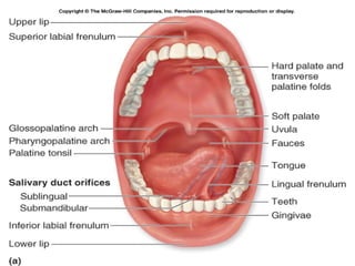
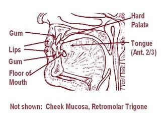
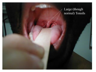
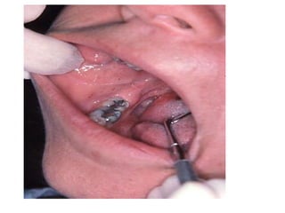
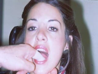
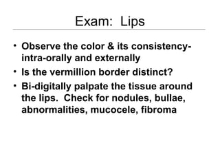
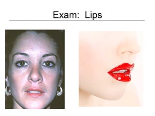
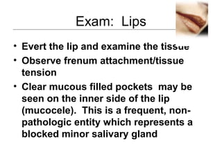
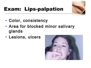
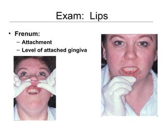
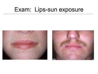
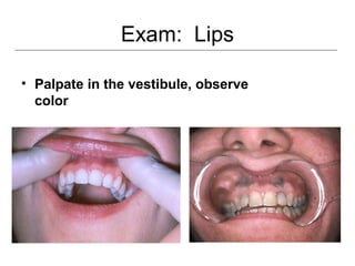
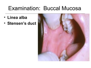
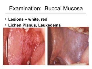
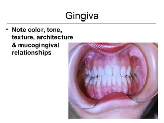
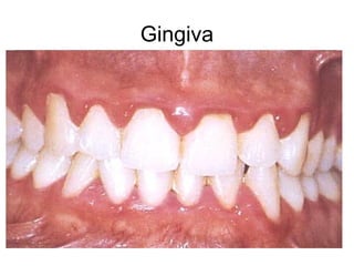
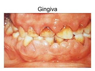
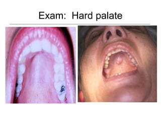
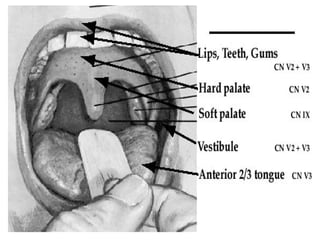
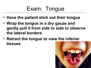
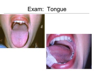
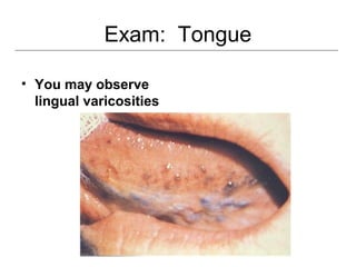
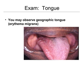
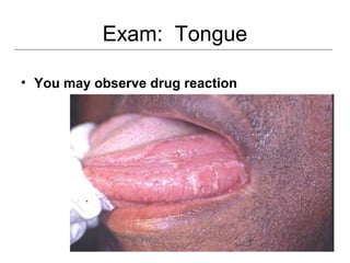
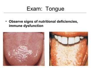
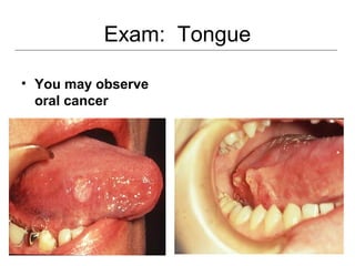
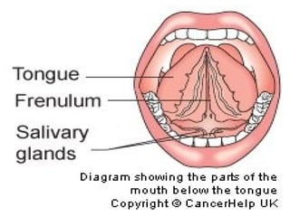
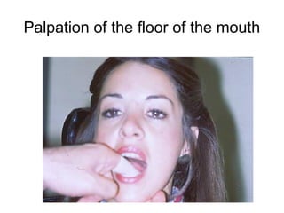
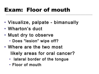
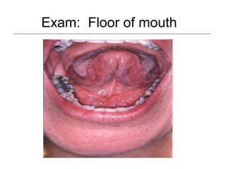
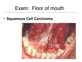
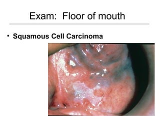
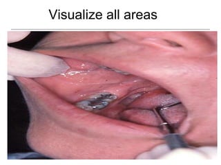
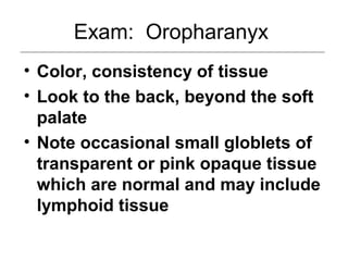
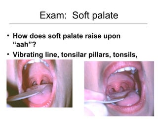
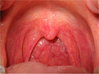
This document provides guidance on examining the oral cavity and oropharynx. Key areas to examine include the lips, checking for color, consistency, lesions or abnormalities; the buccal mucosa, looking for lesions; the gingiva, noting color, tone and architecture; the hard and soft palates; the tongue, checking all surfaces for lesions or signs of nutritional deficiencies; the floor of the mouth, visualizing and palpating for lesions or masses; and the oropharynx, checking for normal color and consistency of tissues. The lateral borders of the tongue and floor of the mouth are the most common sites for oral cancer, so these areas require close examination. A systematic and complete examination of all oral tissues
8. Exam: Lips
• Observe the color & its consistency-
intra-orally and externally
• Is the vermillion border distinct?
• Bi-digitally palpate the tissue around
the lips. Check for nodules, bullae,
abnormalities, mucocele, fibroma
11. Exam: Lips
• Evert the lip and examine the tissue
• Observe frenum attachment/tissue
tension
• Clear mucous filled pockets may be
seen on the inner side of the lip
(mucocele). This is a frequent, non-
pathologic entity which represents a
blocked minor salivary gland
23. Exam: Tongue
• The tongue and the floor of the mouth
are the most common places for oral
cancer to occur
• It can occur other places; so visualize
all areas
• You may observe:
– Circumvalate papillae, epiglottis
24. Exam: Tongue
• Have the patient stick out their tongue
• Wrap the tongue in a dry gauze and
gently pull it from side to side to observe
the lateral borders
• Retract the tongue to view the inferior
tissues
33. Exam: Floor of mouth
• Visualize, palpate - bimanually
• Wharton’s duct
• Must dry to observe
• Does “lesion” wipe off?
• Where are the two most
likely areas for oral cancer?
• lateral border of the tongue
• Floor of mouth
34. Exam: Floor of mouth
• Oral Cancer:
• Red
• White
• Red and White
• Does the patient have important
risk factors for oral cancer?
• Counseling for smoking and alcohol
• Cessation
45. Systematic Oral
Examination
• Done at initial exam & at recalls
unless patient history requires
sooner
• You must visualize all areas of the
oral cavity
46. Systematic Oral
Examination
• Oral cancer can occur in other
places than the lateral borders of
the tongue & the floor of the mouth
• Be complete
• Do good, do no harm, do justice,
respect autonomy
48. Exam: Oropharanyx
• Color, consistency of tissue
• Look to the back, beyond the soft
palate
• Note occasional small globlets of
transparent or pink opaque tissue
which are normal and may include
lymphoid tissue
49. Exam: Soft palate
• How does soft palate raise upon
“aah”?
• Vibrating line, tonsilar pillars, tonsils,
oropharynx