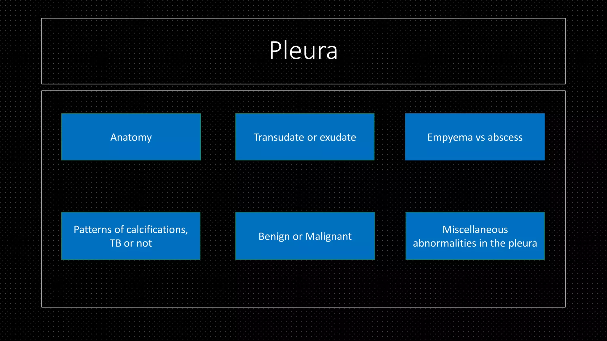
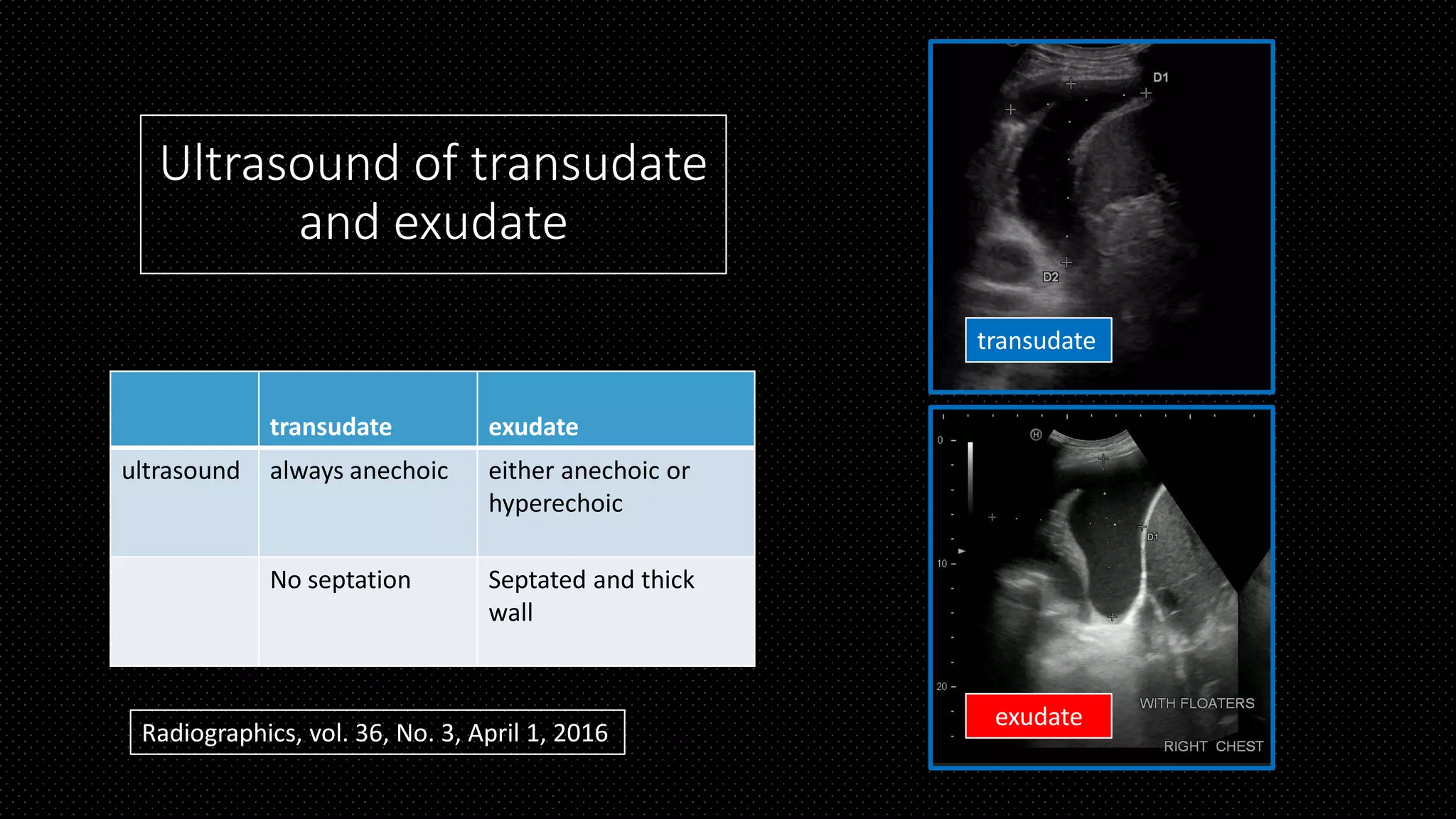
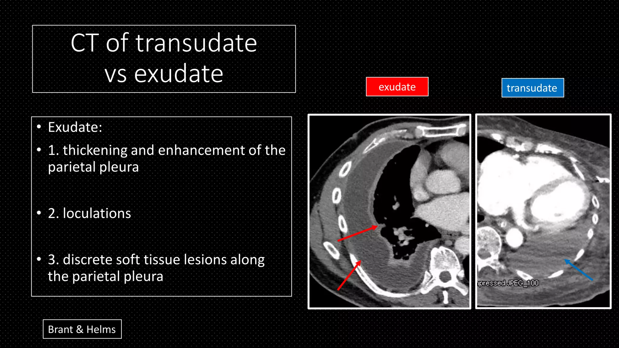
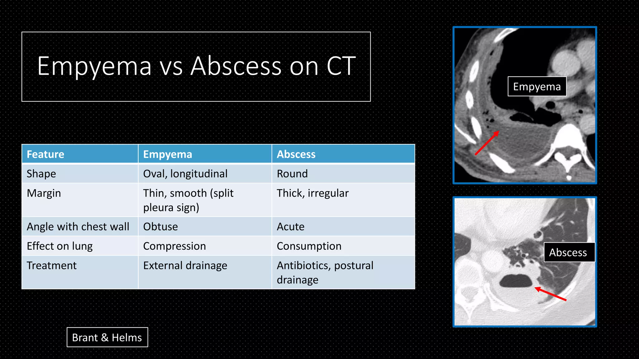
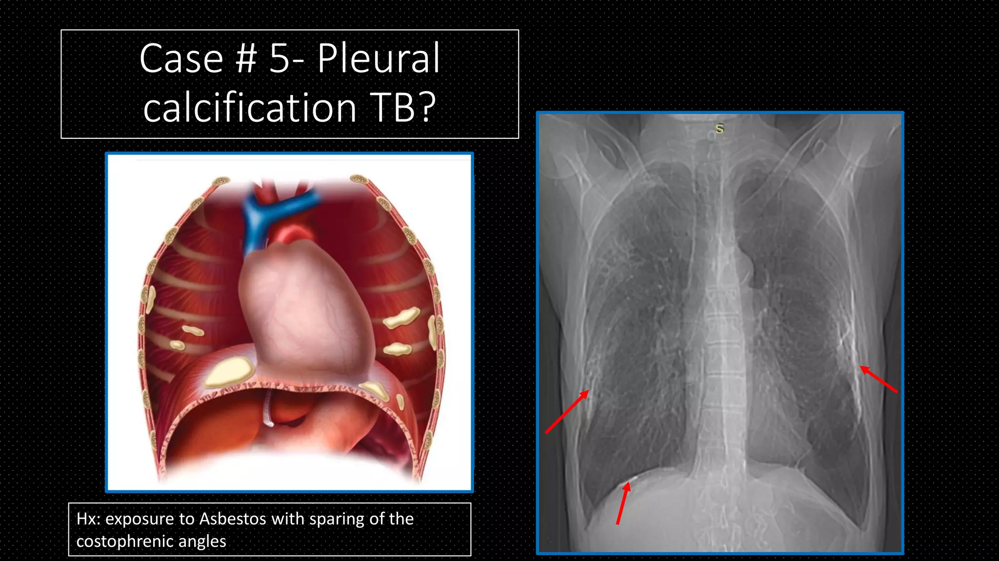
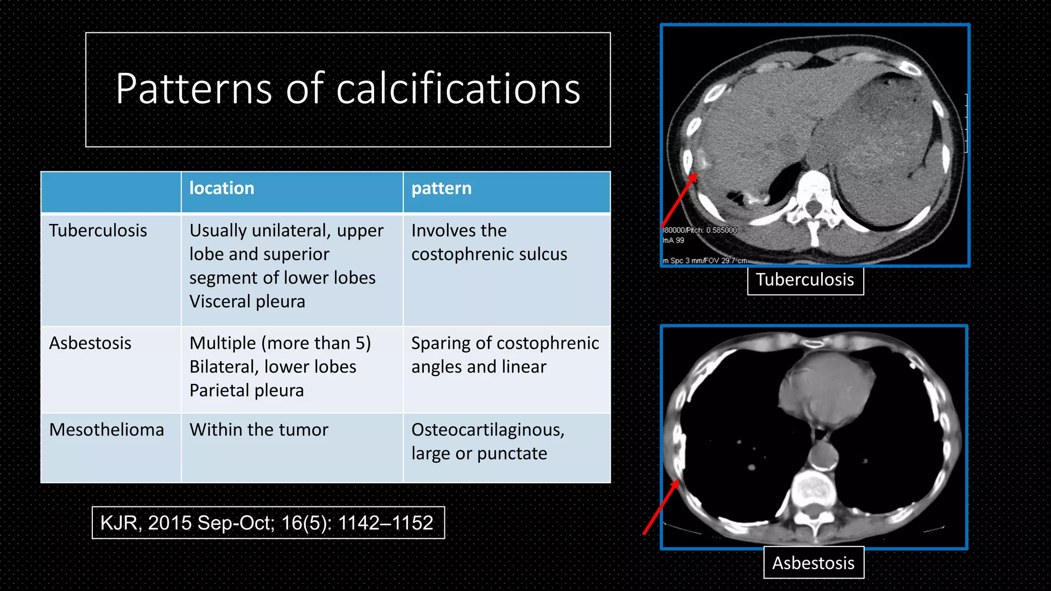
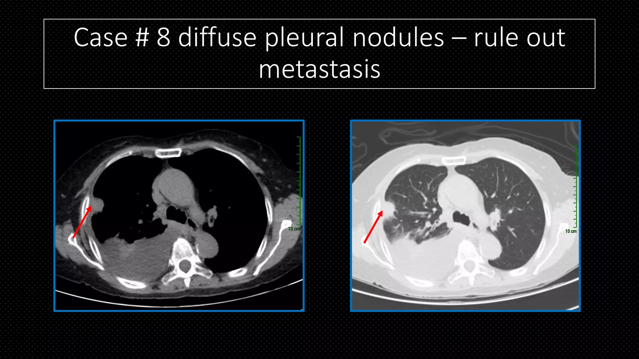
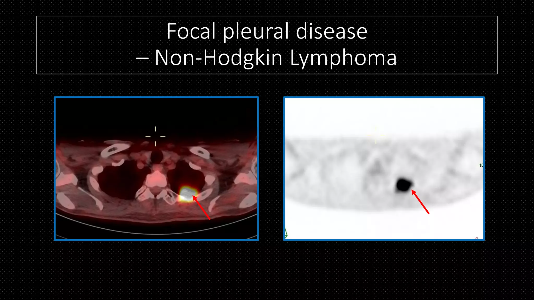
07 histology, cytology and biomarkers in pleural diseasesipmslmc
This document discusses histology, cytology, and biomarkers of pleural diseases. It begins with an overview of pleural histology and then focuses on distinguishing benign from malignant pleural lesions. Key points include identifying lesions as epithelial or mesothelial using immunohistochemistry, assessing features to differentiate benign mesothelial proliferations from malignant mesothelioma, and evaluating biomarkers like BAP1 and p16 for atypical cases. The document also reviews cytology of pleural effusions and summarizes approaches to pleural biopsies showing thickening or atypical proliferations.
Pleural effusion caused by malignancies has been described as malignant pleural effusion. Etiology,pathogenesis,diagnosis and management of malignant pleural effusion has been descibed in this powerpoint presentation.
Abstract
Carotid body tumors are rare, slow-growing, hypervascular neuroendocrine tumors. Although these tumors are benign neoplasm, they also have a tendency to malignant transformation. Complete surgical excision is the gold standard therapeutic modality for the treatment of carotid body tumors. Early surgical removal is recommended to prevent the development of larger and more advanced tumors, which are associated with higher morbidity and mortality. In this report, we presented three cases of carotid body tumor which were successfully treated with complete surgical excision, and reviewed the current literature. Furthermore, it was emphasized the necessity of early surgical management regardless of patient age and tumor size.
The document provides guidelines for managing malignant pleural effusions. It discusses:
- Lung cancer and breast cancer are the most common causes of malignant pleural effusions.
- Effusions are usually symptomatic, with dyspnea being most common. Massive effusions often indicate malignancy.
- Management depends on symptoms, primary tumor type, and lung re-expansion. Options include observation, therapeutic aspiration, tube drainage with sclerosant, or thoracoscopy with pleurodesis. Tube drainage and pleurodesis is preferred to prevent recurrence, unless lung is trapped.
Lung tumors are commonly fatal malignancies that occur most often in adults aged 40-70 years. The main risk factors are cigarette smoking, air pollution, asbestos exposure, and certain industrial chemicals. The most common types are squamous cell carcinoma, adenocarcinoma, large cell carcinoma, and small cell carcinoma. Clinical presentation depends on whether the tumor is localized, invasive, or metastatic. Imaging techniques like chest X-ray, CT, MRI, PET, and PET-CT are used to characterize tumors and detect spread. Treatment options vary based on tumor type and stage.
The document discusses the classification, epidemiology, histology, and clinical features of lung cancer. Lung cancer is classified into non-small cell lung cancer (70-75%), small cell lung cancer (20-25%), and combined patterns (5-10%). Cigarette smoking is the primary risk factor and causes 40-70% of lung cancers, which often present with cough, weight loss, and chest pain.
The document discusses lung cancer, its types, risk factors, diagnosis, and treatment options. It describes that lung cancer is uncontrolled cell growth in lung tissues and the major types are small cell lung cancer and non-small cell lung cancer such as adenocarcinoma and squamous cell carcinoma. Risk factors include smoking, occupational exposures, family history, and asbestos exposure. Diagnosis involves tests such as CT scans, PET scans, biopsies, and biomarker analysis. Treatment depends on cancer type and stage but may include surgery, chemotherapy, radiation therapy, targeted therapy, immunotherapy, or a combination.
This document summarizes the diagnostic imaging characteristics of different types of lung cancer. It discusses:
- Adenocarcinoma typically presents as a peripheral nodule under 4cm in diameter on CT. Bronchoalveolar carcinoma presents as a solitary nodule, multifocal disease, or localized consolidation with bubble-like low attenuation areas.
- Squamous cell carcinoma is often centrally located, larger than 4cm, and cavitates in up to 82%. Small cell lung cancer presents with bulky hilar and mediastinal lymph nodes and rarely cavitates.
- Imaging techniques like chest x-rays, CT, MRI, and PET scans are used to diagnose and stage lung cancers. CT is
Lung cancer accounts for about 6.8% of malignancies in India. Risk factors include cigarette smoking as well as exposure to radioactive isotopes, polycyclic hydrocarbons, and other chemicals. Symptoms may include cough, dyspnea, chest pain, and weight loss. Diagnostic tests include x-ray, CT scan, PET scan, and biopsy. Treatment involves surgery, radiation therapy, chemotherapy, or a combination. Nursing care focuses on managing symptoms, promoting effective breathing and nutrition, providing psychological support, and health education.
While lung cancer remains a very challenging cancer to treat, new treatments that capitalize on advances in our understanding of cancer. It is likely that a more personalized approach to treatment using biological markers and combinations of therapies will provide better results in the future.
There are five main histological types of bronchogenic carcinoma: squamous cell carcinoma, adenocarcinoma, bronchioalveolar carcinoma, small cell carcinoma, and large cell carcinoma. Squamous cell carcinoma typically presents as a central hilar tumor with necrosis and keratinization. Adenocarcinoma usually presents as a peripheral nodule and shows gland formation and mucin production. Bronchioalveolar carcinoma has a lepidic growth pattern along alveoli. Small cell carcinoma appears as a central hilar nodule with sheets of small blue cells. Large cell carcinoma forms a peripheral lobulated mass with large, anaplastic cells showing no clear cell type.
This document summarizes the staging of bronchogenic carcinoma (lung cancer). It discusses the epidemiology, pathology, and histological types of lung cancer. It then covers the tools used to stage lung cancer, including imaging techniques like CT, PET, and MRI. Physical examination, sputum cytology, and bronchoscopy are also described. The TNM staging system is explained along with invasive staging procedures like mediastinoscopy. Finally, it discusses the distinct staging system for small cell lung cancer.
BENIGN AND MALIGNANT LUNG NEOPLASAM MASSESnishit viradia
The document discusses benign and malignant lung neoplastic masses. It begins by stating that a wide variety of neoplasms can arise in the lungs, including both benign and malignant tumors. Carcinoma of the bronchus is the most common and important primary lung tumor. Various types of lung tumors are described such as solitary pulmonary nodules, benign pulmonary tumors, bronchogenic carcinoma, pulmonary sarcoma, and metastases. Diagnostic criteria and imaging features of solitary pulmonary nodules and different types of lung cancers like adenocarcinoma and squamous cell carcinoma are provided. Peripheral and central lung tumors are also discussed.
1. The document discusses the pathology, staging, and management of lung cancer. It covers the classification, diagnosis, and molecular features of small cell lung cancer and non-small cell lung cancers including adenocarcinoma, squamous cell carcinoma, and large cell carcinoma.
2. Staging involves evaluating the extent of disease spread using imaging tests, biopsies and evaluating the patient's health. Treatment options depend on cancer type and stage and may include surgery, radiation, chemotherapy, targeted therapy or palliative care.
3. For early stage disease, surgery is usually recommended but other options like radiation may be considered. Locally advanced or metastatic disease is generally treated with chemotherapy and/or radiation. Management aims to
1. Lung tumors can be classified as primary lung cancers, metastases, or benign tumors. Primary lung cancers include non-small cell lung cancer (NSCLC), small cell lung cancer (SCLC), and others.
2. Imaging plays a key role in evaluating lung tumors by characterizing findings, aiding diagnosis, and staging. Features such as size, margins, enhancement, and growth over time provide clues about benign vs malignant tumors.
3. NSCLC is the most common type and demonstrates varied radiological appearances depending on size, location, and histology. Imaging is also used to guide biopsy and assess treatment response.
Lung cancer accounts for about 6.8% of malignancies in India, with incidence rates of 6.6 per 100,000 males and 1.7 per 100,000 females. Risk factors include cigarette smoking, exposure to radioactive isotopes, polycyclic hydrocarbons, and other chemicals. Symptoms depend on the location and stage of cancer, ranging from cough and breathing difficulties to weight loss and bone pain. Diagnosis involves tests such as x-rays, CT scans, biopsies and PET scans. Treatment may include surgery, radiation, chemotherapy, or a combination. Nursing care focuses on managing symptoms, improving breathing and nutrition, providing education and psychological support, and maintaining health.
Lung cancer is characterized by uncontrolled cell growth in lung tissues. Worldwide, it is the leading cause of cancer death in men and women, responsible for 1.3 million deaths annually. The main causes are smoking and exposure to radon, asbestos, viruses and other particulates. Symptoms include coughing, shortness of breath, and weight loss. Diagnosis involves imaging tests and biopsies. Treatment depends on cancer type and stage but may include surgery, chemotherapy, and radiation therapy.
Lung cancer is classified into two main types - non-small cell lung carcinoma (NSCLC) and small cell lung carcinoma (SCLC). NSCLC makes up about 80% of cases and can be further divided into squamous cell carcinoma, adenocarcinoma, and large cell carcinoma. SCLC accounts for 10-15% of lung cancers and grows more quickly. The main symptoms are cough, chest pain, and coughing up blood. Risk factors include smoking, asbestos exposure, and radiation exposure. Diagnosis involves tests such as sputum analysis, biopsies, CT scans, and PET scans to determine the cancer type and stage. Treatment options depend on the cancer type and stage but may include surgery, chemotherapy
Non–small cell lung cancer (NSCLC) accounts for approximately 85% of all lung cancers. Histologically, NSCLC is divided into adenocarcinoma, squamous cell carcinoma (SCC) (see the image below), and large cell carcinoma. Small cell lung cancer (SCLC), previously known as oat cell carcinoma, is considered distinct from other lung cancers, which are called non–small cell lung cancers (NSCLCs) because of their clinical and biologic characteristics.
Lung tumors can be primary, arising from lung tissue, or secondary, having spread from other sites. Primary lung tumors are mainly bronchogenic carcinoma (95%), which is usually squamous cell carcinoma or adenocarcinoma. Bronchogenic carcinoma is strongly linked to smoking and has a poor prognosis. Small cell lung cancer is very aggressive and has often metastasized by the time of diagnosis. Secondary lung tumors are common, with metastases arriving via blood or lymph vessels from other carcinomas or sarcomas. Pleural effusions are a frequent manifestation of primary or secondary lung malignancies. Malignant mesothelioma is a rare tumor associated with asbestos exposure that arises from the pleural lining.
Radiological approach to lung neoplasmsSnehaMandal5
This document discusses the radiological approach to lung neoplasms. It begins by introducing the epidemiology and risk factors of lung cancer. It then describes the imaging modalities used for detection, characterization, staging and follow-up. Key imaging features of peripheral and central lung tumors on chest radiography and CT are provided. The document outlines the radiological characteristics of lung cancers by cell type and discusses sampling techniques and staging based on tumor size and local invasion.
Lung cancer is the leading cause of cancer death worldwide. The document discusses the classification, risk factors, clinical features, investigations and treatment options for lung cancer. It notes that lung cancer most commonly presents as cough, haemoptysis or breathlessness. Investigations include chest x-ray, CT, PET scans, bronchoscopy and biopsy. Treatment depends on cancer type and stage but may include surgery for early-stage non-small cell lung cancer, radiotherapy for palliation, and platinum-based chemotherapy mainly for small cell lung cancer. Prognosis remains poor with only 15% of patients surviving more than 5 years.
Pulmonary tuberculosis is caused by the bacterium Mycobacterium tuberculosis. It is transmitted via airborne droplets when people with active TB cough, sneeze or spit. Primary tuberculosis occurs when someone is initially infected, usually resulting in a self-limited infection. Post-primary tuberculosis occurs after a latent period and is usually due to reinfection or reactivation of a latent infection. Diagnosis involves microbiological testing of sputum samples, chest imaging, and tuberculin skin testing or interferon-gamma release assays. Radiographic findings can help determine if a case is active or inactive.
A 55-year-old man presented with a cough, blood in his sputum, chest pain, shortness of breath, and a 6 kg weight loss over 3 months. Imaging showed a mass in his right lung. Biopsy confirmed lung cancer. He had a history of smoking for 30 pack years. Despite treatment, he died and autopsy found the cancer had spread within his lungs.
This document provides an overview of pancreatic pathology, focusing on pancreatitis. It describes the anatomy of the pancreas and imaging modalities used to evaluate pancreatic conditions. Acute and chronic pancreatitis are discussed in detail, including causes, classification, imaging features, and complications like fluid collections, pseudocysts, abscesses, necrosis, and hemorrhage. Other pancreatic conditions summarized include autoimmune pancreatitis and hereditary pancreatitis.
This document discusses tumors of the lung, specifically bronchogenic carcinoma. It covers the most common types including squamous cell carcinoma, adenocarcinoma, small cell carcinoma, and large cell carcinoma. It details their causes such as tobacco smoking, air pollution, and genetic factors. It also describes the gross and microscopic appearance of each tumor type and their spread patterns and clinical presentations.
The document discusses management of oropharyngeal cancers. The oropharynx includes areas like the base of the tongue, soft palate, tonsils and posterior pharyngeal walls. Oropharyngeal cancers commonly spread to cervical lymph nodes in levels II, III and IV. Risk factors include age, gender, smoking, alcohol and HPV infection. Treatment may involve surgery, radiation therapy or chemotherapy depending on the stage of cancer. Imaging tests like CT, MRI and PET scans are used to stage the cancer and detect metastases.
METASTATIC NECK DISEASE FOR ENT & HNS.pptxSatishray9
1) Metastatic neck disease is an important prognostic factor in head and neck cancer and is classified by lymph node levels.
2) Imaging techniques like ultrasound, CT, MRI and PET scans are used to assess cervical lymphadenopathy and detect metastatic neck disease.
3) Treatment of metastatic neck disease depends on factors like number of involved nodes, size of nodes, bilateral involvement and previous treatment and may involve surgery, radiation or chemoradiation.
This document discusses the use of various imaging modalities for evaluating neck masses. Ultrasound is useful for differentiating cystic from solid lesions and assessing lymph node size and vascularity. CT provides details of soft tissues and their relationships. MRI is good for lesion detection and involvement of nearby structures but has limitations for nodal assessment. PET/CT is excellent for staging lymphoma and detecting unknown primary cancers. Biopsy is used when malignancy is suspected. The approach depends on whether the mass is in a child or adult, with ultrasound often the initial study. Location provides clues for cystic lesions. Features help characterize solid lesions and lymph nodes. Further tests are guided by ultrasound findings.
1. Lung metastasis is the second most common site of metastasis after the liver for many cancers. Complete resection of pulmonary metastases can improve survival for certain primary cancers like sarcoma, colon cancer, and breast cancer when the metastases are limited in number and size.
2. Pulmonary metastases can present radiographically in different patterns including cannonball lesions, miliary nodules, cavitating lesions, and endobronchial lesions. Diagnosis is typically made through CT scan, and biopsy may be needed to differentiate from a primary lung cancer.
3. Surgical resection is recommended for isolated, resectable metastases to potentially improve survival, with criteria including controlled primary cancer, no other distant metastases, and ability
This document provides information on neuroblastoma and Wilms' tumor, two common pediatric abdominal masses. Neuroblastoma arises from neural crest tissue and is the most common extracranial solid tumor in children. It typically presents as an abdominal mass and can metastasize. Treatment involves chemotherapy, surgery, and sometimes stem cell transplant depending on risk factors. Wilms' tumor arises from the kidneys and is also typically detected as an asymptomatic abdominal mass in young children. Staging involves evaluating the extent of the primary tumor and presence of metastases to determine the appropriate treatment, which usually involves surgery and chemotherapy. Both tumors require a multidisciplinary approach including imaging, biopsy, surgery, and chemotherapy.
1) Cancers of the penis are rare but devastating, accounting for 0.4-0.6% of cancers in men in the US and Europe but up to 10% in some other regions.
2) Risk factors include poor hygiene, phimosis, HPV infection, and lack of circumcision. Over 95% are squamous cell carcinoma.
3) Staging involves physical exam, biopsy, and imaging of lymph nodes and distant organs. Treatment may include organ-sparing surgery or penile amputation depending on size, grade, and extent of invasion.
Testicular tumors-Cassification, Biomarkers and Staging by Dr RajeshRajesh Sinwer
This document discusses testicular tumors, including:
- Germ cell tumors are the most common type, comprising 95% of cases. Seminomas and non-seminomatous germ cell tumors are the main subtypes.
- Important biomarkers for testicular cancer include AFP, HCG, LDH, and PLAP. Elevated levels can indicate the presence of a non-seminoma.
- Staging is important and is based on whether the cancer is confined to the testis or has spread to lymph nodes or other organs. Spread beyond the retroperitoneum is considered stage III.
- Diagnostic workup involves imaging like ultrasound, CT, MRI and PET scans
This document provides an overview of bladder cancer presented by Dr. Vikas Kumar. Some key points:
- Bladder cancer is the 9th most common cancer worldwide and the 13th most common cause of death. Risk factors include smoking, occupational exposures, infections, and genetic factors.
- At initial presentation, 80% of bladder cancers are non-muscle invasive. Staging involves evaluating the extent of primary tumor invasion and spread to lymph nodes and distant organs.
- Diagnosis involves cystoscopy, urine cytology, and imaging tests. Random bladder biopsies are also recommended to detect cancers that cannot be seen.
- For non-muscle invasive cancers, the main treatment is transure
This document provides tips and instructions for using a PowerPoint presentation (PPT) on nasopharyngeal carcinoma. Some key points:
- Slides can be freely edited and modified. Half the slides are blank for active learning exercises.
- The presentation will go through three revisions: showing blank slides to elicit student responses, then showing content slides.
- It is useful for both self-study and active learning sessions.
- Bibliographic references are provided in the notes section.
- The PPT then provides detailed content on the epidemiology, etiology, clinical features, investigations, management and more of nasopharyngeal carcinoma.
The document provides information on evaluating and managing solitary thyroid nodules. It discusses obtaining a detailed history, performing a physical exam focusing on the thyroid and lymph nodes, and using diagnostic tests like ultrasound, biopsy, and bloodwork. Fine-needle aspiration biopsy is the most important test, and can determine if a nodule is benign, suspicious, malignant, or nondiagnostic. Treatment depends on biopsy results and risk factors, and may include observation, surgery, radiation, or medication. The document outlines common thyroid cancers like papillary and follicular carcinoma.
The document discusses carcinoma of the esophagus, including its anatomy, epidemiology, risk factors, staging, types, and management. It covers the various surgical techniques for esophagectomy, such as Ivor Lewis, McKeown, and transhiatal procedures. Post-operative care including drainage tube removal and diet progression is also summarized.
This document provides an overview of the diagnostic workup for breast cancer. It discusses the history, physical examination, investigations including imaging like ultrasound, mammography and MRI, histopathology including biopsy types, and tumor staging. The key points are that the diagnostic workup involves a thorough history, physical exam including breast inspection and palpation, imaging tests like ultrasound and mammography, biopsy for histopathological examination, and tumor staging using the TNM system to determine cancer extent and prognosis. A triple assessment approach combining clinical, radiological and histopathological examination has a high positive predictive value of detecting breast cancer.
This document discusses the ultrasound evaluation of adnexal masses in reproductive age women. It provides clinical background information on adnexal masses and covers ultrasound techniques like 2D, Doppler, 3D and power Doppler imaging. Pattern recognition is emphasized as the best method for distinguishing benign from malignant tumors. The IOTA simple rules classification system is presented as well as newer markers for characterizing adnexal masses with ultrasound like impedance values and vascular patterns.
This document discusses adnexal masses in reproductive age women. It provides clinical background and epidemiology of adnexal masses. It discusses ultrasound techniques for evaluating adnexal masses such as 2D ultrasound, Doppler ultrasound, 3D and power Doppler ultrasound. Pattern recognition of benign disease using ultrasound is covered. The document also discusses predicting malignancy using systems like IOTA simple rules and pattern recognition. Characterization of adnexal masses and predicting histopathology and management are key focuses.
This document discusses advanced (metastatic) breast cancer, including:
- Breast cancer is the most common cancer in women worldwide, with over 22 million new cases diagnosed in 2020.
- If breast cancer spreads to distant organs, the 5-year survival rate drops to 29%. Common sites of metastasis include bone, lung, liver and brain.
- Advanced breast cancer is typically treated through a combination of surgery, chemotherapy, hormone therapy, targeted therapy and radiation depending on the cancer type, location and stage.
- Staging uses the TNM system to classify tumors by size (T), lymph node involvement (N) and metastasis (M), along with other factors like hormone receptor status. This
This document discusses oral squamous cell carcinoma (OSCC). It covers the epidemiology, risk factors, early detection methods, premalignant lesions, investigations, management including surgery and reconstruction, and treatment including radiation and chemotherapy. OSCC is the 6th most common cancer worldwide and the most common cancer in Indian men. Tobacco and alcohol are major risk factors. Detection methods include toluidine blue staining and tissue autofluorescence. Premalignant lesions include leukoplakia and erythroplakia. Management involves wide local excision and neck dissection, with reconstruction options like flaps and grafts. Radiation and chemotherapy may be used as adjuvant or palliative treatment.
Basavarajeeyam is a Sreshta Sangraha grantha (Compiled book ), written by Neelkanta kotturu Basavaraja Virachita. It contains 25 Prakaranas, First 24 Chapters related to Rogas& 25th to Rasadravyas.
Promoting Wellbeing - Applied Social Psychology - Psychology SuperNotesPsychoTech Services
A proprietary approach developed by bringing together the best of learning theories from Psychology, design principles from the world of visualization, and pedagogical methods from over a decade of training experience, that enables you to: Learn better, faster!
Does Over-Masturbation Contribute to Chronic Prostatitis.pptxwalterHu5
In some case, your chronic prostatitis may be related to over-masturbation. Generally, natural medicine Diuretic and Anti-inflammatory Pill can help mee get a cure.
8 Surprising Reasons To Meditate 40 Minutes A Day That Can Change Your Life.pptxHolistified Wellness
We’re talking about Vedic Meditation, a form of meditation that has been around for at least 5,000 years. Back then, the people who lived in the Indus Valley, now known as India and Pakistan, practised meditation as a fundamental part of daily life. This knowledge that has given us yoga and Ayurveda, was known as Veda, hence the name Vedic. And though there are some written records, the practice has been passed down verbally from generation to generation.
Recomendações da OMS sobre cuidados maternos e neonatais para uma experiência pós-natal positiva.
Em consonância com os ODS – Objetivos do Desenvolvimento Sustentável e a Estratégia Global para a Saúde das Mulheres, Crianças e Adolescentes, e aplicando uma abordagem baseada nos direitos humanos, os esforços de cuidados pós-natais devem expandir-se para além da cobertura e da simples sobrevivência, de modo a incluir cuidados de qualidade.
Estas diretrizes visam melhorar a qualidade dos cuidados pós-natais essenciais e de rotina prestados às mulheres e aos recém-nascidos, com o objetivo final de melhorar a saúde e o bem-estar materno e neonatal.
Uma “experiência pós-natal positiva” é um resultado importante para todas as mulheres que dão à luz e para os seus recém-nascidos, estabelecendo as bases para a melhoria da saúde e do bem-estar a curto e longo prazo. Uma experiência pós-natal positiva é definida como aquela em que as mulheres, pessoas que gestam, os recém-nascidos, os casais, os pais, os cuidadores e as famílias recebem informação consistente, garantia e apoio de profissionais de saúde motivados; e onde um sistema de saúde flexível e com recursos reconheça as necessidades das mulheres e dos bebês e respeite o seu contexto cultural.
Estas diretrizes consolidadas apresentam algumas recomendações novas e já bem fundamentadas sobre cuidados pós-natais de rotina para mulheres e neonatos que recebem cuidados no pós-parto em unidades de saúde ou na comunidade, independentemente dos recursos disponíveis.
É fornecido um conjunto abrangente de recomendações para cuidados durante o período puerperal, com ênfase nos cuidados essenciais que todas as mulheres e recém-nascidos devem receber, e com a devida atenção à qualidade dos cuidados; isto é, a entrega e a experiência do cuidado recebido. Estas diretrizes atualizam e ampliam as recomendações da OMS de 2014 sobre cuidados pós-natais da mãe e do recém-nascido e complementam as atuais diretrizes da OMS sobre a gestão de complicações pós-natais.
O estabelecimento da amamentação e o manejo das principais intercorrências é contemplada.
Recomendamos muito.
Vamos discutir essas recomendações no nosso curso de pós-graduação em Aleitamento no Instituto Ciclos.
Esta publicação só está disponível em inglês até o momento.
Prof. Marcus Renato de Carvalho
www.agostodourado.com
Osteoporosis - Definition , Evaluation and Management .pdfJim Jacob Roy
Osteoporosis is an increasing cause of morbidity among the elderly.
In this document , a brief outline of osteoporosis is given , including the risk factors of osteoporosis fractures , the indications for testing bone mineral density and the management of osteoporosis
Here is the updated list of Top Best Ayurvedic medicine for Gas and Indigestion and those are Gas-O-Go Syp for Dyspepsia | Lavizyme Syrup for Acidity | Yumzyme Hepatoprotective Capsules etc
Histololgy of Female Reproductive System.pptxAyeshaZaid1
Dive into an in-depth exploration of the histological structure of female reproductive system with this comprehensive lecture. Presented by Dr. Ayesha Irfan, Assistant Professor of Anatomy, this presentation covers the Gross anatomy and functional histology of the female reproductive organs. Ideal for students, educators, and anyone interested in medical science, this lecture provides clear explanations, detailed diagrams, and valuable insights into female reproductive system. Enhance your knowledge and understanding of this essential aspect of human biology.
1. “Pleural disease: emerging updates
& recent advances”
THORACIC RADIOLOGY & IMAGING
Roy P. Vizcarra, MD
SLMC
April 4, 2019, 9-9:30am
2. Pleura
Anatomy Transudate or exudate Empyema vs abscess
Benign or Malignant
Patterns of calcifications,
TB or not
Miscellaneous
abnormalities in the pleura
6. Ultrasound of transudate
and exudate
transudate exudate
ultrasound always anechoic either anechoic or
hyperechoic
No septation Septated and thick
wall
exudate
transudate
Radiographics, vol. 36, No. 3, April 1, 2016
7. CT of transudate
vs exudate transudateexudate
Brant & Helms
• Exudate:
• 1. thickening and enhancement of the
parietal pleura
• 2. loculations
• 3. discrete soft tissue lesions along
the parietal pleura
15. Patterns of calcifications
Tuberculosis
Asbestosis
location pattern
Tuberculosis Usually unilateral, upper
lobe and superior
segment of lower lobes
Visceral pleura
Involves the
costophrenic sulcus
Asbestosis Multiple (more than 5)
Bilateral, lower lobes
Parietal pleura
Sparing of costophrenic
angles and linear
Mesothelioma Within the tumor Osteocartilaginous,
large or punctate
KJR, 2015 Sep-Oct; 16(5): 1142–1152
17. TB Empyema necessitans
• Spontaneous discharge of
empyema through the parietal
pleura into the chest wall forms
a subcutaneous abscess
Radiographics, vol. 21, No. 4, July 1, 2001
18. Case # 8 diffuse pleural nodules – rule out
metastasis
20. Pleural metastasis
• Usually affect both the visceral and parietal pleura, multiple with
enhancement
• Most common malignancies with pleural metastases:
• 1. lung carcinoma - 40 %
• 2. breast carcinoma - 20 %
• 3. ovarian and lymphomas - 10 %
Insights Imaging. 2015 Dec; 6(6): 729–740.
21. FDG-PET as a highly accurate and noninvasive modality for the
differential diagnosis of pleural diseases in patients with cancer
• 1. sensitivity 53%–100%
• 2. specificity 67%–94%
• 3. accuracy 79%–97%
• 4. PPV 76%–87%
• 5. NPV 78%–100%
Invest Radiol. 2005 Apr; 40(4):204-9.
23. Mesothelioma
• Malignant pleural mesothelioma (MPM) is the most common
primary malignancy of the pleura and is associated with asbestos
exposure in approximately 80% of patients.
• Arises from the mesothelial cells that cover the lung and chest
wall with latency periods ranging from 20 to 50 years. Begins in
the parietal pleura.
24. CT signs of Mesothelioma
• 1. unilateral pleural effusion - seen in up to 74% of patients
• 2. pleural thickening which is nodular or lobular - 92% of cases
• 3. pleural thickening that is nodular, circumferential, and greater than
1 cm in thickness is highly suggestive of malignant pleural disease,
including MPM
25. Case #10 – Focal
pleural disease
November 27, 2018
February 27, 2019
27. Non Hodgkin lymphoma
• 1. soft tissue nodules or masses
• 2. effusion
• 3. broad thickening of the pleural membranes
• 4. majority is seen together with other thoracic abnormalities
Radiographics, Mar 13 2017
28. Malignant from Benign Pleural Disease
• 1. circumferential pleural thickening (sensitivity 41% and specificity 100%)
• 2. nodular pleural thickening (sensitivity 51% and specificity 94%)
• 3. parietal pleural thickening >1 cm (sensitivity 36% and specificity 94%)
• 4. mediastinal pleural involvement. (sensitivity 56% and specificity 88%)
AJR 154:487-492, March 1990
31. April 29, 2014January 15, 2017 September 26, 2017
Non hypermetabolic – Mesothelioma, Cancer
and Neurofibroma
32. Pleural mass
• Focal pleural masses are usually benign neoplasms such as lipomas;
loculated pleural fluid can mimic a pleural mass radiographically.
• Thoracic lipomas may arise in the chest wall or subpleural fat.
Homogeneous fat attenuation on CT scan (-30 to -100 H) is diagnostic
Brant and Helms
35. MRI of the pleura
• Smooth enhancement of the pleura can be seen in both infectious
and neoplastic conditions, but nodular or mass-like enhancement
should raise suspicion for tumor.
Radiographics. Jan 10 2018
37. Case #12 – pneumothorax secondary to
ruptured bleb
38. Case #12 – pneumothorax secondary to
bulla/bleb
39. Spontaneous pneumothorax
• Most often occurs in young or middle-aged men. A familial incidence
and a propensity for tall, thin individuals has been noted.
• Affected patients may have blebs or bullae in the lung apices that are
responsible for the development of recurrent pneumothoraces.
40. • 1. Bleb - cystic space 1 cm or less in diameter
• 2. Bulla - >1cm
• Blebs and bullae are commonly subpleural with coexisting
centrilobular and paraseptal emphysema.
InRadiology, Multiple cystlike lung lesions in the adult
Cystlike lesions in adults