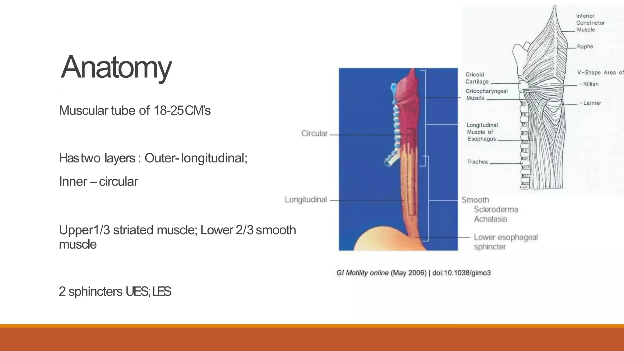
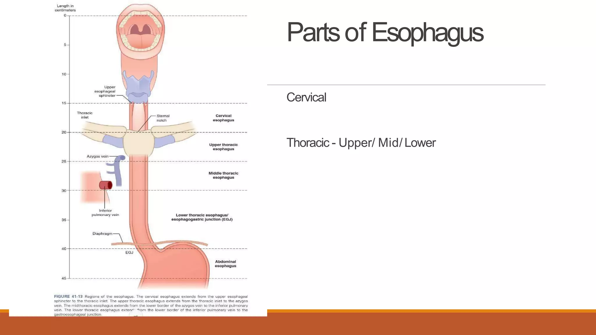
SlideShare a Scribd company logo
EsophagealMotility
Disorders
SIDHARTHAN
Anatomy
Muscular tube of 18-25CM’s
Hastwo layers : Outer-longitudinal;
Inner –circular
Upper1/3 striated muscle; Lower 2/3smooth
muscle
2 sphincters UES;LES
Partsof Esophagus
Cervical
Thoracic - Upper/ Mid/Lower
4 Layersof Esophagus
1. mucosa
2.submucosa
3.muscularis propria
4. adventitia
Peristalsis
Esophageal peristalsis results from sequential contraction of circular muscle, which
serves to push the ingested food bolus toward the stomach. Esophageal longitudinal
muscle may also play a role in peristalsis.
Swallow-induced peristalsis is called primary peristalsis.
Peristalsis elicited by esophageal distention is called secondary peristalsis.
Peristaltic contractions are always preceded by inhibition that, in the case of primary
peristalsis, is called deglutitive inhibition.
Peristalsis in the striated muscle part of the esophagus is dependent on central
mechanisms, involving sequential activation of vagal lower motor neurons in the vagal
nucleus ambiguus.
Peristalsis in the smooth muscle of the esophagus is dependent on both central and
peripheral mechanisms.
Nervesof
Esophagus
Central Neural control forswallowing
The central mechanism involves
patterned activation of the
preganglionic neurons in the dorsal
motor nucleus of the vagus that
project onto inhibitory and excitatory
neurons in the esophageal myenteric
plexus.
Medullary Swallowingcentre
The DSG contains the generator neurons
involved in triggering, shaping, and
timing the sequential or rhythmic
swallowing pattern.
The VSG contains the switching neurons,
which distribute the swallowing drive to
the various pools of motoneurons
involved in swallowing.
The esophageal circuit may involve
a DSG, a VSG, and the motor or
preganglionic nuclei receives information
from the periphery, from the cerebral
cortex, and from various supramedullary
structures.
Peripheral Neural control ofswallowing
The peripheral mechanism involves regional
differences in the inhibitory and excitatory
intramural nerves and intrinsic properties of
the muscle.
Intramural inhibitory nerves act by releasing
nitric oxide (NO) and vasoactive intestinal
peptide, whereas the excitatory nerves
release acetylcholine and substance P.
Central and Enteric nervouscoordination
in EsophagealSmoothMuscles
The nerve fibers that innervate the smooth muscle of the lower esophagus have their
cell bodies in enteric ganglia. Peristalsis in Smooth Muscle is also coordinated from the
CNS.
The enteric ganglia of the smooth muscle esophagus are directly innervated by pre-
enteric neurons of the dorsal motor nucleus of the vagus, and lesion of this nucleus
impairs the motility patterns of the smooth muscle esophagus .
The vagus is involved in relaxing the lower esophageal sphincter (LES), to allow
passage of food, through a descending inhibitory reflex that relaxes the sphincter when a
bolus of food enters the last part of the esophageal body and its intraluminal pressure is
raised.
The reflex relaxation is inhibited by cooling the vagus nerve However, sphincter
relaxation still occurs in response to distension following vagal block, indicating that a
local reflex can be elicited
Smooth muscles: Circularand
Longitudinal musclefunction
Circular smooth muscle (CSM) generates radial closure pressure to create a local
peristaltic closure
Longitudinal smooth muscle (LSM) contracts before circular smooth muscle (CSM)
contraction and ends after it resulting in Local Longitudinal Shortening (LLS)
The mechanical function of LLS is to reduce the level of pressure required to maintain
closure. The combined physiological and mechanical consequences of LLS are to
reduce circular muscle fibre tension and power, by as much as 1/10 that would be
required for peristalsis without Longitudinal Muscle layer.
PrimaryPeristalsis
Primary peristalsis is initiated by motor neurons located in swallowing center of the
brainstem. Once contractile activity starts in the esophageal body it may be modulated
by both the central nervous system and locally mediated influences.
This intrinsic regulation of peristalsis is controlled either by regional differences of the
muscle cells in excitability (myogenic control) or the enteric nervous system (neurogenic
control).
This is compounded with Gravitational action.
The peristaltic wave travels at 2cm/s to 5 cm/s.
SecondaryPeristalsis
activated by esophageal distention.
This can occur physiologically by food left behind after the primary peristaltic
wave has passed, or by refluxed contents from the stomach. House Keeping
reflex.
Unlike primary peristalsis, secondary peristalsis is not accompanied by
deglutition with associated pharyngeal and upper esophageal sphincter motor
function.
Tertiaryperistalsis
Simultaneous, isolated, Dysfunctional contractions
These are non peristaltic and have no physiological role
Noted in elderly and radiologically is described as Presbyesophagus
Velocity and Pressureof Peristalticwaves
The peristaltic velocity averages
3 cm/sec in the upper esophagus,
5 cm/sec in the mid-esophagus,
2.5 cm/sec distally.
Peak pressures average
53+/-9 mmHg in the upper esophagus,
35+/-6 mmHg in the mid portion , and
70+/-12 mmHg in the lower esophagus
Duration and Amplitude of Peristalticwaves
The duration and amplitude of individual pressure waves also varies along the
esophagus.
The duration of the wave is shortest in the proximal esophagus (approximately
2 seconds) and longest distally (approximately 5 to 7 seconds).
Variablesinfluencing Peristalsis
These parameters can be influenced by a number of variables including bolus
size, viscosity, patient position (e.g., upright vs. supine), and bolus
temperature.
large bolus elicits stronger peristaltic contractions that migrate distally at a
slower rate than a small bolus.
Warm boluses tend to enhance, whereas cold boluses inhibit the amplitude of
peristaltic contractions.
These alterations are likely mediated by local neuromuscular reflexes as well
as by vagovagal reflexes.
Important FeaturesofSwallow
•EGJ relaxation
•Esophageal ContractileActivity
•Esophageal Pressurisation
EsophagealMotility Disorders
Primary
Achlasia Cardia
Nutcracker Esophagus
Diffuse EsophagealSpasm
Isolated Hypertension of LowerEsophageal
Sphincter
Ineffective EsophagealMotility
Fragmented EsophagealMotility
Secondary
Scleroderma
Diabetes Mellitus
Alcoholconsumption
Psychiatric disorders
Presbyesophagus
Esophagealmotility disorders-
Classification
Relaxation disorders
Achalasia Cardia
Atypical disorders
Contraction disorders
Hyper contraction
Nut cracker esophagus
Isolated Hypertension LES
Hypo contraction
Ineffective esophageal motility
Fragmented esophageal motility
Important HRManometryParameters
The pressure topographic measurements used are
•integrated relaxation pressure (IRP)
•distal contractile integral (DCI)
•contractile deceleration point (CDP)
•distal latency (DL)
Integrated RelaxationPressure
IRP, defined as the mean pressure for the 4 seconds of maximal deglutitive relaxation in
the 10-second window starting with deglutitive upper esophageal sphincter (UES)
relaxation, is the best metric to differentiate between normal and impaired EGJ
relaxation
Post-Deglutitive esophagealcontraction
•CDP- represents the inflection point in the contractile front propagation velocity in the distal
esophagus.
•DL- represents the interval between UESrelaxation and the CDP.It is considered an important
metric indicating the integrity of the inhibitory pathway in the distal esophagus.Avalue less
than 4.5 secondsdefines apremature contraction, indicative ofspasm.
•DCI - describes the vigor of the distal esophageal contraction. It is measured asthe “volume” of
the esophagealcontraction spanningfrom the transition zoneto the EGJ.
DCI
TheDCIis the product of the integral of the amplitude exceeding 20 mm Hg,the duration, and
the length of the contractile segment between the transition zoneand the EGJ.
Amplitude xDuration xLength
Cutoff values defining different diagnostic categories depend on the type of HRMhardware and
software used. DCIin normal subjects rangesbetween 450 and 8000 mm Hg-s-cm.
Hypercontractility is defined by aDCIgreater than 8000 mm Hg-s-cm.ADCIranging between
100 and 450 mm Hg-s-cmdefines weak peristalsis, whereas aDCIlower than 100 mm Hg-s-cm
identifies failed peristalsis.
Pathogenesisof Esophagealmotility
disorders
Poorly understood- post viral and genetics hypothesised.
Achalasia – loss of intrinsic Ganglion cells
at esophageal body- loss Ach containing Nerves – loss of peristalticcontraction
at LES- loss of inhibitory Neurons secreting NO and VIP - loss of relaxation
In spasmodic disorders – loss of imbalance between excitatory and inhibitory post
ganglionic pathways mostly in distal esophagus
Clinical Featuresof RelaxationDisorders
Dysphagia for solids or/ and liquids
ChokingandAspiration
Dyspepsia
Chestpain
Epigastricpain
Odynophagia
Weight loss / persistentDehydration
Watch for Skinchangesin Scleroderma
Clinical Featuresof ContractionDisorders
Retro sternal pain
Dysphagia intermittent
Vomiting
Dyspeptic symptoms
Choking and aspiration
SwallowingTests
Water swallowtest
Modified Water SwallowTest
Swallowprovocation test
a thin tube inserted through the nose into the oropharynx area followed by injection ofa
small volume of water
measure the time from injection to start of swallowing reflex.
Average time in healthy individuals was 1.7 seconds when using 0.4 mL of
distilled water at normal temperature, and 3 seconds or longer isconsidered
abnormal.
IThis test allows the assessment of sensory input and motor output in the pharynx in the
absence of influence of the oral phase, and therefore can assess the risk of silent
aspiration.
This method requires some experience of tube insertion.
WorkUp
Barium Swallow
Manometry
HR Manometry
Endoscopy
EUS
Endoscopic Biopsy
BariumSwallow
Manometry
HRManometry isGold Standard for
EsophagealMotility Disorders
HRM is coupled with sophisticated algorithms to display the manometric data as pressure
topography plots, esophageal contractility is visualized with isobaric conditions among
sensors indicated by isocoloric regions on the pressure topography plots. Clouse plots
y-axis represents the axial length of the esophageal
body, with the pharynx and upper esophageal
sphincter (UES) at the top of the graph and the
esophagogastric junction (EGJ) and proximal
stomach at the bottom.
x-axis represents time.
Pressure is represented as color, with “hot” colors
(red, orange) representing higher pressures and
“cool” colors (green, blue) depicting lower pressures.
IRP– Relaxation- LESrelaxation after 4sof Wet Swallow
DCI-Contraction – Vigour of Swallow
DL– inhibitory ability of DistalEsophagus
CDP-propagation of wave in DistalEsophagus
AchalasiaCardia
Increases with age, with 65-85 years ; can affect adults of all age groups.
males =females,
genetic etiology : The triple-A syndrome (Allgrove disease) is a rare condition presenting with
achalasia, alacrima, adrenocorticotropic hormone (ACTH)–resistant adrenal insufficiency, and
neurologic disturbances.
ClinicalFeatures
Progressive dysphagia to both solids and liquids is the most common presenting
symptom (90%),
followed by regurgitation of undigested food (76% to 91%),
respiratory complications (nocturnal cough [30%] and aspiration [8%]),
chest pain (25% to 64%),
heartburn (18% to 52%), and
weight loss (35% to 91%)
Endoscopicfindings
in the more advanced stage of achalasia include a dilated esophagus containing
retained saliva or food residue, stasis changes in the mucosa, and occasionally the
presence of candidiasis
early on endoscopic findings may be unremarkable.
The puckered appearing gastroesophageal junction presents with mild resistance when
attempting to intubate the stomach.
The feeling of a stronger resistance should raise suspicion for pseudoachalasia, and the
need for further evaluation.
Careful inspection of the gastroesophageal junction (GEJ) and cardia, including a
retroflex view, should help exclude infiltrating lesions.
BariumStudies
Typical findings on barium esophagram in advanced disease are a dilated esophagus
with food and contrast retention, lack of peristaltic stripping waves, and a narrowed GEJ
(the so-called bird beak).
Time barium esophagram (TBE) - to assess esophageal emptying
This simple technique involves drinking a large bolus of barium in an upright position
and obtaining a radiograph after 1 and 5 minutes and assessing the height of the barium
column in the esophagus
Lack of adequate reduction in the height of the barium column after therapy is
associated with higher risk of treatment failure during follow-up of ballon dilatation or
myotomy.
Manometry
impaired lower esophageal sphincter (LES) relaxation + apersiatalsis, in the absence
of obstructive lesions involving the gastroesophageal junction.
Esophageal pressure topography has allowed for the classification of achalasia into
three variants on the basis of the contractile pattern of the esophagealbody.
Type I achalasia (classic achalasia), no contractile activity is detected and no significant
pressurization is within the esophageal body.
Type II achalasia, swallowing water results in panesophageal pressurization,
Type III achalasia (spastic/vigorous achalasia) is associated with premature
contractions (characterized by short distal latency)
AchalasiaTypeI
UESopened
Aperistalsis in body
LESnot relaxed
Bolus accumulation in lower end
Type I achalasia (classic)Median IRP>15 mm Hg;
100% failed peristalsis (DCI <100 mm Hg•s •cm);
premature contractions with DCI<450 mm Hg•s
•cm satisfy criteria for failedperistalsis
AchalasiaType2
TypeII achalasia (with esophageal
compression)Median IRP>15mm Hg;100%
failed peristalsis, panesophageal
pressurization with ≥20%of swallows
AchalasiaType3
TypeIII achalasia (spastic achalasia)Median
IRP>15mm Hg;no normalperistalsis,
spastic contractions with DCI>450 mmHg•s
•cm with ≥20%ofswallows
FLIP
Functional lumen imaging probe (FLIP),hasdetected esophageal contractile activity in achalasianot
seenwith the standard thin intraluminal manometriccatheter
Medical Management
NITRATES and CALCIUM CHANNEL BLOCKERS
Isosorbide dinitrate 5 mg, taken 10 to 15 minutes before meals
Nifedipine sublingually, at a dose of 10 to 30 mg - 30 to 45 minutes before meals.
Limited symptomatic response and side effects such as hypotension, dizziness,
and headaches
BOTULINUM
injected into the LES endoscopically by a sclerotherapy needle, at a total dose of
100 U, usually divided in four aliquots, injected to the four quadrants of the LES,
just above the squamocolumnar junction.
relapse is observed in approximately 50% of patients by 1 year, and repeat injections
are required
PneumaticDilatation
comes in three sizes (30, 35, and 40 mm diameters).
positioned across the LES, commonly under fluoroscopic control and with the aid of a
guidewire, and is inflated with air to forcefully stretch the LES muscle.
The balloon is kept inflated for about 15 to 60 seconds, while straightening of the
balloon waist at the level of the LES is observed.
Following dilation, patients are evaluated by a Gastrografin study followed by a barium
esophagram to exclude esophageal perforation.
Graded dilation, starting with the smallest diameter, is the recommended approach,with
symptomatic and radiographic assessment within 4 to 6 weeks after treatment,
Esophagealstents
Diameter varied between 20 and 30 mm, and
stents were removed after 6 or 30 days,
respectively. Follow-up of up to 36 months
showed good remission rates with low
morbidity related to stent migration in a few
patients. This treatment modality may be
attractive in high-risk patients, but further data
are needed for better assessment of its role
Heller’sMyotomy
•Ernest Heller, a German surgeon did first cardiomyotomy based on Gottstein’s idea
through abdominal approach. He did myotomies at both anterior and posterior walls. It
is an extramucosal oesophagomyotomy. Double myotomies.
•many modifications were tried on this procedure.
•. Groeneveldt modified it as only anterior myotomy which was popularised by Zaaiijer
(1923).
•Myotomy facilitates gravity induced swallowing. Incidence of reflux oesophagitis is very
high (20%) and so partial or often total Nissen’s fundoplication is added to prevent reflux.
Pressure of the lower oesophageal sphincter (LES) is completely reduced but not
eliminated by myotomy.
Abdominal approach
• Patient is in head up (reverse Trendelenburg)
position.
• self-retaining retractor is placed. Sternal self-
retaining chain retractor is better if available.
• Left lobe is mobilised by placing surgeon’s left
hand over the left lobe retracting it below and
towards right.
• Left triangular ligament is divided using cautery
and scissor.
• Thus left lobe is mobilised and retraced by folding
it inwards to expose hiatus fairly adequately.
• Wet mop should be placed under the retractor to
avoid injury to left lobe of liver during retraction.
• Stomach is displaced below by downward
traction by first assistant using fingers or
Babcocks.
• Peritoneal reflection over the OG junction is lifted
using long dissecting forceps and using long
curved scissor it is cut horizontally to push it
upwards towards hiatus.
• Anterior left vagus should be safeguarded, gently
dissected and kept aside using smooth sling
Entire abdominal oesophagus is mobilised using right index finger with blunt dissection.
Dissection facilitates separation of the abdominal oesophagus from behind away from aorta.
Mobilised lower oesophagus is hooked with right index finger and long gauze or tape or infant
feeding tube or Penrose drain is passed around
Phrenooesophageal membrane is divided to allow the dissection into the peritoneum. Right
pleura may get injured while dividing this membrane/ligament and so care should be taken to
avoid it. Oesophagus is dissected by blunt and sharp dissection further upwards across the
hiatus
Oesophagus is held under tension by giving traction on the sling. Constriction part can be felt
with this method. Using left index finger lower oesophagus is encircled; longitudinal incision is
made on the anterior left lateral aspect of the oesophagus over the muscle; both longitudinal and
circular muscles are cut; mucosa can be visualised
Both muscles are cut even though only circular muscle is hypertrophied and thickened due to the
disease. Careshould be taken not to injure it. Using right angles Mixter forceps dissectionbetween
mucosaand muscular layers is carried out upwards creating separation; separated muscle is cut
using curved scissor; bipolar cautery can be used for small bleeder with care to avoid injury to
mucosa.
Cranially myotomy is done 8-10 cm. Often ametal clip is placed
at this point for future identifi cation of uppermost point of
myotomy. Upper myotomy should go to the proximal 2 cmdilated
segment of oesophagus. Myotomy is also done downwards over
the stomach wall for lessthan 1 cm. It should never exceed more
than 1cm.
Edgeof the cut musculature is held using dissecting forceps and
using fi ne scissor it is raised from the deeper mucosal plane for
half the circumference. It prevents from muscle reuniting again
leading to stenosis. Raisingof the muscle is done on bothedges.
Metal clip is also placed over the lowermost cut edge foridentifi
cation. 200 ml methylene blue instillation is done to check for
perforation.
Dor anterior fundoplication: Here right margin of the fundus is sutured to left margin of the
oesophagus; front part of fundus is sutured to right margin of the oesophagus; 2nd row is
also sutured to right crus. It is useful after Heller’s cardiomyotomy if perforation of
oesophagus has occurred.
Thoracicapproach
POEM
The principles of POEM procedure are
(1) mucosal incision and submucosal access
(2) submucosal tunnel creation
(3) esophageal myotomy, and
(4) mucosal incision closure.
POEM is usually performed in the operating room under generalAnesthesia
Indications for POEM
• classic achalasia,
•complicated achalasia (like a
dilated “sigmoid” esophagus and
failure of previous myotomy),
• as well as other spastic
esophageal motility disorders.
Contraindications to POEM
• severe pulmonary disease,
• coagulopathy, and
•prior interventions that compromise
esophageal mucosal integrity like
an endoscopic mucosal resection,
radiofrequency ablation, and
radiation therapy.
A submucosal bleb is created in the mid-
esophagus using saline solution mixed with
indigo carmine.
A 1.5–2 cm longitudinal mucosal incision is
made with an endoscopic submucosal dissection
knife.
The submucosal space is dissected, and the
submucosal tunnel is extended until passing
LES and at least 2–3 cm into the stomach
Subsequently, an anterior or posterior myotomy
of the inner circular muscle bundles is performed
starting 2 cm distal to the mucosal entry point
and extending distally 2–3 cm into the gastric
cardia.
After completion of the myotomy, the
gastroscope is introduced into the esophageal
lumen, and smooth passage into the stomach
through the GEJ is confirmed.
The mucosal incision site is closed using
endoscopic clips.
Esophagectomy
Failed Myotomy and Endoscopic procedures
Megaesophagus
Sigmoid Esophagus
EGJoutflow obstruction
characterized by impaired EGJ relaxation in the presence of preserved peristaltic
contractions
Esophagogastric junction outflow obstruction (achalasia in
evolution)Median IRP >15 mm Hg; sufficient evidence of
peristalsis such that criteria for types I-III are not met
Botox injections alleviate symptoms temporarily, and
hydrostatic balloon dilation may provide long-term
symptomatic relief.
Surgery is indicated for patients who fail to respond to
interventional treatments and those with significant
symptoms.
A laparoscopic modified Heller esophagomyotomy is the
operation of choice. In patients with normal esophageal
motility, a partial antireflux procedure (e.g., Dor or
Toupet fundoplication) is added
Diffuse EsophagealSpasm
The basic pathology is related to a motor abnormality of the esophageal body thatis
most notable in the lower two thirds of the esophagus.
Muscular hypertrophy and degeneration of the branches of the vagus nerve in the
esophagus have been observed. As a result, the esophageal contractions arerepetitive,
simultaneous , and of high amplitude.
chest pain and dysphagia mimic those of angina.
Patients will complain of a squeezing pressure in the chest that may radiate to thejaw,
arms, and upper back.
Regurgitation of esophageal contents and saliva is common but acid reflux is not.
However, acid reflux can aggravate the symptoms.
Workup
The classic picture of the corkscrew esophagus or pseudo-diverticulosis on an
esophagram is caused by the presence of tertiary contractions and indicates advanced
disease.
The classic manometry findings in DES are simultaneous multipeaked contractions of
high amplitude (>120 mm Hg) or long duration (>2.5 seconds).
These erratic contractions occur after more than 10% of wetswallows.
Because of the spontaneous contractions and intermittent normal peristalsis, standard
Manometry may not be enough to identify DES.
HRManometry
Distal esophageal spasm
Normal median IRP; ≥20%
premature
contractions DL <4 sec with
DCI >450 mm Hg•s•cm
Medical management
•If dysphagia is a component of a patient’s symptoms, steps must be taken to eliminate
trigger foods or drinks from the diet.
•If reflux is a component, acid suppression medications are helpful.
•Nitrates, calcium channel blockers, sedatives, and anticholinergics may be effectivein
some
•Peppermint may also provide temporary symptomatic relief.
•Bougie dilation of the esophagus up to 50 or 60 Fr provides relief for severe dysphagia
and is 70% to 80% effective.
•Botulinum toxin injections have also been tried with some success, but the results are
not sustainable.
Surgicalmanagement
A long esophageal myotomy extending from thoracic inlet toLES
Thoracic approach
Plus Dor’s for reflux symptoms
NutcrackerEsophagus
esophagus with hypertensive peristalsis or high-amplitude peristaltic contractions. all
ages, with equal gender predilection,
most common of all esophageal hypermotility disorders.
Like DES, the pathophysiologic process is not well understood. It is associated with
hypertrophic musculature that results in high amplitude contractions of the
esophagus and is the most painful of all esophageal motility disorders.
Patients with nutcracker esophagus present in a similar fashion to those with
DES and frequently complain of chest pain and dysphagia. Odynophagia is also
noted, but regurgitation and reflux are uncommon.
An esophagram may or may not reveal any abnormalities, depending on how
well “behaved” the esophagus is during the examination.
Hypercontractile esophagus (jackhammer)At least 2
swallows with DCI >8000 mm Hg•cm •s
Ambulatory monitoring can help distinguish this
disorder from DES. This is of critical importance because
a subset of DES patients with dysphagia can
be helped with esophagomyotomy, but surgery is of
questionable value in patients with a nutcracker
esophagus.
Calcium channel blockers, nitrates, and antispasmodics
may offer temporary relief during acute spasms.
Bougie dilation may offer some temporary relief of
severe discomfort but has no long-term benefits.
Patients with nutcracker esophagus may have triggers
and are counseled to avoid caffeine, cold, and hot
foods.
Ineffective peristalsis
contraction abnormality of the distal esophagus and is usually associated with
GERD. It may be secondary to inflammatory injury of the esophageal body
because of increased exposure to gastric contents.
Dampened motility of the esophageal body leads to poor acid clearance in the
lower esophagus. Once altered motility is present, the condition appears to be
irreversible.
symptoms of IEM are mixed, but patients usually present with symptoms of reflux
and dysphagia. Heartburn, chest pain, and regurgitation are noted.
A barium esophagram demonstrates nonspecific abnormalities of esophageal
contraction but will not further distinguish IEM from other motor disorders.
Diagnosis made by manometry. IEM is defined
by greater than 50% of swallows being deemed
ineffective (distal contractile integral <450 mm
Hg).
best treatment of IEM is prevention, which is
associated with effective treatment of GERD
FragmentedPeristalsis
Fragmented peristalsis≥50% fragmented contractions with
DCI >450 mm Hg•cm
References
1. Physiology of the Gastrointestinal Tract (Sixth Edition) 2018, Pages 485-516
2.Furness J.B., Callaghan B.P., Rivera L.R., Cho HJ. (2014) The Enteric Nervous System and
Gastrointestinal Innervation: Integrated Local and Central Control. In: Lyte M., Cryan J. (eds) Microbial
Endocrinology: The Microbiota-Gut-Brain Axis in Health and Disease. Advances inExperimental
Medicine and Biology, vol 817. Springer, New York, NY
3. Esophageal peristalsis William G. Paterson, M.D. GI Motility online (2006) doi:10.1038/gimo13
4.Screening Tests in Evaluating Swallowing FunctionJMAJ 54(1): 31–34, 2011 Satoshi HORIGUCHI,
Yasushi SUZUKI
5. Debi, U., SharInoueHetalma, M., Singh, L., & Sinha, A. (2019). Barium esophagogram in various
esophageal diseases: A pictorial essay. The Indian journal of radiology & imaging, 29(2), 141–154.
https://doi.org/10.4103/ijri.IJRI_465_18
6.InoueHetal. POEMforesophagealachalasia... Endoscopy2010;42:265–271
7. Shackelford 8th Edition
8. Sabiston 20th Edition

More Related Content

What's hot

Journal club oesophageal motility disorders and manometry
Journal club oesophageal motility disorders and manometryJournal club oesophageal motility disorders and manometry
Journal club oesophageal motility disorders and manometry
Aravind Endamu
 
Gastric outlet obstruction
Gastric outlet obstruction Gastric outlet obstruction
Gastric outlet obstruction
Prakat Aryal
 
Motility disorders of esophagus
Motility disorders of esophagusMotility disorders of esophagus
Motility disorders of esophagus
Sabyasachi Saha
 
Carcinoma oesophagus
Carcinoma oesophagusCarcinoma oesophagus
Carcinoma oesophagus
Dr Vandana Singh Kushwaha
 
LOWER GI BLEEDING
LOWER GI BLEEDINGLOWER GI BLEEDING
LOWER GI BLEEDING
Arkaprovo Roy
 
ACUTE MESENTERIC ISCHAEMIA
ACUTE MESENTERIC ISCHAEMIAACUTE MESENTERIC ISCHAEMIA
ACUTE MESENTERIC ISCHAEMIA
Arkaprovo Roy
 
Esophageal ca
Esophageal caEsophageal ca
Esophageal ca
Uday Sankar Reddy
 
Zenkers DIverticulum
Zenkers DIverticulumZenkers DIverticulum
Zenkers DIverticulum
Shravan Nadkarni
 
Rif mass
Rif massRif mass
Rif mass
drvijayabhasker
 
Esophageal perforation
Esophageal perforation Esophageal perforation
Esophageal perforation
Jamsheer Vt
 
Post cholecystectomy syndrome
Post cholecystectomy syndromePost cholecystectomy syndrome
Post cholecystectomy syndrome
Nuwan Gunapala
 
Abdiminal tuberculosis
Abdiminal tuberculosisAbdiminal tuberculosis
Abdiminal tuberculosis
Thorlikonda Sasidhar
 
Dysphagia (Surgery) - causes, Types and Approach
Dysphagia (Surgery) - causes, Types and ApproachDysphagia (Surgery) - causes, Types and Approach
Dysphagia (Surgery) - causes, Types and Approach
Kabilan Selvan
 
Classification of esophageal motility disorders
Classification of esophageal motility disordersClassification of esophageal motility disorders
Classification of esophageal motility disordersSamir Haffar
 
Esophageal Perforation
Esophageal PerforationEsophageal Perforation
Esophageal Perforation
Dr. Md. Abir Tazim Chowdhury
 
Gastric outlet obstruction
Gastric outlet obstructionGastric outlet obstruction
Gastric outlet obstruction
ikramdr01
 
Obstructive jaundice
Obstructive jaundiceObstructive jaundice
Obstructive jaundice
MohammedAlHinai18
 
Approach to dysphagia
Approach to dysphagiaApproach to dysphagia
Approach to dysphagia
Ruhul Amin
 
Hydatid disease of liver
Hydatid disease of liverHydatid disease of liver
Hydatid disease of liver
Summu Thakur
 
Internal hernia
Internal herniaInternal hernia
Internal hernia
Kutty Saravanan
 

What's hot (20)

Journal club oesophageal motility disorders and manometry
Journal club oesophageal motility disorders and manometryJournal club oesophageal motility disorders and manometry
Journal club oesophageal motility disorders and manometry
 
Gastric outlet obstruction
Gastric outlet obstruction Gastric outlet obstruction
Gastric outlet obstruction
 
Motility disorders of esophagus
Motility disorders of esophagusMotility disorders of esophagus
Motility disorders of esophagus
 
Carcinoma oesophagus
Carcinoma oesophagusCarcinoma oesophagus
Carcinoma oesophagus
 
LOWER GI BLEEDING
LOWER GI BLEEDINGLOWER GI BLEEDING
LOWER GI BLEEDING
 
ACUTE MESENTERIC ISCHAEMIA
ACUTE MESENTERIC ISCHAEMIAACUTE MESENTERIC ISCHAEMIA
ACUTE MESENTERIC ISCHAEMIA
 
Esophageal ca
Esophageal caEsophageal ca
Esophageal ca
 
Zenkers DIverticulum
Zenkers DIverticulumZenkers DIverticulum
Zenkers DIverticulum
 
Rif mass
Rif massRif mass
Rif mass
 
Esophageal perforation
Esophageal perforation Esophageal perforation
Esophageal perforation
 
Post cholecystectomy syndrome
Post cholecystectomy syndromePost cholecystectomy syndrome
Post cholecystectomy syndrome
 
Abdiminal tuberculosis
Abdiminal tuberculosisAbdiminal tuberculosis
Abdiminal tuberculosis
 
Dysphagia (Surgery) - causes, Types and Approach
Dysphagia (Surgery) - causes, Types and ApproachDysphagia (Surgery) - causes, Types and Approach
Dysphagia (Surgery) - causes, Types and Approach
 
Classification of esophageal motility disorders
Classification of esophageal motility disordersClassification of esophageal motility disorders
Classification of esophageal motility disorders
 
Esophageal Perforation
Esophageal PerforationEsophageal Perforation
Esophageal Perforation
 
Gastric outlet obstruction
Gastric outlet obstructionGastric outlet obstruction
Gastric outlet obstruction
 
Obstructive jaundice
Obstructive jaundiceObstructive jaundice
Obstructive jaundice
 
Approach to dysphagia
Approach to dysphagiaApproach to dysphagia
Approach to dysphagia
 
Hydatid disease of liver
Hydatid disease of liverHydatid disease of liver
Hydatid disease of liver
 
Internal hernia
Internal herniaInternal hernia
Internal hernia
 

Similar to Esophageal motility disorders

Neurogenic bowel by Dr shyam sunder sharma
Neurogenic bowel by Dr shyam sunder sharmaNeurogenic bowel by Dr shyam sunder sharma
Neurogenic bowel by Dr shyam sunder sharma
drshyamsundersharma
 
dysphagia disease.ppt
dysphagia disease.pptdysphagia disease.ppt
dysphagia disease.ppt
AadeshSinghHaihaywan1
 
Git 4th 2nd.
Git 4th 2nd.Git 4th 2nd.
Git 4th 2nd.
Shaikhani.
 
D.s.d.
D.s.d.D.s.d.
D.s.d.
anchal8203
 
Achalasia
AchalasiaAchalasia
Achalasia
Ayub Abdi
 
Achalasia
Achalasia Achalasia
Achalasia
Vu Huong
 
Gut motility lecture
Gut motility lectureGut motility lecture
Gut motility lecture
Antonia Jameson Jordan
 
Achalasia cardia
Achalasia cardiaAchalasia cardia
Detrusor Sphincter Dyssynergia
Detrusor Sphincter DyssynergiaDetrusor Sphincter Dyssynergia
Detrusor Sphincter Dyssynergia
GAURAV NAHAR
 
Gastrointestinal physiology
Gastrointestinal physiologyGastrointestinal physiology
Gastrointestinal physiology
Chipego benkele
 
Esophageal motility
Esophageal motilityEsophageal motility
Esophageal motility
SanjogBam
 
Luto
LutoLuto
PHYSIOLOGY AND DIAGNOSTICS IN ESOPHAGEAL DISORDERS.pptx
PHYSIOLOGY AND DIAGNOSTICS IN ESOPHAGEAL DISORDERS.pptxPHYSIOLOGY AND DIAGNOSTICS IN ESOPHAGEAL DISORDERS.pptx
PHYSIOLOGY AND DIAGNOSTICS IN ESOPHAGEAL DISORDERS.pptx
SofiaJohn5
 
Physio Gi 1,2.
Physio Gi 1,2.Physio Gi 1,2.
Physio Gi 1,2.
Shaikhani.
 
Motility OF GIT.pdf
Motility OF GIT.pdfMotility OF GIT.pdf
Motility OF GIT.pdf
elphaswalela
 
INTESTINE MOTILITY
INTESTINE MOTILITYINTESTINE MOTILITY
INTESTINE MOTILITY
Dr Nilesh Kate
 
Gastrointestinal physiology
Gastrointestinal physiologyGastrointestinal physiology
Gastrointestinal physiology
Jonathan Bwalya
 
Physiology of defecation
Physiology of defecationPhysiology of defecation
Physiology of defecation
Mayuri Mane
 

Similar to Esophageal motility disorders (20)

Neurogenic bowel by Dr shyam sunder sharma
Neurogenic bowel by Dr shyam sunder sharmaNeurogenic bowel by Dr shyam sunder sharma
Neurogenic bowel by Dr shyam sunder sharma
 
dysphagia disease.ppt
dysphagia disease.pptdysphagia disease.ppt
dysphagia disease.ppt
 
Git 4th 2nd.
Git 4th 2nd.Git 4th 2nd.
Git 4th 2nd.
 
D.s.d.
D.s.d.D.s.d.
D.s.d.
 
Achalasia
AchalasiaAchalasia
Achalasia
 
Achalasia
Achalasia Achalasia
Achalasia
 
Gut motility lecture
Gut motility lectureGut motility lecture
Gut motility lecture
 
Achalasia cardia
Achalasia cardiaAchalasia cardia
Achalasia cardia
 
Detrusor Sphincter Dyssynergia
Detrusor Sphincter DyssynergiaDetrusor Sphincter Dyssynergia
Detrusor Sphincter Dyssynergia
 
Gastrointestinal physiology
Gastrointestinal physiologyGastrointestinal physiology
Gastrointestinal physiology
 
Esophageal motility
Esophageal motilityEsophageal motility
Esophageal motility
 
Luto
LutoLuto
Luto
 
PHYSIOLOGY AND DIAGNOSTICS IN ESOPHAGEAL DISORDERS.pptx
PHYSIOLOGY AND DIAGNOSTICS IN ESOPHAGEAL DISORDERS.pptxPHYSIOLOGY AND DIAGNOSTICS IN ESOPHAGEAL DISORDERS.pptx
PHYSIOLOGY AND DIAGNOSTICS IN ESOPHAGEAL DISORDERS.pptx
 
Physio Gi 1,2.
Physio Gi 1,2.Physio Gi 1,2.
Physio Gi 1,2.
 
EG
EGEG
EG
 
Motility OF GIT.pdf
Motility OF GIT.pdfMotility OF GIT.pdf
Motility OF GIT.pdf
 
INTESTINE MOTILITY
INTESTINE MOTILITYINTESTINE MOTILITY
INTESTINE MOTILITY
 
Gastrointestinal physiology
Gastrointestinal physiologyGastrointestinal physiology
Gastrointestinal physiology
 
Physiology of defecation
Physiology of defecationPhysiology of defecation
Physiology of defecation
 
Sccl 1
Sccl 1Sccl 1
Sccl 1
 

More from rks sivasankar

Carcinoma esophagus
Carcinoma esophagusCarcinoma esophagus
Carcinoma esophagus
rks sivasankar
 
Acute pancreatitis
Acute pancreatitis Acute pancreatitis
Acute pancreatitis
rks sivasankar
 
Chronic pancreatitis and its surgical management
Chronic pancreatitis and its surgical managementChronic pancreatitis and its surgical management
Chronic pancreatitis and its surgical management
rks sivasankar
 
Facio maxillary injuries
Facio maxillary injuriesFacio maxillary injuries
Facio maxillary injuries
rks sivasankar
 
Malignant Melanoma
Malignant MelanomaMalignant Melanoma
Malignant Melanoma
rks sivasankar
 
Salivary neoplasm
Salivary neoplasmSalivary neoplasm
Salivary neoplasm
rks sivasankar
 
Perioperative care
Perioperative carePerioperative care
Perioperative care
rks sivasankar
 
Surgical anatomy of inguinal canal
Surgical anatomy of inguinal canalSurgical anatomy of inguinal canal
Surgical anatomy of inguinal canal
rks sivasankar
 
Management of fistula in ano recent advances
Management of fistula in ano recent advancesManagement of fistula in ano recent advances
Management of fistula in ano recent advances
rks sivasankar
 

More from rks sivasankar (9)

Carcinoma esophagus
Carcinoma esophagusCarcinoma esophagus
Carcinoma esophagus
 
Acute pancreatitis
Acute pancreatitis Acute pancreatitis
Acute pancreatitis
 
Chronic pancreatitis and its surgical management
Chronic pancreatitis and its surgical managementChronic pancreatitis and its surgical management
Chronic pancreatitis and its surgical management
 
Facio maxillary injuries
Facio maxillary injuriesFacio maxillary injuries
Facio maxillary injuries
 
Malignant Melanoma
Malignant MelanomaMalignant Melanoma
Malignant Melanoma
 
Salivary neoplasm
Salivary neoplasmSalivary neoplasm
Salivary neoplasm
 
Perioperative care
Perioperative carePerioperative care
Perioperative care
 
Surgical anatomy of inguinal canal
Surgical anatomy of inguinal canalSurgical anatomy of inguinal canal
Surgical anatomy of inguinal canal
 
Management of fistula in ano recent advances
Management of fistula in ano recent advancesManagement of fistula in ano recent advances
Management of fistula in ano recent advances
 

Recently uploaded

RPMS TEMPLATE FOR SCHOOL YEAR 2023-2024 FOR TEACHER 1 TO TEACHER 3
RPMS TEMPLATE FOR SCHOOL YEAR 2023-2024 FOR TEACHER 1 TO TEACHER 3RPMS TEMPLATE FOR SCHOOL YEAR 2023-2024 FOR TEACHER 1 TO TEACHER 3
RPMS TEMPLATE FOR SCHOOL YEAR 2023-2024 FOR TEACHER 1 TO TEACHER 3
IreneSebastianRueco1
 
Digital Artifact 1 - 10VCD Environments Unit
Digital Artifact 1 - 10VCD Environments UnitDigital Artifact 1 - 10VCD Environments Unit
Digital Artifact 1 - 10VCD Environments Unit
chanes7
 
MATATAG CURRICULUM: ASSESSING THE READINESS OF ELEM. PUBLIC SCHOOL TEACHERS I...
MATATAG CURRICULUM: ASSESSING THE READINESS OF ELEM. PUBLIC SCHOOL TEACHERS I...MATATAG CURRICULUM: ASSESSING THE READINESS OF ELEM. PUBLIC SCHOOL TEACHERS I...
MATATAG CURRICULUM: ASSESSING THE READINESS OF ELEM. PUBLIC SCHOOL TEACHERS I...
NelTorrente
 
Normal Labour/ Stages of Labour/ Mechanism of Labour
Normal Labour/ Stages of Labour/ Mechanism of LabourNormal Labour/ Stages of Labour/ Mechanism of Labour
Normal Labour/ Stages of Labour/ Mechanism of Labour
Wasim Ak
 
Best Digital Marketing Institute In NOIDA
Best Digital Marketing Institute In NOIDABest Digital Marketing Institute In NOIDA
Best Digital Marketing Institute In NOIDA
deeptiverma2406
 
Advantages and Disadvantages of CMS from an SEO Perspective
Advantages and Disadvantages of CMS from an SEO PerspectiveAdvantages and Disadvantages of CMS from an SEO Perspective
Advantages and Disadvantages of CMS from an SEO Perspective
Krisztián Száraz
 
Aficamten in HCM (SEQUOIA HCM TRIAL 2024)
Aficamten in HCM (SEQUOIA HCM TRIAL 2024)Aficamten in HCM (SEQUOIA HCM TRIAL 2024)
Aficamten in HCM (SEQUOIA HCM TRIAL 2024)
Ashish Kohli
 
TESDA TM1 REVIEWER FOR NATIONAL ASSESSMENT WRITTEN AND ORAL QUESTIONS WITH A...
TESDA TM1 REVIEWER  FOR NATIONAL ASSESSMENT WRITTEN AND ORAL QUESTIONS WITH A...TESDA TM1 REVIEWER  FOR NATIONAL ASSESSMENT WRITTEN AND ORAL QUESTIONS WITH A...
TESDA TM1 REVIEWER FOR NATIONAL ASSESSMENT WRITTEN AND ORAL QUESTIONS WITH A...
EugeneSaldivar
 
World environment day ppt For 5 June 2024
World environment day ppt For 5 June 2024World environment day ppt For 5 June 2024
World environment day ppt For 5 June 2024
ak6969907
 
How to Build a Module in Odoo 17 Using the Scaffold Method
How to Build a Module in Odoo 17 Using the Scaffold MethodHow to Build a Module in Odoo 17 Using the Scaffold Method
How to Build a Module in Odoo 17 Using the Scaffold Method
Celine George
 
S1-Introduction-Biopesticides in ICM.pptx
S1-Introduction-Biopesticides in ICM.pptxS1-Introduction-Biopesticides in ICM.pptx
S1-Introduction-Biopesticides in ICM.pptx
tarandeep35
 
June 3, 2024 Anti-Semitism Letter Sent to MIT President Kornbluth and MIT Cor...
June 3, 2024 Anti-Semitism Letter Sent to MIT President Kornbluth and MIT Cor...June 3, 2024 Anti-Semitism Letter Sent to MIT President Kornbluth and MIT Cor...
June 3, 2024 Anti-Semitism Letter Sent to MIT President Kornbluth and MIT Cor...
Levi Shapiro
 
Biological Screening of Herbal Drugs in detailed.
Biological Screening of Herbal Drugs in detailed.Biological Screening of Herbal Drugs in detailed.
Biological Screening of Herbal Drugs in detailed.
Ashokrao Mane college of Pharmacy Peth-Vadgaon
 
A Survey of Techniques for Maximizing LLM Performance.pptx
A Survey of Techniques for Maximizing LLM Performance.pptxA Survey of Techniques for Maximizing LLM Performance.pptx
A Survey of Techniques for Maximizing LLM Performance.pptx
thanhdowork
 
Pride Month Slides 2024 David Douglas School District
Pride Month Slides 2024 David Douglas School DistrictPride Month Slides 2024 David Douglas School District
Pride Month Slides 2024 David Douglas School District
David Douglas School District
 
Acetabularia Information For Class 9 .docx
Acetabularia Information For Class 9  .docxAcetabularia Information For Class 9  .docx
Acetabularia Information For Class 9 .docx
vaibhavrinwa19
 
MASS MEDIA STUDIES-835-CLASS XI Resource Material.pdf
MASS MEDIA STUDIES-835-CLASS XI Resource Material.pdfMASS MEDIA STUDIES-835-CLASS XI Resource Material.pdf
MASS MEDIA STUDIES-835-CLASS XI Resource Material.pdf
goswamiyash170123
 
PIMS Job Advertisement 2024.pdf Islamabad
PIMS Job Advertisement 2024.pdf IslamabadPIMS Job Advertisement 2024.pdf Islamabad
PIMS Job Advertisement 2024.pdf Islamabad
AyyanKhan40
 
ANATOMY AND BIOMECHANICS OF HIP JOINT.pdf
ANATOMY AND BIOMECHANICS OF HIP JOINT.pdfANATOMY AND BIOMECHANICS OF HIP JOINT.pdf
ANATOMY AND BIOMECHANICS OF HIP JOINT.pdf
Priyankaranawat4
 
2024.06.01 Introducing a competency framework for languag learning materials ...
2024.06.01 Introducing a competency framework for languag learning materials ...2024.06.01 Introducing a competency framework for languag learning materials ...
2024.06.01 Introducing a competency framework for languag learning materials ...
Sandy Millin
 

Recently uploaded (20)

RPMS TEMPLATE FOR SCHOOL YEAR 2023-2024 FOR TEACHER 1 TO TEACHER 3
RPMS TEMPLATE FOR SCHOOL YEAR 2023-2024 FOR TEACHER 1 TO TEACHER 3RPMS TEMPLATE FOR SCHOOL YEAR 2023-2024 FOR TEACHER 1 TO TEACHER 3
RPMS TEMPLATE FOR SCHOOL YEAR 2023-2024 FOR TEACHER 1 TO TEACHER 3
 
Digital Artifact 1 - 10VCD Environments Unit
Digital Artifact 1 - 10VCD Environments UnitDigital Artifact 1 - 10VCD Environments Unit
Digital Artifact 1 - 10VCD Environments Unit
 
MATATAG CURRICULUM: ASSESSING THE READINESS OF ELEM. PUBLIC SCHOOL TEACHERS I...
MATATAG CURRICULUM: ASSESSING THE READINESS OF ELEM. PUBLIC SCHOOL TEACHERS I...MATATAG CURRICULUM: ASSESSING THE READINESS OF ELEM. PUBLIC SCHOOL TEACHERS I...
MATATAG CURRICULUM: ASSESSING THE READINESS OF ELEM. PUBLIC SCHOOL TEACHERS I...
 
Normal Labour/ Stages of Labour/ Mechanism of Labour
Normal Labour/ Stages of Labour/ Mechanism of LabourNormal Labour/ Stages of Labour/ Mechanism of Labour
Normal Labour/ Stages of Labour/ Mechanism of Labour
 
Best Digital Marketing Institute In NOIDA
Best Digital Marketing Institute In NOIDABest Digital Marketing Institute In NOIDA
Best Digital Marketing Institute In NOIDA
 
Advantages and Disadvantages of CMS from an SEO Perspective
Advantages and Disadvantages of CMS from an SEO PerspectiveAdvantages and Disadvantages of CMS from an SEO Perspective
Advantages and Disadvantages of CMS from an SEO Perspective
 
Aficamten in HCM (SEQUOIA HCM TRIAL 2024)
Aficamten in HCM (SEQUOIA HCM TRIAL 2024)Aficamten in HCM (SEQUOIA HCM TRIAL 2024)
Aficamten in HCM (SEQUOIA HCM TRIAL 2024)
 
TESDA TM1 REVIEWER FOR NATIONAL ASSESSMENT WRITTEN AND ORAL QUESTIONS WITH A...
TESDA TM1 REVIEWER  FOR NATIONAL ASSESSMENT WRITTEN AND ORAL QUESTIONS WITH A...TESDA TM1 REVIEWER  FOR NATIONAL ASSESSMENT WRITTEN AND ORAL QUESTIONS WITH A...
TESDA TM1 REVIEWER FOR NATIONAL ASSESSMENT WRITTEN AND ORAL QUESTIONS WITH A...
 
World environment day ppt For 5 June 2024
World environment day ppt For 5 June 2024World environment day ppt For 5 June 2024
World environment day ppt For 5 June 2024
 
How to Build a Module in Odoo 17 Using the Scaffold Method
How to Build a Module in Odoo 17 Using the Scaffold MethodHow to Build a Module in Odoo 17 Using the Scaffold Method
How to Build a Module in Odoo 17 Using the Scaffold Method
 
S1-Introduction-Biopesticides in ICM.pptx
S1-Introduction-Biopesticides in ICM.pptxS1-Introduction-Biopesticides in ICM.pptx
S1-Introduction-Biopesticides in ICM.pptx
 
June 3, 2024 Anti-Semitism Letter Sent to MIT President Kornbluth and MIT Cor...
June 3, 2024 Anti-Semitism Letter Sent to MIT President Kornbluth and MIT Cor...June 3, 2024 Anti-Semitism Letter Sent to MIT President Kornbluth and MIT Cor...
June 3, 2024 Anti-Semitism Letter Sent to MIT President Kornbluth and MIT Cor...
 
Biological Screening of Herbal Drugs in detailed.
Biological Screening of Herbal Drugs in detailed.Biological Screening of Herbal Drugs in detailed.
Biological Screening of Herbal Drugs in detailed.
 
A Survey of Techniques for Maximizing LLM Performance.pptx
A Survey of Techniques for Maximizing LLM Performance.pptxA Survey of Techniques for Maximizing LLM Performance.pptx
A Survey of Techniques for Maximizing LLM Performance.pptx
 
Pride Month Slides 2024 David Douglas School District
Pride Month Slides 2024 David Douglas School DistrictPride Month Slides 2024 David Douglas School District
Pride Month Slides 2024 David Douglas School District
 
Acetabularia Information For Class 9 .docx
Acetabularia Information For Class 9  .docxAcetabularia Information For Class 9  .docx
Acetabularia Information For Class 9 .docx
 
MASS MEDIA STUDIES-835-CLASS XI Resource Material.pdf
MASS MEDIA STUDIES-835-CLASS XI Resource Material.pdfMASS MEDIA STUDIES-835-CLASS XI Resource Material.pdf
MASS MEDIA STUDIES-835-CLASS XI Resource Material.pdf
 
PIMS Job Advertisement 2024.pdf Islamabad
PIMS Job Advertisement 2024.pdf IslamabadPIMS Job Advertisement 2024.pdf Islamabad
PIMS Job Advertisement 2024.pdf Islamabad
 
ANATOMY AND BIOMECHANICS OF HIP JOINT.pdf
ANATOMY AND BIOMECHANICS OF HIP JOINT.pdfANATOMY AND BIOMECHANICS OF HIP JOINT.pdf
ANATOMY AND BIOMECHANICS OF HIP JOINT.pdf
 
2024.06.01 Introducing a competency framework for languag learning materials ...
2024.06.01 Introducing a competency framework for languag learning materials ...2024.06.01 Introducing a competency framework for languag learning materials ...
2024.06.01 Introducing a competency framework for languag learning materials ...
 

Esophageal motility disorders

  • 2. Anatomy Muscular tube of 18-25CM’s Hastwo layers : Outer-longitudinal; Inner –circular Upper1/3 striated muscle; Lower 2/3smooth muscle 2 sphincters UES;LES
  • 4. 4 Layersof Esophagus 1. mucosa 2.submucosa 3.muscularis propria 4. adventitia
  • 5. Peristalsis Esophageal peristalsis results from sequential contraction of circular muscle, which serves to push the ingested food bolus toward the stomach. Esophageal longitudinal muscle may also play a role in peristalsis. Swallow-induced peristalsis is called primary peristalsis. Peristalsis elicited by esophageal distention is called secondary peristalsis. Peristaltic contractions are always preceded by inhibition that, in the case of primary peristalsis, is called deglutitive inhibition. Peristalsis in the striated muscle part of the esophagus is dependent on central mechanisms, involving sequential activation of vagal lower motor neurons in the vagal nucleus ambiguus. Peristalsis in the smooth muscle of the esophagus is dependent on both central and peripheral mechanisms.
  • 7. Central Neural control forswallowing The central mechanism involves patterned activation of the preganglionic neurons in the dorsal motor nucleus of the vagus that project onto inhibitory and excitatory neurons in the esophageal myenteric plexus.
  • 8. Medullary Swallowingcentre The DSG contains the generator neurons involved in triggering, shaping, and timing the sequential or rhythmic swallowing pattern. The VSG contains the switching neurons, which distribute the swallowing drive to the various pools of motoneurons involved in swallowing. The esophageal circuit may involve a DSG, a VSG, and the motor or preganglionic nuclei receives information from the periphery, from the cerebral cortex, and from various supramedullary structures.
  • 9. Peripheral Neural control ofswallowing The peripheral mechanism involves regional differences in the inhibitory and excitatory intramural nerves and intrinsic properties of the muscle. Intramural inhibitory nerves act by releasing nitric oxide (NO) and vasoactive intestinal peptide, whereas the excitatory nerves release acetylcholine and substance P.
  • 10. Central and Enteric nervouscoordination in EsophagealSmoothMuscles The nerve fibers that innervate the smooth muscle of the lower esophagus have their cell bodies in enteric ganglia. Peristalsis in Smooth Muscle is also coordinated from the CNS. The enteric ganglia of the smooth muscle esophagus are directly innervated by pre- enteric neurons of the dorsal motor nucleus of the vagus, and lesion of this nucleus impairs the motility patterns of the smooth muscle esophagus . The vagus is involved in relaxing the lower esophageal sphincter (LES), to allow passage of food, through a descending inhibitory reflex that relaxes the sphincter when a bolus of food enters the last part of the esophageal body and its intraluminal pressure is raised. The reflex relaxation is inhibited by cooling the vagus nerve However, sphincter relaxation still occurs in response to distension following vagal block, indicating that a local reflex can be elicited
  • 11. Smooth muscles: Circularand Longitudinal musclefunction Circular smooth muscle (CSM) generates radial closure pressure to create a local peristaltic closure Longitudinal smooth muscle (LSM) contracts before circular smooth muscle (CSM) contraction and ends after it resulting in Local Longitudinal Shortening (LLS) The mechanical function of LLS is to reduce the level of pressure required to maintain closure. The combined physiological and mechanical consequences of LLS are to reduce circular muscle fibre tension and power, by as much as 1/10 that would be required for peristalsis without Longitudinal Muscle layer.
  • 12. PrimaryPeristalsis Primary peristalsis is initiated by motor neurons located in swallowing center of the brainstem. Once contractile activity starts in the esophageal body it may be modulated by both the central nervous system and locally mediated influences. This intrinsic regulation of peristalsis is controlled either by regional differences of the muscle cells in excitability (myogenic control) or the enteric nervous system (neurogenic control). This is compounded with Gravitational action. The peristaltic wave travels at 2cm/s to 5 cm/s.
  • 13. SecondaryPeristalsis activated by esophageal distention. This can occur physiologically by food left behind after the primary peristaltic wave has passed, or by refluxed contents from the stomach. House Keeping reflex. Unlike primary peristalsis, secondary peristalsis is not accompanied by deglutition with associated pharyngeal and upper esophageal sphincter motor function.
  • 14. Tertiaryperistalsis Simultaneous, isolated, Dysfunctional contractions These are non peristaltic and have no physiological role Noted in elderly and radiologically is described as Presbyesophagus
  • 15.
  • 16.
  • 17.
  • 18.
  • 19. Velocity and Pressureof Peristalticwaves The peristaltic velocity averages 3 cm/sec in the upper esophagus, 5 cm/sec in the mid-esophagus, 2.5 cm/sec distally. Peak pressures average 53+/-9 mmHg in the upper esophagus, 35+/-6 mmHg in the mid portion , and 70+/-12 mmHg in the lower esophagus
  • 20. Duration and Amplitude of Peristalticwaves The duration and amplitude of individual pressure waves also varies along the esophagus. The duration of the wave is shortest in the proximal esophagus (approximately 2 seconds) and longest distally (approximately 5 to 7 seconds).
  • 21. Variablesinfluencing Peristalsis These parameters can be influenced by a number of variables including bolus size, viscosity, patient position (e.g., upright vs. supine), and bolus temperature. large bolus elicits stronger peristaltic contractions that migrate distally at a slower rate than a small bolus. Warm boluses tend to enhance, whereas cold boluses inhibit the amplitude of peristaltic contractions. These alterations are likely mediated by local neuromuscular reflexes as well as by vagovagal reflexes.
  • 22. Important FeaturesofSwallow •EGJ relaxation •Esophageal ContractileActivity •Esophageal Pressurisation
  • 23. EsophagealMotility Disorders Primary Achlasia Cardia Nutcracker Esophagus Diffuse EsophagealSpasm Isolated Hypertension of LowerEsophageal Sphincter Ineffective EsophagealMotility Fragmented EsophagealMotility Secondary Scleroderma Diabetes Mellitus Alcoholconsumption Psychiatric disorders Presbyesophagus
  • 24. Esophagealmotility disorders- Classification Relaxation disorders Achalasia Cardia Atypical disorders Contraction disorders Hyper contraction Nut cracker esophagus Isolated Hypertension LES Hypo contraction Ineffective esophageal motility Fragmented esophageal motility
  • 25.
  • 26.
  • 27. Important HRManometryParameters The pressure topographic measurements used are •integrated relaxation pressure (IRP) •distal contractile integral (DCI) •contractile deceleration point (CDP) •distal latency (DL)
  • 28. Integrated RelaxationPressure IRP, defined as the mean pressure for the 4 seconds of maximal deglutitive relaxation in the 10-second window starting with deglutitive upper esophageal sphincter (UES) relaxation, is the best metric to differentiate between normal and impaired EGJ relaxation
  • 29. Post-Deglutitive esophagealcontraction •CDP- represents the inflection point in the contractile front propagation velocity in the distal esophagus. •DL- represents the interval between UESrelaxation and the CDP.It is considered an important metric indicating the integrity of the inhibitory pathway in the distal esophagus.Avalue less than 4.5 secondsdefines apremature contraction, indicative ofspasm. •DCI - describes the vigor of the distal esophageal contraction. It is measured asthe “volume” of the esophagealcontraction spanningfrom the transition zoneto the EGJ.
  • 30. DCI TheDCIis the product of the integral of the amplitude exceeding 20 mm Hg,the duration, and the length of the contractile segment between the transition zoneand the EGJ. Amplitude xDuration xLength Cutoff values defining different diagnostic categories depend on the type of HRMhardware and software used. DCIin normal subjects rangesbetween 450 and 8000 mm Hg-s-cm. Hypercontractility is defined by aDCIgreater than 8000 mm Hg-s-cm.ADCIranging between 100 and 450 mm Hg-s-cmdefines weak peristalsis, whereas aDCIlower than 100 mm Hg-s-cm identifies failed peristalsis.
  • 31. Pathogenesisof Esophagealmotility disorders Poorly understood- post viral and genetics hypothesised. Achalasia – loss of intrinsic Ganglion cells at esophageal body- loss Ach containing Nerves – loss of peristalticcontraction at LES- loss of inhibitory Neurons secreting NO and VIP - loss of relaxation In spasmodic disorders – loss of imbalance between excitatory and inhibitory post ganglionic pathways mostly in distal esophagus
  • 32.
  • 33. Clinical Featuresof RelaxationDisorders Dysphagia for solids or/ and liquids ChokingandAspiration Dyspepsia Chestpain Epigastricpain Odynophagia Weight loss / persistentDehydration Watch for Skinchangesin Scleroderma
  • 34. Clinical Featuresof ContractionDisorders Retro sternal pain Dysphagia intermittent Vomiting Dyspeptic symptoms Choking and aspiration
  • 36.
  • 37.
  • 39.
  • 41.
  • 42.
  • 43. Swallowprovocation test a thin tube inserted through the nose into the oropharynx area followed by injection ofa small volume of water measure the time from injection to start of swallowing reflex. Average time in healthy individuals was 1.7 seconds when using 0.4 mL of distilled water at normal temperature, and 3 seconds or longer isconsidered abnormal. IThis test allows the assessment of sensory input and motor output in the pharynx in the absence of influence of the oral phase, and therefore can assess the risk of silent aspiration. This method requires some experience of tube insertion.
  • 46.
  • 47.
  • 48.
  • 49.
  • 51.
  • 52. HRManometry isGold Standard for EsophagealMotility Disorders HRM is coupled with sophisticated algorithms to display the manometric data as pressure topography plots, esophageal contractility is visualized with isobaric conditions among sensors indicated by isocoloric regions on the pressure topography plots. Clouse plots y-axis represents the axial length of the esophageal body, with the pharynx and upper esophageal sphincter (UES) at the top of the graph and the esophagogastric junction (EGJ) and proximal stomach at the bottom. x-axis represents time. Pressure is represented as color, with “hot” colors (red, orange) representing higher pressures and “cool” colors (green, blue) depicting lower pressures.
  • 53.
  • 54.
  • 55.
  • 56.
  • 57. IRP– Relaxation- LESrelaxation after 4sof Wet Swallow DCI-Contraction – Vigour of Swallow DL– inhibitory ability of DistalEsophagus CDP-propagation of wave in DistalEsophagus
  • 58.
  • 59. AchalasiaCardia Increases with age, with 65-85 years ; can affect adults of all age groups. males =females, genetic etiology : The triple-A syndrome (Allgrove disease) is a rare condition presenting with achalasia, alacrima, adrenocorticotropic hormone (ACTH)–resistant adrenal insufficiency, and neurologic disturbances.
  • 60. ClinicalFeatures Progressive dysphagia to both solids and liquids is the most common presenting symptom (90%), followed by regurgitation of undigested food (76% to 91%), respiratory complications (nocturnal cough [30%] and aspiration [8%]), chest pain (25% to 64%), heartburn (18% to 52%), and weight loss (35% to 91%)
  • 61. Endoscopicfindings in the more advanced stage of achalasia include a dilated esophagus containing retained saliva or food residue, stasis changes in the mucosa, and occasionally the presence of candidiasis early on endoscopic findings may be unremarkable. The puckered appearing gastroesophageal junction presents with mild resistance when attempting to intubate the stomach. The feeling of a stronger resistance should raise suspicion for pseudoachalasia, and the need for further evaluation. Careful inspection of the gastroesophageal junction (GEJ) and cardia, including a retroflex view, should help exclude infiltrating lesions.
  • 62. BariumStudies Typical findings on barium esophagram in advanced disease are a dilated esophagus with food and contrast retention, lack of peristaltic stripping waves, and a narrowed GEJ (the so-called bird beak). Time barium esophagram (TBE) - to assess esophageal emptying This simple technique involves drinking a large bolus of barium in an upright position and obtaining a radiograph after 1 and 5 minutes and assessing the height of the barium column in the esophagus Lack of adequate reduction in the height of the barium column after therapy is associated with higher risk of treatment failure during follow-up of ballon dilatation or myotomy.
  • 63. Manometry impaired lower esophageal sphincter (LES) relaxation + apersiatalsis, in the absence of obstructive lesions involving the gastroesophageal junction. Esophageal pressure topography has allowed for the classification of achalasia into three variants on the basis of the contractile pattern of the esophagealbody. Type I achalasia (classic achalasia), no contractile activity is detected and no significant pressurization is within the esophageal body. Type II achalasia, swallowing water results in panesophageal pressurization, Type III achalasia (spastic/vigorous achalasia) is associated with premature contractions (characterized by short distal latency)
  • 64. AchalasiaTypeI UESopened Aperistalsis in body LESnot relaxed Bolus accumulation in lower end Type I achalasia (classic)Median IRP>15 mm Hg; 100% failed peristalsis (DCI <100 mm Hg•s •cm); premature contractions with DCI<450 mm Hg•s •cm satisfy criteria for failedperistalsis
  • 65. AchalasiaType2 TypeII achalasia (with esophageal compression)Median IRP>15mm Hg;100% failed peristalsis, panesophageal pressurization with ≥20%of swallows
  • 66. AchalasiaType3 TypeIII achalasia (spastic achalasia)Median IRP>15mm Hg;no normalperistalsis, spastic contractions with DCI>450 mmHg•s •cm with ≥20%ofswallows
  • 67. FLIP Functional lumen imaging probe (FLIP),hasdetected esophageal contractile activity in achalasianot seenwith the standard thin intraluminal manometriccatheter
  • 68. Medical Management NITRATES and CALCIUM CHANNEL BLOCKERS Isosorbide dinitrate 5 mg, taken 10 to 15 minutes before meals Nifedipine sublingually, at a dose of 10 to 30 mg - 30 to 45 minutes before meals. Limited symptomatic response and side effects such as hypotension, dizziness, and headaches BOTULINUM injected into the LES endoscopically by a sclerotherapy needle, at a total dose of 100 U, usually divided in four aliquots, injected to the four quadrants of the LES, just above the squamocolumnar junction. relapse is observed in approximately 50% of patients by 1 year, and repeat injections are required
  • 69. PneumaticDilatation comes in three sizes (30, 35, and 40 mm diameters). positioned across the LES, commonly under fluoroscopic control and with the aid of a guidewire, and is inflated with air to forcefully stretch the LES muscle. The balloon is kept inflated for about 15 to 60 seconds, while straightening of the balloon waist at the level of the LES is observed. Following dilation, patients are evaluated by a Gastrografin study followed by a barium esophagram to exclude esophageal perforation. Graded dilation, starting with the smallest diameter, is the recommended approach,with symptomatic and radiographic assessment within 4 to 6 weeks after treatment,
  • 70.
  • 71.
  • 72. Esophagealstents Diameter varied between 20 and 30 mm, and stents were removed after 6 or 30 days, respectively. Follow-up of up to 36 months showed good remission rates with low morbidity related to stent migration in a few patients. This treatment modality may be attractive in high-risk patients, but further data are needed for better assessment of its role
  • 73. Heller’sMyotomy •Ernest Heller, a German surgeon did first cardiomyotomy based on Gottstein’s idea through abdominal approach. He did myotomies at both anterior and posterior walls. It is an extramucosal oesophagomyotomy. Double myotomies. •many modifications were tried on this procedure. •. Groeneveldt modified it as only anterior myotomy which was popularised by Zaaiijer (1923). •Myotomy facilitates gravity induced swallowing. Incidence of reflux oesophagitis is very high (20%) and so partial or often total Nissen’s fundoplication is added to prevent reflux. Pressure of the lower oesophageal sphincter (LES) is completely reduced but not eliminated by myotomy.
  • 74. Abdominal approach • Patient is in head up (reverse Trendelenburg) position. • self-retaining retractor is placed. Sternal self- retaining chain retractor is better if available. • Left lobe is mobilised by placing surgeon’s left hand over the left lobe retracting it below and towards right. • Left triangular ligament is divided using cautery and scissor. • Thus left lobe is mobilised and retraced by folding it inwards to expose hiatus fairly adequately. • Wet mop should be placed under the retractor to avoid injury to left lobe of liver during retraction. • Stomach is displaced below by downward traction by first assistant using fingers or Babcocks. • Peritoneal reflection over the OG junction is lifted using long dissecting forceps and using long curved scissor it is cut horizontally to push it upwards towards hiatus. • Anterior left vagus should be safeguarded, gently dissected and kept aside using smooth sling
  • 75. Entire abdominal oesophagus is mobilised using right index finger with blunt dissection. Dissection facilitates separation of the abdominal oesophagus from behind away from aorta. Mobilised lower oesophagus is hooked with right index finger and long gauze or tape or infant feeding tube or Penrose drain is passed around Phrenooesophageal membrane is divided to allow the dissection into the peritoneum. Right pleura may get injured while dividing this membrane/ligament and so care should be taken to avoid it. Oesophagus is dissected by blunt and sharp dissection further upwards across the hiatus Oesophagus is held under tension by giving traction on the sling. Constriction part can be felt with this method. Using left index finger lower oesophagus is encircled; longitudinal incision is made on the anterior left lateral aspect of the oesophagus over the muscle; both longitudinal and circular muscles are cut; mucosa can be visualised
  • 76. Both muscles are cut even though only circular muscle is hypertrophied and thickened due to the disease. Careshould be taken not to injure it. Using right angles Mixter forceps dissectionbetween mucosaand muscular layers is carried out upwards creating separation; separated muscle is cut using curved scissor; bipolar cautery can be used for small bleeder with care to avoid injury to mucosa.
  • 77. Cranially myotomy is done 8-10 cm. Often ametal clip is placed at this point for future identifi cation of uppermost point of myotomy. Upper myotomy should go to the proximal 2 cmdilated segment of oesophagus. Myotomy is also done downwards over the stomach wall for lessthan 1 cm. It should never exceed more than 1cm. Edgeof the cut musculature is held using dissecting forceps and using fi ne scissor it is raised from the deeper mucosal plane for half the circumference. It prevents from muscle reuniting again leading to stenosis. Raisingof the muscle is done on bothedges. Metal clip is also placed over the lowermost cut edge foridentifi cation. 200 ml methylene blue instillation is done to check for perforation.
  • 78. Dor anterior fundoplication: Here right margin of the fundus is sutured to left margin of the oesophagus; front part of fundus is sutured to right margin of the oesophagus; 2nd row is also sutured to right crus. It is useful after Heller’s cardiomyotomy if perforation of oesophagus has occurred.
  • 80. POEM The principles of POEM procedure are (1) mucosal incision and submucosal access (2) submucosal tunnel creation (3) esophageal myotomy, and (4) mucosal incision closure. POEM is usually performed in the operating room under generalAnesthesia
  • 81. Indications for POEM • classic achalasia, •complicated achalasia (like a dilated “sigmoid” esophagus and failure of previous myotomy), • as well as other spastic esophageal motility disorders. Contraindications to POEM • severe pulmonary disease, • coagulopathy, and •prior interventions that compromise esophageal mucosal integrity like an endoscopic mucosal resection, radiofrequency ablation, and radiation therapy.
  • 82. A submucosal bleb is created in the mid- esophagus using saline solution mixed with indigo carmine. A 1.5–2 cm longitudinal mucosal incision is made with an endoscopic submucosal dissection knife. The submucosal space is dissected, and the submucosal tunnel is extended until passing LES and at least 2–3 cm into the stomach Subsequently, an anterior or posterior myotomy of the inner circular muscle bundles is performed starting 2 cm distal to the mucosal entry point and extending distally 2–3 cm into the gastric cardia. After completion of the myotomy, the gastroscope is introduced into the esophageal lumen, and smooth passage into the stomach through the GEJ is confirmed. The mucosal incision site is closed using endoscopic clips.
  • 83. Esophagectomy Failed Myotomy and Endoscopic procedures Megaesophagus Sigmoid Esophagus
  • 84. EGJoutflow obstruction characterized by impaired EGJ relaxation in the presence of preserved peristaltic contractions Esophagogastric junction outflow obstruction (achalasia in evolution)Median IRP >15 mm Hg; sufficient evidence of peristalsis such that criteria for types I-III are not met Botox injections alleviate symptoms temporarily, and hydrostatic balloon dilation may provide long-term symptomatic relief. Surgery is indicated for patients who fail to respond to interventional treatments and those with significant symptoms. A laparoscopic modified Heller esophagomyotomy is the operation of choice. In patients with normal esophageal motility, a partial antireflux procedure (e.g., Dor or Toupet fundoplication) is added
  • 85. Diffuse EsophagealSpasm The basic pathology is related to a motor abnormality of the esophageal body thatis most notable in the lower two thirds of the esophagus. Muscular hypertrophy and degeneration of the branches of the vagus nerve in the esophagus have been observed. As a result, the esophageal contractions arerepetitive, simultaneous , and of high amplitude. chest pain and dysphagia mimic those of angina. Patients will complain of a squeezing pressure in the chest that may radiate to thejaw, arms, and upper back. Regurgitation of esophageal contents and saliva is common but acid reflux is not. However, acid reflux can aggravate the symptoms.
  • 86. Workup The classic picture of the corkscrew esophagus or pseudo-diverticulosis on an esophagram is caused by the presence of tertiary contractions and indicates advanced disease. The classic manometry findings in DES are simultaneous multipeaked contractions of high amplitude (>120 mm Hg) or long duration (>2.5 seconds). These erratic contractions occur after more than 10% of wetswallows. Because of the spontaneous contractions and intermittent normal peristalsis, standard Manometry may not be enough to identify DES.
  • 87. HRManometry Distal esophageal spasm Normal median IRP; ≥20% premature contractions DL <4 sec with DCI >450 mm Hg•s•cm
  • 88. Medical management •If dysphagia is a component of a patient’s symptoms, steps must be taken to eliminate trigger foods or drinks from the diet. •If reflux is a component, acid suppression medications are helpful. •Nitrates, calcium channel blockers, sedatives, and anticholinergics may be effectivein some •Peppermint may also provide temporary symptomatic relief. •Bougie dilation of the esophagus up to 50 or 60 Fr provides relief for severe dysphagia and is 70% to 80% effective. •Botulinum toxin injections have also been tried with some success, but the results are not sustainable.
  • 89. Surgicalmanagement A long esophageal myotomy extending from thoracic inlet toLES Thoracic approach Plus Dor’s for reflux symptoms
  • 90. NutcrackerEsophagus esophagus with hypertensive peristalsis or high-amplitude peristaltic contractions. all ages, with equal gender predilection, most common of all esophageal hypermotility disorders. Like DES, the pathophysiologic process is not well understood. It is associated with hypertrophic musculature that results in high amplitude contractions of the esophagus and is the most painful of all esophageal motility disorders. Patients with nutcracker esophagus present in a similar fashion to those with DES and frequently complain of chest pain and dysphagia. Odynophagia is also noted, but regurgitation and reflux are uncommon. An esophagram may or may not reveal any abnormalities, depending on how well “behaved” the esophagus is during the examination.
  • 91. Hypercontractile esophagus (jackhammer)At least 2 swallows with DCI >8000 mm Hg•cm •s Ambulatory monitoring can help distinguish this disorder from DES. This is of critical importance because a subset of DES patients with dysphagia can be helped with esophagomyotomy, but surgery is of questionable value in patients with a nutcracker esophagus. Calcium channel blockers, nitrates, and antispasmodics may offer temporary relief during acute spasms. Bougie dilation may offer some temporary relief of severe discomfort but has no long-term benefits. Patients with nutcracker esophagus may have triggers and are counseled to avoid caffeine, cold, and hot foods.
  • 92. Ineffective peristalsis contraction abnormality of the distal esophagus and is usually associated with GERD. It may be secondary to inflammatory injury of the esophageal body because of increased exposure to gastric contents. Dampened motility of the esophageal body leads to poor acid clearance in the lower esophagus. Once altered motility is present, the condition appears to be irreversible. symptoms of IEM are mixed, but patients usually present with symptoms of reflux and dysphagia. Heartburn, chest pain, and regurgitation are noted. A barium esophagram demonstrates nonspecific abnormalities of esophageal contraction but will not further distinguish IEM from other motor disorders.
  • 93. Diagnosis made by manometry. IEM is defined by greater than 50% of swallows being deemed ineffective (distal contractile integral <450 mm Hg). best treatment of IEM is prevention, which is associated with effective treatment of GERD
  • 94. FragmentedPeristalsis Fragmented peristalsis≥50% fragmented contractions with DCI >450 mm Hg•cm
  • 95. References 1. Physiology of the Gastrointestinal Tract (Sixth Edition) 2018, Pages 485-516 2.Furness J.B., Callaghan B.P., Rivera L.R., Cho HJ. (2014) The Enteric Nervous System and Gastrointestinal Innervation: Integrated Local and Central Control. In: Lyte M., Cryan J. (eds) Microbial Endocrinology: The Microbiota-Gut-Brain Axis in Health and Disease. Advances inExperimental Medicine and Biology, vol 817. Springer, New York, NY 3. Esophageal peristalsis William G. Paterson, M.D. GI Motility online (2006) doi:10.1038/gimo13 4.Screening Tests in Evaluating Swallowing FunctionJMAJ 54(1): 31–34, 2011 Satoshi HORIGUCHI, Yasushi SUZUKI 5. Debi, U., SharInoueHetalma, M., Singh, L., & Sinha, A. (2019). Barium esophagogram in various esophageal diseases: A pictorial essay. The Indian journal of radiology & imaging, 29(2), 141–154. https://doi.org/10.4103/ijri.IJRI_465_18 6.InoueHetal. POEMforesophagealachalasia... Endoscopy2010;42:265–271 7. Shackelford 8th Edition 8. Sabiston 20th Edition