
Download to read offline
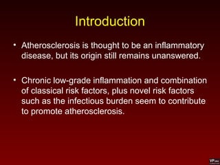


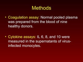

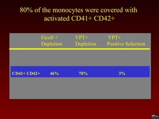


Virus-infected monocytes can initiate coagulation and promote inflammation. An in vitro study found that monocytes infected with cytomegalovirus, influenza A virus, or Chlamydia pneumoniae expressed tissue factor and reduced clotting time, indicating initiation of coagulation. The viruses also stimulated monocytes to produce inflammatory cytokines like IL-6 and IL-8. While all three viruses infected monocytes, influenza A most strongly induced cytokine production. This suggests virus-infected monocytes may contribute to both acute coronary events by promoting plaque instability and the chronic atherosclerosis process through sustained inflammation and coagulation.


