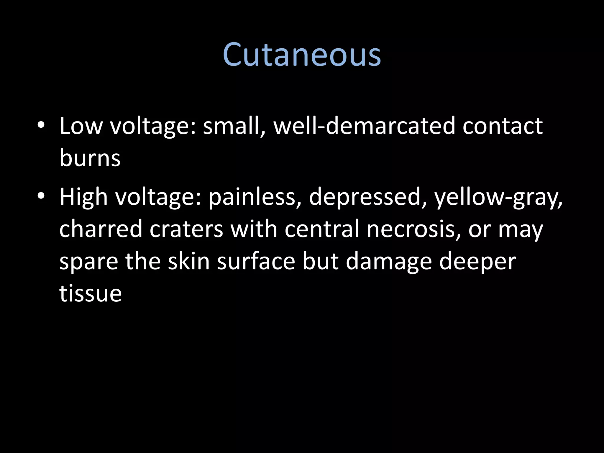
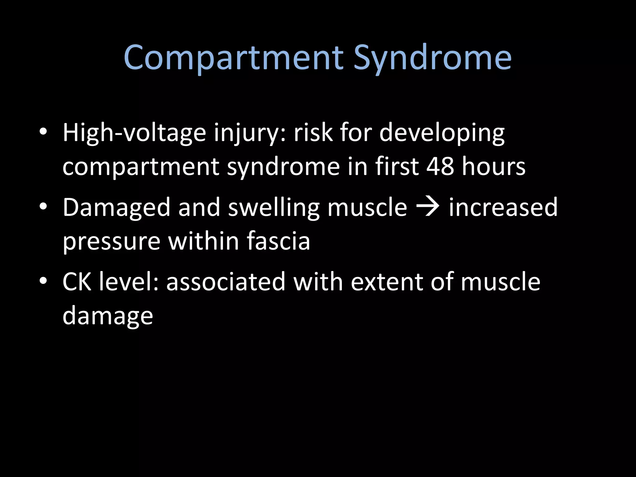
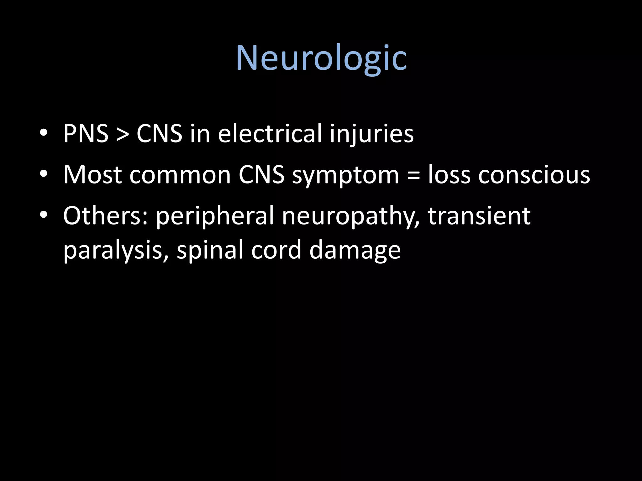
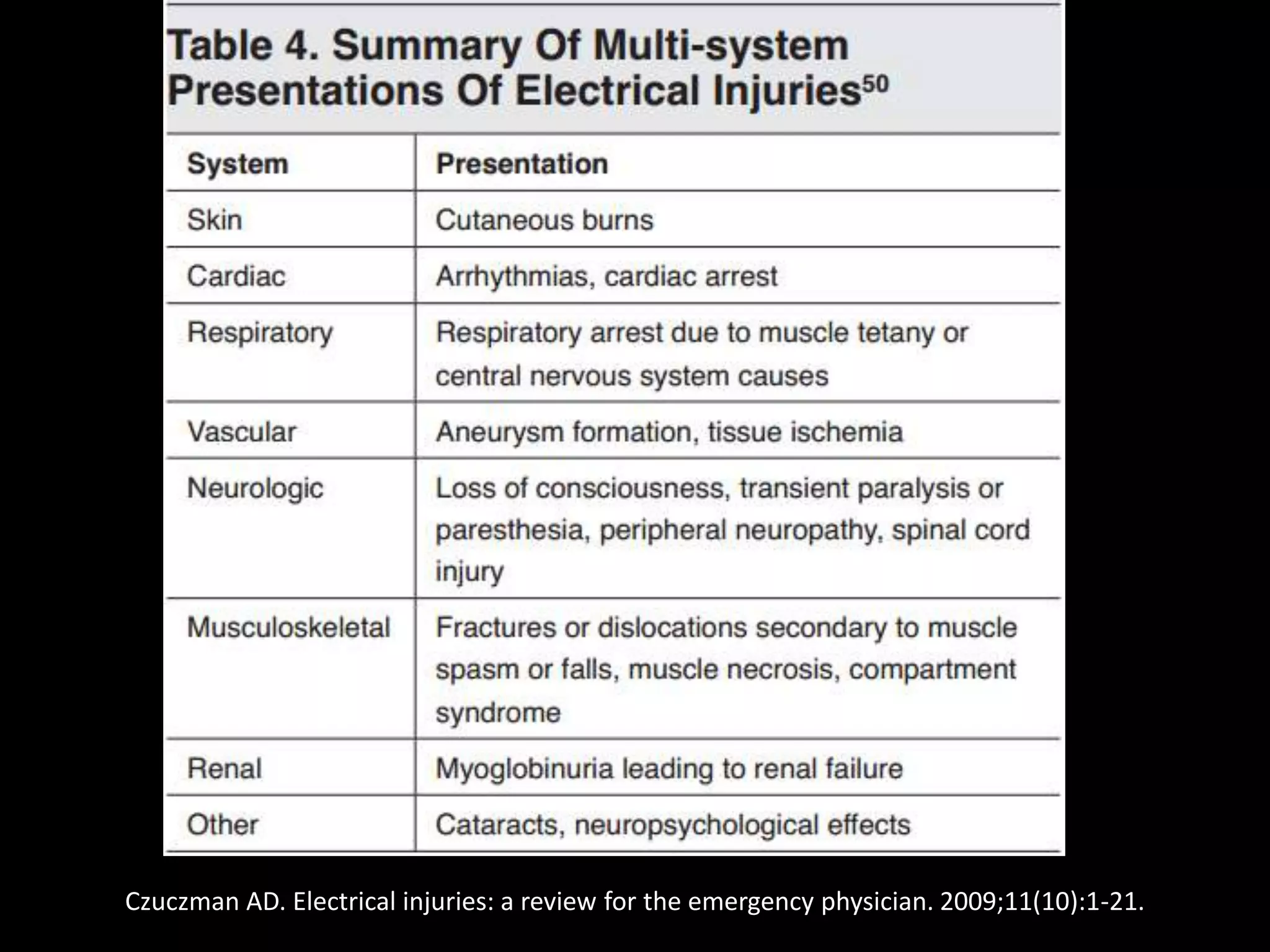
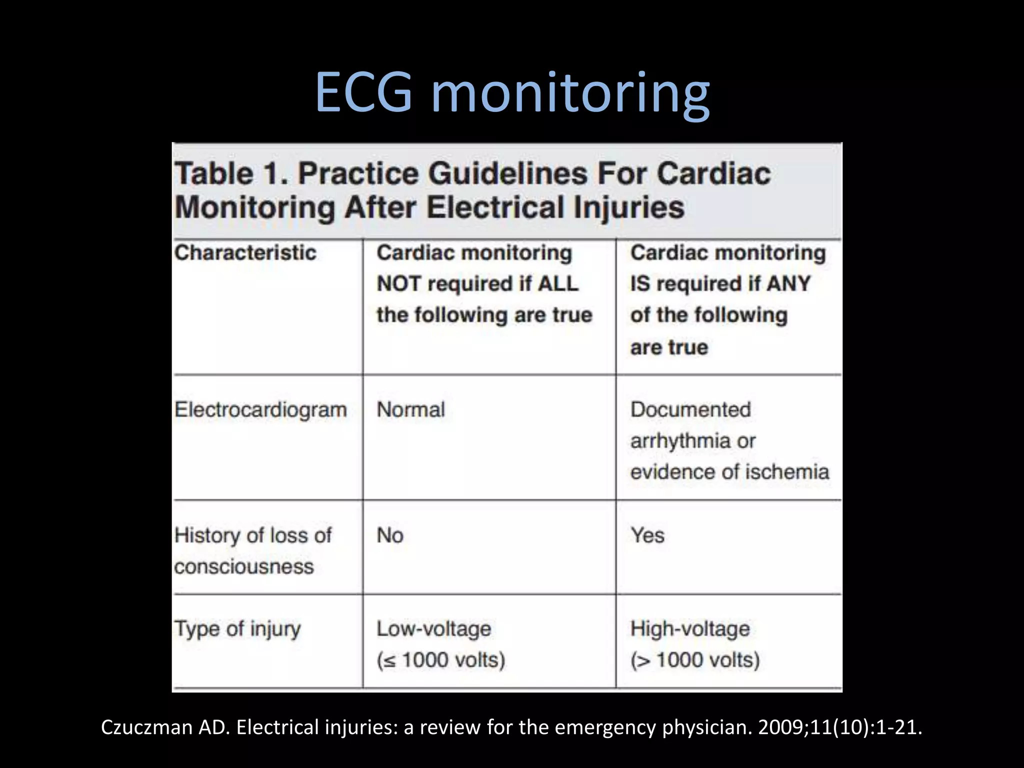
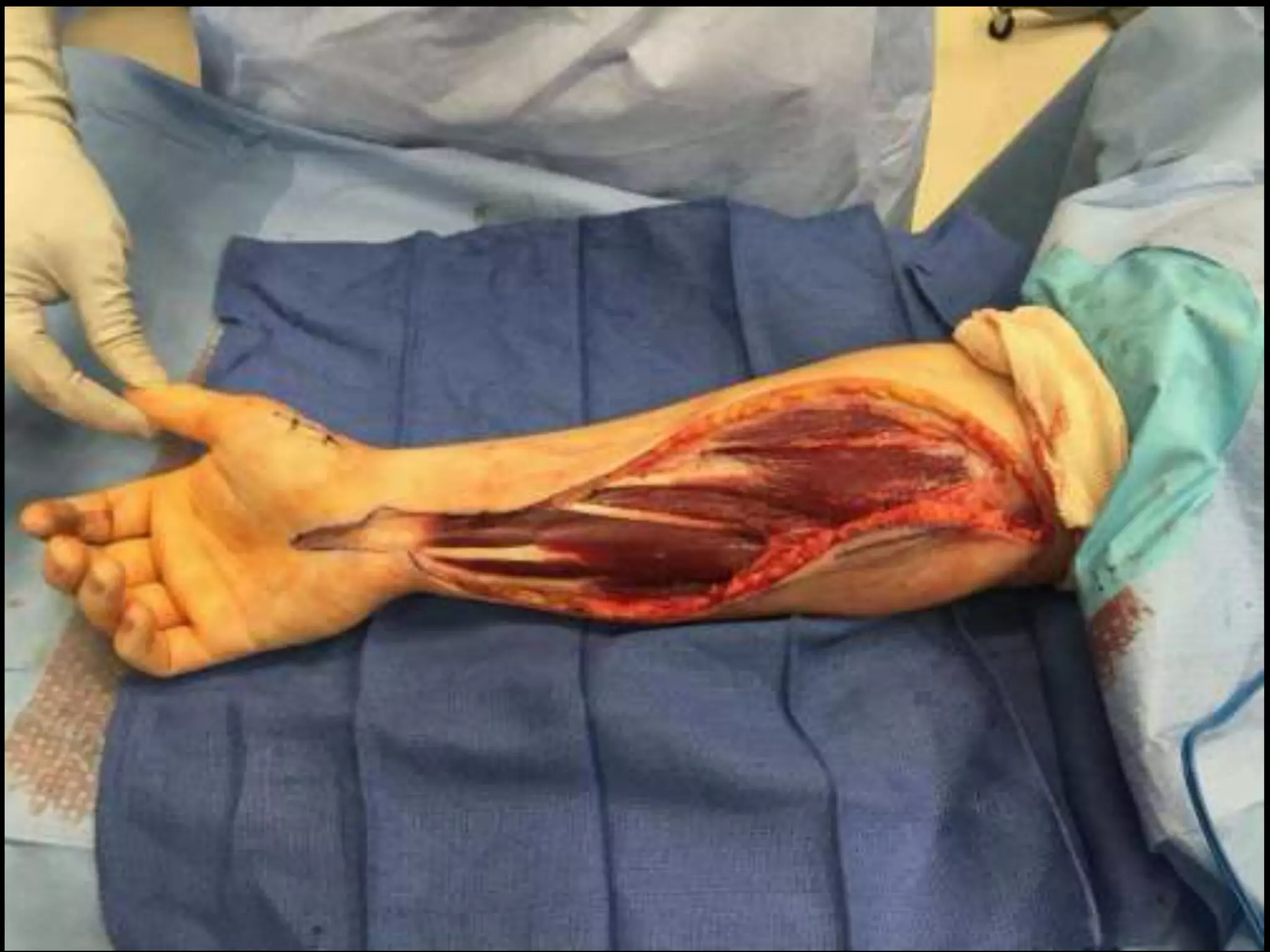
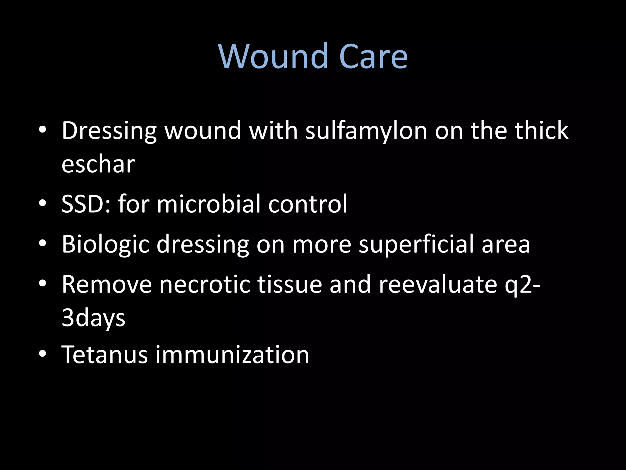
Introduction to
Electrical Injury
Facebook: Happy Friday Knight
• Electrical burns: a small part of major burn
centers  5-7%
• Most devastating of thermal injuries on size-
to-size basis: involving both skin and deeper
tissue
• Most frequent cause of amputation in burn
service
PATHOPHYSIOLOGY
• Exact pathophysiology is unknown
• Numerous variables cannot be measured
when an electrical currents pass through the
tissue
• With high voltage (≥1000V), most of injury is
thermal and resulting in coagulation necrosis
• The injury can be explained by Ohm’s Law
I = V/R
P = I2R
I = current, in amperes (A)
V = potential, in volts (V)
R = resistance, in ohms (Ω)
P = power, in Joules (J)
Factors Determining Severity
• Type of circuit
• Duration
• Resistance of tissue
• Voltage
• Amperage
• Pathway of current
Type of Circuit
• Direct (DC) or alternating current (AC)
• DC: Cause single spasm and throw the victim
from the source  increased traumatic blunt
injury
• AC: cause continuous muscle contraction or
tetany  no-let-go phenomenon and creating
potential for continually increasing severity
– Occur when both flexor and extensor are stimulated
but flexor is stronger making victim unable to let go
voluntarily
Duration of Contact
• Longer duration with high-voltage current 
greater tissue destruction
Tissue Resistance
• The word entrance and exit  replaced by
contact points
• The higher resistance, the greater
transformation electrical injury to thermal
injury
• Least: nerve, blood, mucous membrane,
muscle
• Intermediate: dry skin
• Most: tendon, fat, bone
Tissue Resistance: skin
• Primary resistor
• Thick and harden skin = greater resistance
• Sweating: decrease resistance
Voltage
• Low voltage < 1000V
• High voltage ≥ 1000V
• Thailand domestic wiring 220V
• Higher voltage, higher morbidity
Amperage
• P = I2R and I = V/R
• More amperage, more heat
• The voltage of source is often known but not
for resistance
• Physical effects vary with different amperage
Price TG and Cooper MA. Electrical and lightning injuries: in Rosen’s emergency
medicine. Access via PDF
Pathway
• Where the current passes: between the
contact points
MECHANISMS OF INJURY
• Primary is burn
• Secondary: fall or being thrown from electrical
source by muscle contractioin
• 4 types of electrical burn
– Direct contact: electrothermal heating
– indirect contact: arc, flame
• Electrical arc:
– A current spark between 2 objects differing
potential not contact to each other
– Most destructive indirect injury
– Usually highly charged source and a ground
– Temperature of the arc reaches 2500oC
• Flame: result from ignition of clothing
Heart
• Most serious: cardiac arrest
• Dysrhythmia: AF
• ECG abnormality: non-specific ST-T change
• Can occur both low and high voltage
• ECG monitoring is mandatory in patients with
ECG abnormality
Respiratory
• May result from:
– Tetanic contraction of thoracic musculature
– Injury to respiratory control center of CNS
– Combined cardiopulmonary arrest secondary to
asystole or VF
– Blunt chest trauma from being thrown
Cutaneous
• Low voltage: small, well-demarcated contact
burns
• High voltage: painless, depressed, yellow-gray,
charred craters with central necrosis, or may
spare the skin surface but damage deeper
tissue
Czuczman AD. Electrical injuries: a review for the emergency physician. 2009;11(10):1-21
Muscle
• Coagulation necrosis  becoming edematous
and necrotic 
• Myoglobinuria, rhabdomyolysis, and acute
renal failure
• Compartment syndrome
Myoglobinuria
• Dark urine, mahogany-colored
• Significant muscle damage with potentially
ischemia
• UA: urine dipstick +ve for blood with few RBC
• Risk of acute renal failure
• Elevated CPK
Compartment Syndrome
• High-voltage injury: risk for developing
compartment syndrome in first 48 hours
• Damaged and swelling muscle  increased
pressure within fascia
• CK level: associated with extent of muscle
damage
Vascular
• Greatest damage to media layer  delayed
aneurysm formation
• Intima: thrombosis and occlusion
• Most severe in small muscle branch: tissue
necrosis
• Any vascular injury can cause compartment
syndrome
Neurologic
• PNS > CNS in electrical injuries
• Most common CNS symptom = loss conscious
• Others: peripheral neuropathy, transient
paralysis, spinal cord damage
Gastrointestinal injuries
• Suspected in burn at abdominal wall
• History of fall, blast, blunt trauma
Oral Burn
• Children who put a power cord in the mouth
• Labial artery bleeding: occur up to 2 weeks
after injury
• Eye injury: cataracts
• Auditory system: path of current may be
damaged
Czuczman AD. Electrical injuries: a review for the emergency physician. 2009;11(10):1-21.
TREATMENT
Prehospital Care
• Secure the scene
• Ensure that the power source has been turned
off
• CPR + follow ACLS
• Protect cervical spine and splint fracture
• Close burn wound with clean dry dressing
ER care
• Follow ACLS and ATLS
• Resuscitation
• Complete lab
• Remove constricting objects
• ECG
Fluid Resuscitation
• Parkland formula: only rough starting point
• In absence of gross myo/hemoglobinuria, goal
is to maintain v/s: 0.5 ml/kg/hr
• In children:
– 10% burn: resuscitation
– Fluid of choice: LRS
– <2years: 5%DLR
– Maintenance fluid: 5%DN/2
ECG monitoring
Czuczman AD. Electrical injuries: a review for the emergency physician. 2009;11(10):1-21.
ECG monitoring
• Duration: 24-48 hours
• Low voltage + normal ECG: can be discharged
• Utility of CK-MB:
– Not reliable due to other muscle injury
– Use of troponin: insufficient data
Treatment of Myoglobinuria
• Monitoring urine for myoglobin and serum for
CPK
• Maintain a urine output “double” goal rate: 1
– 1.5 ml/kg/hr
• Fluid should be NSS 1000 ml + NaHCO3 50
mEq
• pH of ABG: > 7.45
• Mannitol: 25 g, then 12.5 g/h
Compartment Syndrome
• 4 compartment fasciotomies for lower
extremities
• Anterior and posterior fasciotomies with
carpal tunnel release for upper extremities
• Primary amputations are not generally
performed
Wound Care
• Dressing wound with sulfamylon on the thick
eschar
• SSD: for microbial control
• Biologic dressing on more superficial area
• Remove necrotic tissue and reevaluate q2-
3days
• Tetanus immunization
Pain Management
• Acute pain: opioids and paracetamol
• Rehabilitation phase: NSAIDs, antidepressant,
massage therapy, cognitive behavioral therapy
References
ชัยรัตน์ บุรุษพัฒน์. Electrical injuries: principle and management. เวชสารแพทย์
ทหารบก.2011;4:207-10.
Herndon DN. Total burn care. 4th ed. China: Elsevier, 2012.
Arnoldo B, Klein M, Gibran NS. Practice guidelines for the management of
electrical injuries. Journal of burn care and research. 2006;27(4):439-46.
Czuczman AD. Electrical injuries: a review for the emergency physician.
2009;11(10):1-21.
Fish RM. Electric injury, part II: specific injuries. The journal of emergency
medicine. 2000;18(1):27-34.
References
Price TG and Cooper MA. Electrical and lightning injuries: in Rosen’s
emergency medicine. Access via PDF
Guidelines for paediatric burn resuscitation

Electrical injury and burn

  • 1.
  • 2.
    • Electrical burns:a small part of major burn centers  5-7% • Most devastating of thermal injuries on size- to-size basis: involving both skin and deeper tissue • Most frequent cause of amputation in burn service
  • 3.
  • 4.
    • Exact pathophysiologyis unknown • Numerous variables cannot be measured when an electrical currents pass through the tissue • With high voltage (≥1000V), most of injury is thermal and resulting in coagulation necrosis • The injury can be explained by Ohm’s Law
  • 5.
    I = V/R P= I2R I = current, in amperes (A) V = potential, in volts (V) R = resistance, in ohms (Ω) P = power, in Joules (J)
  • 6.
    Factors Determining Severity •Type of circuit • Duration • Resistance of tissue • Voltage • Amperage • Pathway of current
  • 7.
    Type of Circuit •Direct (DC) or alternating current (AC) • DC: Cause single spasm and throw the victim from the source  increased traumatic blunt injury • AC: cause continuous muscle contraction or tetany  no-let-go phenomenon and creating potential for continually increasing severity – Occur when both flexor and extensor are stimulated but flexor is stronger making victim unable to let go voluntarily
  • 8.
    Duration of Contact •Longer duration with high-voltage current  greater tissue destruction
  • 9.
    Tissue Resistance • Theword entrance and exit  replaced by contact points • The higher resistance, the greater transformation electrical injury to thermal injury • Least: nerve, blood, mucous membrane, muscle • Intermediate: dry skin • Most: tendon, fat, bone
  • 10.
    Tissue Resistance: skin •Primary resistor • Thick and harden skin = greater resistance • Sweating: decrease resistance
  • 11.
    Voltage • Low voltage< 1000V • High voltage ≥ 1000V • Thailand domestic wiring 220V • Higher voltage, higher morbidity
  • 12.
    Amperage • P =I2R and I = V/R • More amperage, more heat • The voltage of source is often known but not for resistance • Physical effects vary with different amperage
  • 13.
    Price TG andCooper MA. Electrical and lightning injuries: in Rosen’s emergency medicine. Access via PDF
  • 14.
    Pathway • Where thecurrent passes: between the contact points
  • 15.
  • 16.
    • Primary isburn • Secondary: fall or being thrown from electrical source by muscle contractioin • 4 types of electrical burn – Direct contact: electrothermal heating – indirect contact: arc, flame
  • 17.
    • Electrical arc: –A current spark between 2 objects differing potential not contact to each other – Most destructive indirect injury – Usually highly charged source and a ground – Temperature of the arc reaches 2500oC • Flame: result from ignition of clothing
  • 18.
    Heart • Most serious:cardiac arrest • Dysrhythmia: AF • ECG abnormality: non-specific ST-T change • Can occur both low and high voltage • ECG monitoring is mandatory in patients with ECG abnormality
  • 19.
    Respiratory • May resultfrom: – Tetanic contraction of thoracic musculature – Injury to respiratory control center of CNS – Combined cardiopulmonary arrest secondary to asystole or VF – Blunt chest trauma from being thrown
  • 20.
    Cutaneous • Low voltage:small, well-demarcated contact burns • High voltage: painless, depressed, yellow-gray, charred craters with central necrosis, or may spare the skin surface but damage deeper tissue
  • 21.
    Czuczman AD. Electricalinjuries: a review for the emergency physician. 2009;11(10):1-21
  • 22.
    Muscle • Coagulation necrosis becoming edematous and necrotic  • Myoglobinuria, rhabdomyolysis, and acute renal failure • Compartment syndrome
  • 23.
    Myoglobinuria • Dark urine,mahogany-colored • Significant muscle damage with potentially ischemia • UA: urine dipstick +ve for blood with few RBC • Risk of acute renal failure • Elevated CPK
  • 24.
    Compartment Syndrome • High-voltageinjury: risk for developing compartment syndrome in first 48 hours • Damaged and swelling muscle  increased pressure within fascia • CK level: associated with extent of muscle damage
  • 25.
    Vascular • Greatest damageto media layer  delayed aneurysm formation • Intima: thrombosis and occlusion • Most severe in small muscle branch: tissue necrosis • Any vascular injury can cause compartment syndrome
  • 26.
    Neurologic • PNS >CNS in electrical injuries • Most common CNS symptom = loss conscious • Others: peripheral neuropathy, transient paralysis, spinal cord damage
  • 27.
    Gastrointestinal injuries • Suspectedin burn at abdominal wall • History of fall, blast, blunt trauma
  • 28.
    Oral Burn • Childrenwho put a power cord in the mouth • Labial artery bleeding: occur up to 2 weeks after injury
  • 29.
    • Eye injury:cataracts • Auditory system: path of current may be damaged
  • 30.
    Czuczman AD. Electricalinjuries: a review for the emergency physician. 2009;11(10):1-21.
  • 31.
  • 32.
    Prehospital Care • Securethe scene • Ensure that the power source has been turned off • CPR + follow ACLS • Protect cervical spine and splint fracture • Close burn wound with clean dry dressing
  • 33.
    ER care • FollowACLS and ATLS • Resuscitation • Complete lab • Remove constricting objects • ECG
  • 34.
    Fluid Resuscitation • Parklandformula: only rough starting point • In absence of gross myo/hemoglobinuria, goal is to maintain v/s: 0.5 ml/kg/hr • In children: – 10% burn: resuscitation – Fluid of choice: LRS – <2years: 5%DLR – Maintenance fluid: 5%DN/2
  • 35.
    ECG monitoring Czuczman AD.Electrical injuries: a review for the emergency physician. 2009;11(10):1-21.
  • 36.
    ECG monitoring • Duration:24-48 hours • Low voltage + normal ECG: can be discharged • Utility of CK-MB: – Not reliable due to other muscle injury – Use of troponin: insufficient data
  • 37.
    Treatment of Myoglobinuria •Monitoring urine for myoglobin and serum for CPK • Maintain a urine output “double” goal rate: 1 – 1.5 ml/kg/hr • Fluid should be NSS 1000 ml + NaHCO3 50 mEq • pH of ABG: > 7.45 • Mannitol: 25 g, then 12.5 g/h
  • 38.
    Compartment Syndrome • 4compartment fasciotomies for lower extremities • Anterior and posterior fasciotomies with carpal tunnel release for upper extremities • Primary amputations are not generally performed
  • 40.
    Wound Care • Dressingwound with sulfamylon on the thick eschar • SSD: for microbial control • Biologic dressing on more superficial area • Remove necrotic tissue and reevaluate q2- 3days • Tetanus immunization
  • 41.
    Pain Management • Acutepain: opioids and paracetamol • Rehabilitation phase: NSAIDs, antidepressant, massage therapy, cognitive behavioral therapy
  • 42.
    References ชัยรัตน์ บุรุษพัฒน์. Electricalinjuries: principle and management. เวชสารแพทย์ ทหารบก.2011;4:207-10. Herndon DN. Total burn care. 4th ed. China: Elsevier, 2012. Arnoldo B, Klein M, Gibran NS. Practice guidelines for the management of electrical injuries. Journal of burn care and research. 2006;27(4):439-46. Czuczman AD. Electrical injuries: a review for the emergency physician. 2009;11(10):1-21. Fish RM. Electric injury, part II: specific injuries. The journal of emergency medicine. 2000;18(1):27-34.
  • 43.
    References Price TG andCooper MA. Electrical and lightning injuries: in Rosen’s emergency medicine. Access via PDF Guidelines for paediatric burn resuscitation

Editor's Notes

  • #3 โรคที่มาพร้อมกับ civilization
  • #10 Nerve เพราะ carry electrical signal Muscle, blood: high electrolyte and water content: good conductor
  • #18 Ignition = starting to burn
  • #21 Crater = หลุม
  • #22 Current arc across both flexor surface
  • #24 CPK  > 1000U/L, half life 20hr, peak 24hr + return to normal 48-72hr
  • #35 Remove constricting object เช่นแหวน ก่อนที่เนื้อเยื่อจะบวมและเอาออกไม่ได้ ECG ทำทุกคนเพื่อหา arrythmia
  • #38 Duration ยังไม่มี study ไหนศึกษาเกี่ยวกับเรื่องนี้โดยตรง แต่ว่าหลายๆอันบอกว่าถ้าไม่มี ECG abn 24hr ปลอดภัย ในขณะที่ dysrhythmia จะ resolve 48hr TnI: ขึ้นตั้งแต่ 2-3 hr แรก และอยู่ต่อหลายวัน
  • #39 Urine pH ไม่ precise ถ้ามี hemochromogen อย่าลืม monitor elyte
  • #40 แผลปิดด้วย sulfamylon q4-6hr, or biologic dressing, keep elevate