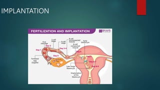
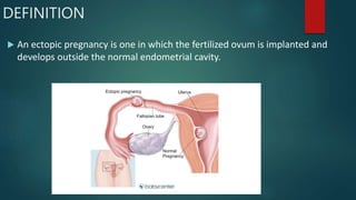
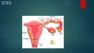
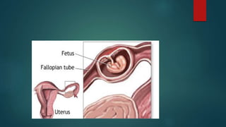
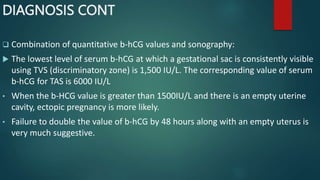
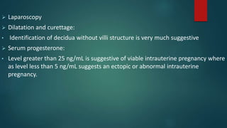
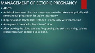
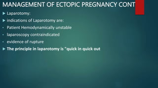
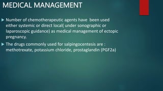
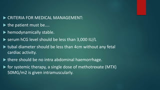
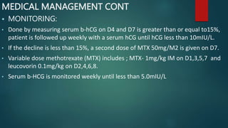
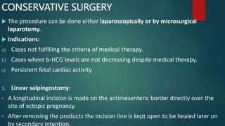
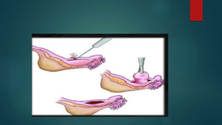
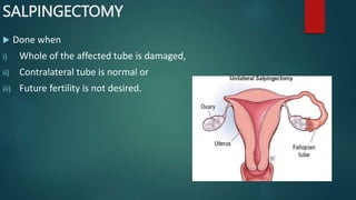
ECTOPIC PREGNANCY
BY
DR FADILA LAWAL
Approval page
PRESENTER DR FADEELA LAWAL
REVIEWED BY DR ADEYEMI
CLINICAL SUPERVISOR
APPROVED BY DR MUHAMMAD M.M.S
LEARNING OBJECTIVES
 TO UNDERSTAND WHAT IS ECTOPIC PREGNANCY
 TO IDENTIFY THE DIFFERENT SITES AND HIGH RISK
GROUPS OF ECTOPIC PREGNANCY
 TO DISCUSS THE OUTCOME OF TUBAL PREGNANCY
OUTLINE
 INTRODUCTION
 DEFINITION
 SITES OF ECTOPIC PREGNANCY
 ETIOLOGY
 RISK FACTORS
 MORBID ANATOMY
 CLINICAL FEATURES
 MANAGEMENT
INTRODUCTION
Ectopic pregnancy is one of the direct causes of maternal
death and is an important causes of maternal mortality in
the first trimester, it is the 8th out 129 direct cause of
death, and account for 9% of all maternal death
IMPLANTATION
DEFINITION
 An ectopic pregnancy is one in which the fertilized ovum is implanted and
develops outside the normal endometrial cavity.
SITES
INCIDENCE
 The incidence of ectopic pregnancies in Nigeria is 1.5–
2.7/100 deliveries
 In 2020 the prevalence of ectopic pregnancy in Kaduna
state was 0.89% of all deliveries and 2.74% of all the
gynaecological admissions
ETIOLOGY
:
 Salpingitis and pelvic inflammatory disease (PID)
 Contraception failure
• There is relative increase in tubal pregnancy (7 times more)
should pregnancy occur with IUD in situ
• After sterilization operation there is 15-50% chance of being
ectopic if pregnancy occurs
 Tubal surgery
 Artificial reproductive technology
 Previous ectopic pregnancy: There is 10-15% chances of repeat
ectopic pregnancy.
 Prior induced abortion significantly increases the risk
 Developmental defects of the tube:
• Elongation
• Diverticulum
RISK FACTORS
 Maternal age of 35 years or above
 History of pelvic surgery
 History of endometriosis
 History of previous ectopic pregnancy
 Smoking
 History of STI
MORBID ANATOMY
 CHANES IN THE TUBE:
• Implantation in the tube occurs more commonly intercolumnar fashion, i.e. in
between two mucosal folds.
• Decidua change at the site of implantation is minimal , the muscles undergo
limited hyperplasia and hypertrophy.
• The blastocyst burrows through the mucous membrane and lies between the
lumen and the peritoneal covering (intramuscular implantation)
• The tube on the implantation site is distended and the wall is thinned out.
MORBID ANATOMY CONT
• The stretching of the peritoneum over the site of implantation results in
episodic pain
• Finally, tubal rapture occurs when the muscles and the serosa are maximally
stretched and undergo necrosis.
• Hemoperitoneum is found in all cases of raptured tubal ectopic pregnancy.
 The trophoblasts of ectopic pregnancy do not usually grow as that of a normal
pregnancy. As a result, hCG production is inadequate compared to a normal
pregnancy.
CHANGES IN THE UTERUS
 Under the influence of estrogen, progesterone form corpus luteum and
chorionic gonadotropin, there is varying amount of enlargement of the uterus
with increased vascularity.
 The decidua develops all the characteristics of intrauterine pregnancy except
that it contains no evidence of chorionic villi.
 When progesterone level falls due to fall in the level of hCG, endometrial
growth is no longer maintained. Endometrium sloughs out causing uterine
bleeding.
 Sometimes entire decidua expelled as a single piece through the cervix. This is
known as decidua cast that may be confused with spontaneous abortion.
Heterotopic pregnancy
 its defined as the presence of multiple gestation with one
being present in the uterine cavity and the other outside
the uterus ,commonly in the fallopian tube and
uncommonly in the cervix or ovary
 Its more common following ART procedures
CLINCAL FEATURES OF ECTOPIC PREGNANCY
 Clinically 3 distinct types are described as per the duration of manifestation:
 Acute/Ruptured (obstetric) emergency
 Unruptured
 Chronic
ACUTE/RUPTURED
 Less common (30%)- associated with cases of tubal rupture or tubal abortion
along with massive intraperitoneal hemorrhage.
 Usually young patient (20-30 years).
 Mostly Nulliparous
 Alter infertility treatment.
CLINICAL PRESENTATION IN RUPTURED
 Symptoms:
• Amenorrhea of short period 6-8 weeks or a delayed period or slight spotting.
• Abdominal pain.
• Pain is agonizing or colicky.
• Vaginal bleeding-slight, sanguineous or dark coloured.
• Nausea, vomiting, fainting attacks, syncope (10%)
CLINICAL PRESENTATION IN RUPTURED CONT
 On examination:
• Tachycardia
• Hypotension
• Pallor
 On per abdomen examination:
• Tenderness and muscle guard on lower abdomen on affected side.
• An irregular and tender mass in the lower abdomen
• Cullen’s sign: dark discoloration surrounding umbilicus suggesting
intraperitoneal hemorrhage
CLINICAL PRESENTATION IN RUPTURED CONT
 On Bimanual examination:
• Vaginal mucosa pale
• Uterus normal or bulky
• Cervical movement tenderness
• Tender fornices with or without palpable mass
• An ill defined boggy and tender mass felt through the posterolateral fornix
extending to the POD
UNRUPTURED
 Clinical symptoms:
• Delayed period or spotting
• Uneasiness on one side of the flank
 Signs:
• Uterus slightly smaller than the period of amenorrhea- evidence of early
pregnancy.
• A small pulsatile, tender, well circumscribed mass felt in one fornix separated
from the uterus
CHRONIC ECTOPIC
 Clinical symptoms:
• Amenorrhea for 6-8 weeks
• Lower abdominal pain-starts as acute and gradually becomes dull and colicky
• Vaginal bleeding
• symptoms- bladder irritation, dysuria frequency, retention of urine, rectal
tenesmus following infected hematocele.
 On examination:
• Patient looks ill with varying degree of pallor not proportionate to vaginal
bleeding
• Persistent high pulse rate even during rest
• Features of shock are absent
DIAGNOSIS
 INVESTIGATIONS:
• Blood examination
 ABO and Rh grouping
 Culdocentesis:
• A 18-guage lumbar puncture needle fitted with a syringe, the posterior fornix is
punctured to gain access to the pouch of Douglas
• Aspiration of non-clothing blood with hematocrit greater than 15% signifies
ruptured ectopic pregnancy
DIAGNOSIS CONT
 Examination of beta hCG:
 A single estimation of beta hCG level either in the scrum or in the urine confirms
pregnancy but cannot determine its location.
 The suspicious findings are:
 Lower concentration compared of normal intrauterine pregnancy
 Doubling time in plasma fails to occur in 2days (less than 66% rise in 48 hours)
 Sonography
 Transvaginal sonography (TVS):
 The diagnostic features are:
• Absence of intrauterine pregnancy with a positive
pregnancy test.
• Fluid in the pouch of Douglas.
• Adnexal mass clearly separated from the ovary
• Rarely cardiac motion may be seen in an unruptured
tubal ectopic pregnancy
 Colour Doppler Sonography (TV-CDS).
DIAGNOSIS CONT
 Combination of quantitative b-hCG values and sonography:
 The lowest level of serum b-hCG at which a gestational sac is consistently visible
using TVS (discriminatory zone) is 1,500 IU/L. The corresponding value of serum
b-hCG for TAS is 6000 IU/L
• When the b-HCG value is greater than 1500IU/L and there is an empty uterine
cavity, ectopic pregnancy is more likely.
• Failure to double the value of b-hCG by 48 hours along with an empty uterus is
very much suggestive.
 Laparoscopy
 Dilatation and curettage:
• Identification of decidua without villi structure is very much suggestive
 Serum progesterone:
• Level greater than 25 ng/mL is suggestive of viable intrauterine pregnancy where
as level less than 5 ng/mL suggests an ectopic or abnormal intrauterine
pregnancy.
 Laparotomy:
• The old axiom ‘’open and see’’ holds good especially when the patient is
hemodynamically unstable.
MANAGEMENT OF ECTOPIC PREGNANCY
 ACUTE:
 Antishock treatment: Antishock measures are to be taken energetically with
simultaneous preparation for urgent laparotomy.
 Ringers solution (crystalloid) is started , if necessary with venesection
 Arrangement is made for blood transfusion.
 After drawing the blood samples for grouping and cross matching, volume
replacement with colloids s to be done.
MANAGEMENT OF ECTOPIC PREGNANCY CONT
 Laparotomy:
 indications of Laparotomy are:
• Patient Hemodynamically unstable
• laparoscopy contraindicated
• evidence of rupture
 The principle in laparotomy is "quick in quick out
CHRONIC ECTOPIC
 All cases of chronic or suspected ectopic are to be admitted as an emergency.
 the patient is kept under observation, investigations are done and the patient is
put up for laparotomy at the earliest convenient time.
 Usually a pelvic hematocele is found, blood clots are removed. the affected tube
is identified and salpingectomy is commonly done.
 Resumption of Ovulation and contraception:
 About 15% of women ovulate by 19 days and about 25% ovulate by the 30th
postoperative day.
MANAGEMENT OF UNRUPTURED TUBAL PREGNANCY
 Expectant
 Medical
 Surgical
 Conservative
EXPECTANT MANAGEMENT
 Indications are:
• Initial serum hCG level less than 1,000 IU/L and the subsequent levels are falling.
• Gestation sac size level less than 4cm
• No fetal heart beat on TVS
• No evidence of bleeding or rupture on TVS.
CONSERVATIVE MANAGEMENT
 may be either medical or surgical.
 Otherwise salpingectomy is done.
 The advantages of conservative management are:
• Significant reduction in operative morbidity, hospital stay as well as cost.
• Improved chance of subsequent intrauterine pregnancy.
• Less risk of recurrence.
MEDICAL MANAGEMENT
 Number of chemotherapeutic agents have been used
either systemic or direct local( under sonographic or
laparoscopic guidance) as medical management of ectopic
pregnancy.
 The drugs commonly used for salpingocentesis are :
methotrexate, potassium chloride, prostaglandin (PGF2a)
 CRITERIA FOR MEDICAL MANAGEMENT:
 the patient must be....
 hemodynamically stable.
 serum hCG level should be less than 3,000 IU/L
 tubal diameter should be less than 4cm without any fetal
cardiac activity.
 there should be no intra abdominal haemorrhage.
 for systemic therapy, a single dose of methotrexate (MTX)
50MG/m2 is given intramuscularly.
MEDICAL MANAGEMENT CONT
 MONITORING:
• Done by measuring serum b-hCG on D4 and D7 is greater than or equal to15%,
patient is followed up weekly with a serum hCG until hCG less than 10mIU/L.
• If the decline is less than 15%, a second dose of MTX 50mg/M2 is given on D7.
• Variable dose methotrexate (MTX) includes ; MTX- 1mg/kg IM on D1,3,5,7 and
leucovorin 0.1mg/kg on D2,4,6,8.
• Serum b-HCG is monitored weekly until less than 5.0mIU/L
CONSERVATIVE SURGERY
 The procedure can be done either laparoscopically or by microsurgical
laparotomy.
 Indications:
a) Cases not fulfilling the criteria of medical therapy.
b) Cases where b-HCG levels are not decreasing despite medical therapy.
c) Persistent fetal cardiac activity
1. Linear salpingostomy:
• A longitudinal incision is made on the antimesenteric border directly over the
site of ectopic pregnancy.
• After removing the products the incision line is kept open to be healed later on
by secondary intention.
CONSERVATIVE SURGERY CONT
2. Linear salpingotomy:
 The procedure are the same as those of salpingostomy. But the incision line is
closed in two layers with interrupted vicryl sutures. This is not commonly done.
3. Fimbria Expression:
 This is ideal in cases of distal ampulla (fimbria) pregnancy and is done digitally.
PERSISTENT ECTOPIC PREGNANCY
 Due to incomplete removal of trophoblast.
 Prophylactic single dose MTX (1 mg/kg) IM is effective to resolve the problem.
 Following conservative surgery or medical treatment,
estimation of b-hCG should be done weekly till the value
becomes less than 5.0mlU/mL. additional monitoring by
TVS is preferred.
 Following laparoscopic salpingostomy, persistent ectopic
pregnancy ranges between 4% and 20%.
SALPINGECTOMY
 Done when
i) Whole of the affected tube is damaged,
ii) Contralateral tube is normal or
iii) Future fertility is not desired.
CONCLUSION
 Ectopic pregnancy is still the leading cause of death in the
first trimester pregnancy
 A high index of suspicion is required for an early diagnosis
because sign and symptoms are not specific
 With the use of transvaginal ultrasound and serial ß-hCG
levels, and in some cases uterine curretage, most ectopic
pregnancies can be diagnosed and treated at an early stage
 Medical and surgical treatment of ectopic pregnancy have
similar success rates.
THANK YOU

ectopic pregnancy 2 copy.pptx

  • 1.
  • 2.
    Approval page PRESENTER DRFADEELA LAWAL REVIEWED BY DR ADEYEMI CLINICAL SUPERVISOR APPROVED BY DR MUHAMMAD M.M.S
  • 3.
    LEARNING OBJECTIVES  TOUNDERSTAND WHAT IS ECTOPIC PREGNANCY  TO IDENTIFY THE DIFFERENT SITES AND HIGH RISK GROUPS OF ECTOPIC PREGNANCY  TO DISCUSS THE OUTCOME OF TUBAL PREGNANCY
  • 4.
    OUTLINE  INTRODUCTION  DEFINITION SITES OF ECTOPIC PREGNANCY  ETIOLOGY  RISK FACTORS  MORBID ANATOMY  CLINICAL FEATURES  MANAGEMENT
  • 5.
    INTRODUCTION Ectopic pregnancy isone of the direct causes of maternal death and is an important causes of maternal mortality in the first trimester, it is the 8th out 129 direct cause of death, and account for 9% of all maternal death
  • 6.
  • 7.
    DEFINITION  An ectopicpregnancy is one in which the fertilized ovum is implanted and develops outside the normal endometrial cavity.
  • 8.
  • 9.
    INCIDENCE  The incidenceof ectopic pregnancies in Nigeria is 1.5– 2.7/100 deliveries  In 2020 the prevalence of ectopic pregnancy in Kaduna state was 0.89% of all deliveries and 2.74% of all the gynaecological admissions
  • 10.
    ETIOLOGY :  Salpingitis andpelvic inflammatory disease (PID)  Contraception failure • There is relative increase in tubal pregnancy (7 times more) should pregnancy occur with IUD in situ • After sterilization operation there is 15-50% chance of being ectopic if pregnancy occurs  Tubal surgery  Artificial reproductive technology
  • 11.
     Previous ectopicpregnancy: There is 10-15% chances of repeat ectopic pregnancy.  Prior induced abortion significantly increases the risk  Developmental defects of the tube: • Elongation • Diverticulum
  • 12.
    RISK FACTORS  Maternalage of 35 years or above  History of pelvic surgery  History of endometriosis  History of previous ectopic pregnancy  Smoking  History of STI
  • 13.
    MORBID ANATOMY  CHANESIN THE TUBE: • Implantation in the tube occurs more commonly intercolumnar fashion, i.e. in between two mucosal folds. • Decidua change at the site of implantation is minimal , the muscles undergo limited hyperplasia and hypertrophy. • The blastocyst burrows through the mucous membrane and lies between the lumen and the peritoneal covering (intramuscular implantation) • The tube on the implantation site is distended and the wall is thinned out.
  • 14.
    MORBID ANATOMY CONT •The stretching of the peritoneum over the site of implantation results in episodic pain • Finally, tubal rapture occurs when the muscles and the serosa are maximally stretched and undergo necrosis. • Hemoperitoneum is found in all cases of raptured tubal ectopic pregnancy.  The trophoblasts of ectopic pregnancy do not usually grow as that of a normal pregnancy. As a result, hCG production is inadequate compared to a normal pregnancy.
  • 16.
    CHANGES IN THEUTERUS  Under the influence of estrogen, progesterone form corpus luteum and chorionic gonadotropin, there is varying amount of enlargement of the uterus with increased vascularity.  The decidua develops all the characteristics of intrauterine pregnancy except that it contains no evidence of chorionic villi.  When progesterone level falls due to fall in the level of hCG, endometrial growth is no longer maintained. Endometrium sloughs out causing uterine bleeding.  Sometimes entire decidua expelled as a single piece through the cervix. This is known as decidua cast that may be confused with spontaneous abortion.
  • 17.
    Heterotopic pregnancy  itsdefined as the presence of multiple gestation with one being present in the uterine cavity and the other outside the uterus ,commonly in the fallopian tube and uncommonly in the cervix or ovary  Its more common following ART procedures
  • 18.
    CLINCAL FEATURES OFECTOPIC PREGNANCY  Clinically 3 distinct types are described as per the duration of manifestation:  Acute/Ruptured (obstetric) emergency  Unruptured  Chronic
  • 19.
    ACUTE/RUPTURED  Less common(30%)- associated with cases of tubal rupture or tubal abortion along with massive intraperitoneal hemorrhage.  Usually young patient (20-30 years).  Mostly Nulliparous  Alter infertility treatment.
  • 20.
    CLINICAL PRESENTATION INRUPTURED  Symptoms: • Amenorrhea of short period 6-8 weeks or a delayed period or slight spotting. • Abdominal pain. • Pain is agonizing or colicky. • Vaginal bleeding-slight, sanguineous or dark coloured. • Nausea, vomiting, fainting attacks, syncope (10%)
  • 21.
    CLINICAL PRESENTATION INRUPTURED CONT  On examination: • Tachycardia • Hypotension • Pallor  On per abdomen examination: • Tenderness and muscle guard on lower abdomen on affected side. • An irregular and tender mass in the lower abdomen • Cullen’s sign: dark discoloration surrounding umbilicus suggesting intraperitoneal hemorrhage
  • 22.
    CLINICAL PRESENTATION INRUPTURED CONT  On Bimanual examination: • Vaginal mucosa pale • Uterus normal or bulky • Cervical movement tenderness • Tender fornices with or without palpable mass • An ill defined boggy and tender mass felt through the posterolateral fornix extending to the POD
  • 23.
    UNRUPTURED  Clinical symptoms: •Delayed period or spotting • Uneasiness on one side of the flank  Signs: • Uterus slightly smaller than the period of amenorrhea- evidence of early pregnancy. • A small pulsatile, tender, well circumscribed mass felt in one fornix separated from the uterus
  • 24.
    CHRONIC ECTOPIC  Clinicalsymptoms: • Amenorrhea for 6-8 weeks • Lower abdominal pain-starts as acute and gradually becomes dull and colicky • Vaginal bleeding • symptoms- bladder irritation, dysuria frequency, retention of urine, rectal tenesmus following infected hematocele.  On examination: • Patient looks ill with varying degree of pallor not proportionate to vaginal bleeding • Persistent high pulse rate even during rest • Features of shock are absent
  • 25.
    DIAGNOSIS  INVESTIGATIONS: • Bloodexamination  ABO and Rh grouping  Culdocentesis: • A 18-guage lumbar puncture needle fitted with a syringe, the posterior fornix is punctured to gain access to the pouch of Douglas • Aspiration of non-clothing blood with hematocrit greater than 15% signifies ruptured ectopic pregnancy
  • 26.
    DIAGNOSIS CONT  Examinationof beta hCG:  A single estimation of beta hCG level either in the scrum or in the urine confirms pregnancy but cannot determine its location.  The suspicious findings are:  Lower concentration compared of normal intrauterine pregnancy  Doubling time in plasma fails to occur in 2days (less than 66% rise in 48 hours)
  • 27.
     Sonography  Transvaginalsonography (TVS):  The diagnostic features are: • Absence of intrauterine pregnancy with a positive pregnancy test. • Fluid in the pouch of Douglas. • Adnexal mass clearly separated from the ovary • Rarely cardiac motion may be seen in an unruptured tubal ectopic pregnancy  Colour Doppler Sonography (TV-CDS).
  • 28.
    DIAGNOSIS CONT  Combinationof quantitative b-hCG values and sonography:  The lowest level of serum b-hCG at which a gestational sac is consistently visible using TVS (discriminatory zone) is 1,500 IU/L. The corresponding value of serum b-hCG for TAS is 6000 IU/L • When the b-HCG value is greater than 1500IU/L and there is an empty uterine cavity, ectopic pregnancy is more likely. • Failure to double the value of b-hCG by 48 hours along with an empty uterus is very much suggestive.
  • 29.
     Laparoscopy  Dilatationand curettage: • Identification of decidua without villi structure is very much suggestive  Serum progesterone: • Level greater than 25 ng/mL is suggestive of viable intrauterine pregnancy where as level less than 5 ng/mL suggests an ectopic or abnormal intrauterine pregnancy.
  • 30.
     Laparotomy: • Theold axiom ‘’open and see’’ holds good especially when the patient is hemodynamically unstable.
  • 31.
    MANAGEMENT OF ECTOPICPREGNANCY  ACUTE:  Antishock treatment: Antishock measures are to be taken energetically with simultaneous preparation for urgent laparotomy.  Ringers solution (crystalloid) is started , if necessary with venesection  Arrangement is made for blood transfusion.  After drawing the blood samples for grouping and cross matching, volume replacement with colloids s to be done.
  • 32.
    MANAGEMENT OF ECTOPICPREGNANCY CONT  Laparotomy:  indications of Laparotomy are: • Patient Hemodynamically unstable • laparoscopy contraindicated • evidence of rupture  The principle in laparotomy is "quick in quick out
  • 33.
    CHRONIC ECTOPIC  Allcases of chronic or suspected ectopic are to be admitted as an emergency.  the patient is kept under observation, investigations are done and the patient is put up for laparotomy at the earliest convenient time.  Usually a pelvic hematocele is found, blood clots are removed. the affected tube is identified and salpingectomy is commonly done.  Resumption of Ovulation and contraception:  About 15% of women ovulate by 19 days and about 25% ovulate by the 30th postoperative day.
  • 34.
    MANAGEMENT OF UNRUPTUREDTUBAL PREGNANCY  Expectant  Medical  Surgical  Conservative
  • 35.
    EXPECTANT MANAGEMENT  Indicationsare: • Initial serum hCG level less than 1,000 IU/L and the subsequent levels are falling. • Gestation sac size level less than 4cm • No fetal heart beat on TVS • No evidence of bleeding or rupture on TVS.
  • 36.
    CONSERVATIVE MANAGEMENT  maybe either medical or surgical.  Otherwise salpingectomy is done.  The advantages of conservative management are: • Significant reduction in operative morbidity, hospital stay as well as cost. • Improved chance of subsequent intrauterine pregnancy. • Less risk of recurrence.
  • 37.
    MEDICAL MANAGEMENT  Numberof chemotherapeutic agents have been used either systemic or direct local( under sonographic or laparoscopic guidance) as medical management of ectopic pregnancy.  The drugs commonly used for salpingocentesis are : methotrexate, potassium chloride, prostaglandin (PGF2a)
  • 38.
     CRITERIA FORMEDICAL MANAGEMENT:  the patient must be....  hemodynamically stable.  serum hCG level should be less than 3,000 IU/L  tubal diameter should be less than 4cm without any fetal cardiac activity.  there should be no intra abdominal haemorrhage.  for systemic therapy, a single dose of methotrexate (MTX) 50MG/m2 is given intramuscularly.
  • 39.
    MEDICAL MANAGEMENT CONT MONITORING: • Done by measuring serum b-hCG on D4 and D7 is greater than or equal to15%, patient is followed up weekly with a serum hCG until hCG less than 10mIU/L. • If the decline is less than 15%, a second dose of MTX 50mg/M2 is given on D7. • Variable dose methotrexate (MTX) includes ; MTX- 1mg/kg IM on D1,3,5,7 and leucovorin 0.1mg/kg on D2,4,6,8. • Serum b-HCG is monitored weekly until less than 5.0mIU/L
  • 40.
    CONSERVATIVE SURGERY  Theprocedure can be done either laparoscopically or by microsurgical laparotomy.  Indications: a) Cases not fulfilling the criteria of medical therapy. b) Cases where b-HCG levels are not decreasing despite medical therapy. c) Persistent fetal cardiac activity 1. Linear salpingostomy: • A longitudinal incision is made on the antimesenteric border directly over the site of ectopic pregnancy. • After removing the products the incision line is kept open to be healed later on by secondary intention.
  • 42.
    CONSERVATIVE SURGERY CONT 2.Linear salpingotomy:  The procedure are the same as those of salpingostomy. But the incision line is closed in two layers with interrupted vicryl sutures. This is not commonly done. 3. Fimbria Expression:  This is ideal in cases of distal ampulla (fimbria) pregnancy and is done digitally.
  • 43.
    PERSISTENT ECTOPIC PREGNANCY Due to incomplete removal of trophoblast.  Prophylactic single dose MTX (1 mg/kg) IM is effective to resolve the problem.
  • 44.
     Following conservativesurgery or medical treatment, estimation of b-hCG should be done weekly till the value becomes less than 5.0mlU/mL. additional monitoring by TVS is preferred.  Following laparoscopic salpingostomy, persistent ectopic pregnancy ranges between 4% and 20%.
  • 45.
    SALPINGECTOMY  Done when i)Whole of the affected tube is damaged, ii) Contralateral tube is normal or iii) Future fertility is not desired.
  • 46.
    CONCLUSION  Ectopic pregnancyis still the leading cause of death in the first trimester pregnancy  A high index of suspicion is required for an early diagnosis because sign and symptoms are not specific  With the use of transvaginal ultrasound and serial ß-hCG levels, and in some cases uterine curretage, most ectopic pregnancies can be diagnosed and treated at an early stage  Medical and surgical treatment of ectopic pregnancy have similar success rates.
  • 47.