ECG
•Download as PPT, PDF•
0 likes•332 views

An ECG is a graphic recording of the electrical activity of the heart over time captured by electrodes placed on the skin. It detects the impulse as it travels through the heart and provides information about heart rate and rhythm, as well as the size and position of the chambers, any damage to the heart, and the effects of drugs or devices. The standard ECG uses 12 leads attached at specific positions on the limbs and chest to detect the P, QRS, and T waves representing atrial depolarization, ventricular depolarization, and ventricular repolarization respectively. The ECG is useful for diagnosing heart conditions like heart attacks, arrhythmias, and damage as well as monitoring effects of drugs.

Recommended
More Related Content
What's hot
What's hot (20)
Similar to ECG
Similar to ECG (20)
More from DR. SUNIL KUMAR
Recently uploaded
Recently uploaded (20)
ECG
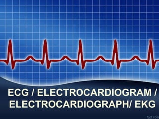
- 1. ECG / ELECTROCARDIOGRAM / ELECTROCARDIOGRAPH/ EKG
- 2. DEFINITION • ECG Is a graphic representation of electrical activity produced by heart beat. • The electrical activity spreads from heart to the body surface from where it is amplified by ECG machine and can be graphically recorded.
- 3. Parts of an ECG • 1) ECG Machine • 2) Electrodes- The standard ECG has 12 leads. – a) 6 leads are “limb leads” – b) 6 leads are “precordial leads”
- 4. • A) 6 leads are “limb leads” – The six limb leads are called lead I, II, III, aVL, aVR and aVF. – The letter “a” stands for “augmented".
- 5. • B) 6 leads are “precordial leads” • The six precordial leads are called leads V1, V2, V3, V4, V5 and V6
- 6. • V1- In the fourth intercostal space (between ribs 4 and 5) just to the right of the sternum (breastbone) • V2- In the fourth intercostal space (between ribs 4 and 5) just to the left of the sternum. • V3- Between leads V2 and V4. • V4- In the fifth intercostal space (between ribs 5 and 6) in the mid-clavicular line. • V5- Horizontally even with V4, in the left anterior axillary line. • V6- Horizontally even with V4 and V5 in the mid-axillary
- 8. • P-wave: – It is a small upward wave that appears first. – It indicates atrial depolarization (systole), during which excitation spreads from SA node to all over atrium. • QRS wave: – It represents the ventricular depolarization (systole)Just after QRS wave begins, ventricles starts to contracts. – Hence QRS wave represents ventricular systole • T- wave: – It is third small wave in the form of a dome-shaped upward deflection. – It indicates ventricular repolarization (diastole)
- 10. • P-R interval: –It represents the time required for an impulse to travel through the atria, AV node and Bundle of His to reach ventricles. • S-T segment: –It is measured from the end of S to the beginning of T- wave –It represents the time when ventricular fibers are fully depolarized
- 11. USES OF ECG • 1) Diagnosis of heart disease like Ischemic heart disease, Myocardial infarction • 2) Hypertrophy of atria and ventricles • 3) Electrolyte imbalance like potassium cause T wave depression