Downloaded 160 times
![Insulin Tolerance Test (ITT)
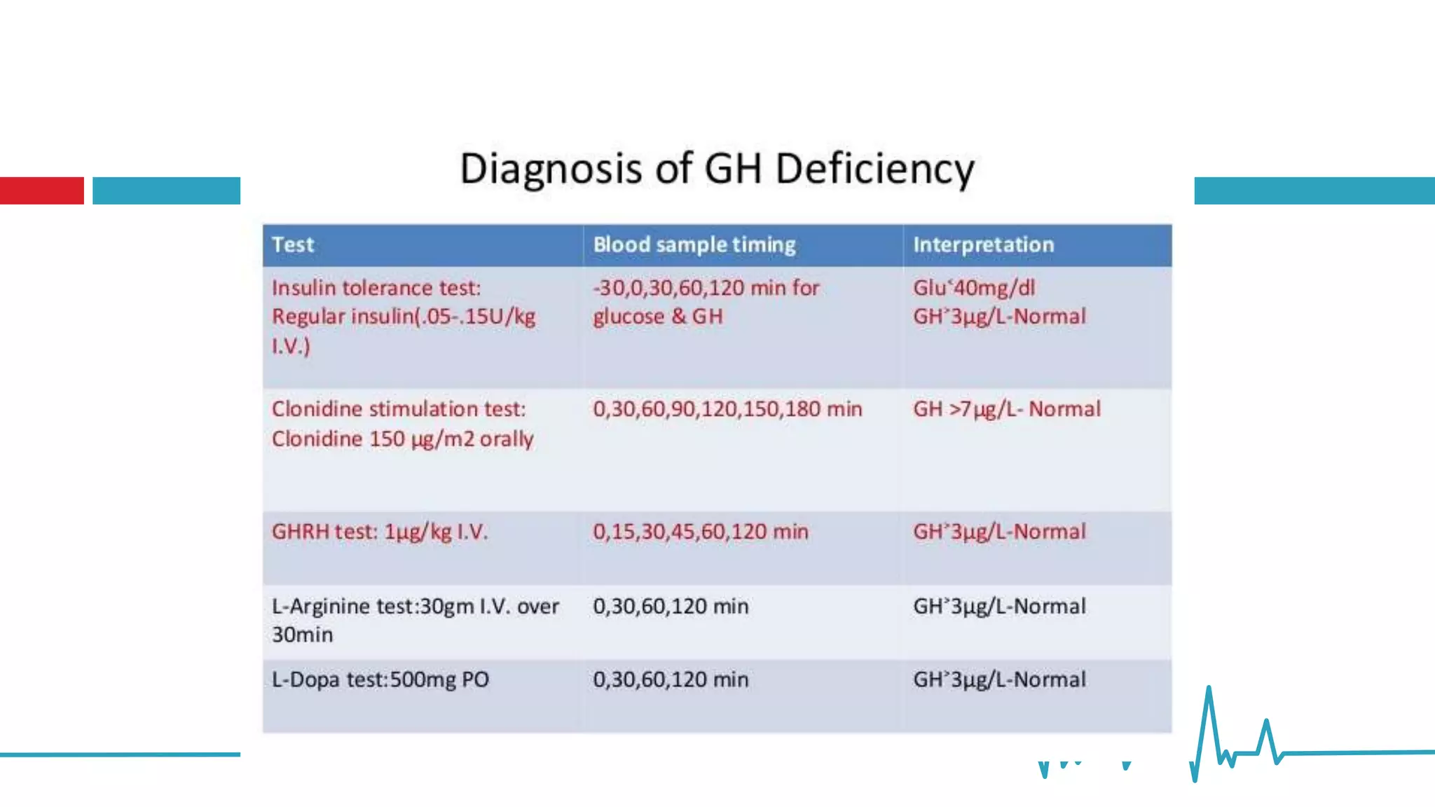
Indication: This test is performed to evaluate
patients with suspected hypothalamic-pituitary-
adrenal (HPA) axis or growth hormone (GH) axis
deficiency. Hypoglycemia causes a major stress
response, with an increase in plasma ACTH,
serum cortisol, and growth hormone. Insulin
Tolerance Test (ITT) is considered the gold
standard test to evaluate the integrity of HPA.
Contraindication: History of coronary artery
disease, seizure disorder, or stroke. Age more than
65 is a relative contraindication.
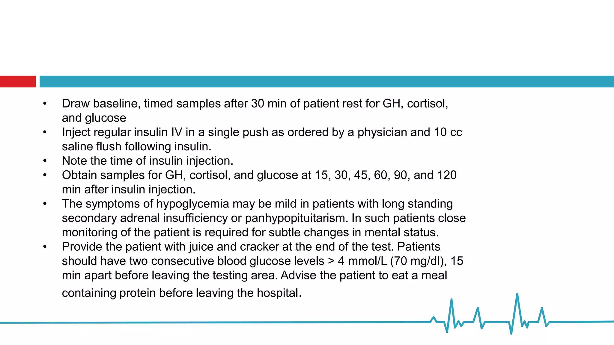
Preparation: NPO except water after midnight and
during test Confirm patient medications and NPO
status with a physician prior to proceeding.
Interpretation:
• A cortisol level <500 nmol/L (18
µg/dL) is consistent with an
abnormal HPA axis [1].
• A serum growth hormone <5
ng/ml (5 mcg/L) is consistent
with severe growth hormone
deficiency].](https://image.slidesharecdn.com/dynamicendocrinetests-190415205146/75/Dynamic-endocrine-tests-7-2048.jpg)
![Growth Hormone Suppression Test (Post-
Glucose Administration)
Indication: To establish the diagnosis of acromegaly
when there is modest elevation of IGF-1 (< 2-fold upper
limit of normal) with absent or equivocal clinical features
[1]
Preparation: 10 h fasting
Draw baseline growth hormone. 4. Give glucose 75 g
orally (glucose drink). 5. Draw glucose and growth
hormone levels at 30, 60, 90, and 120 min [7]. 6.
(Include insulin levels at baseline, 30, 60, 90, and 120
min only if requested).
Diagnosis of acromegaly:
Using ultrasensitive
assays, a GH suppression
to < 0.4 ng/ml is
considered the gold
standard test to rule out
acromegaly [2].
A GH level < 1 ng/mL early
after surgery, in the
absence of presurgical
usage of somatostatin
analogs, predict long-term
remission .](https://image.slidesharecdn.com/dynamicendocrinetests-190415205146/75/Dynamic-endocrine-tests-9-2048.jpg)
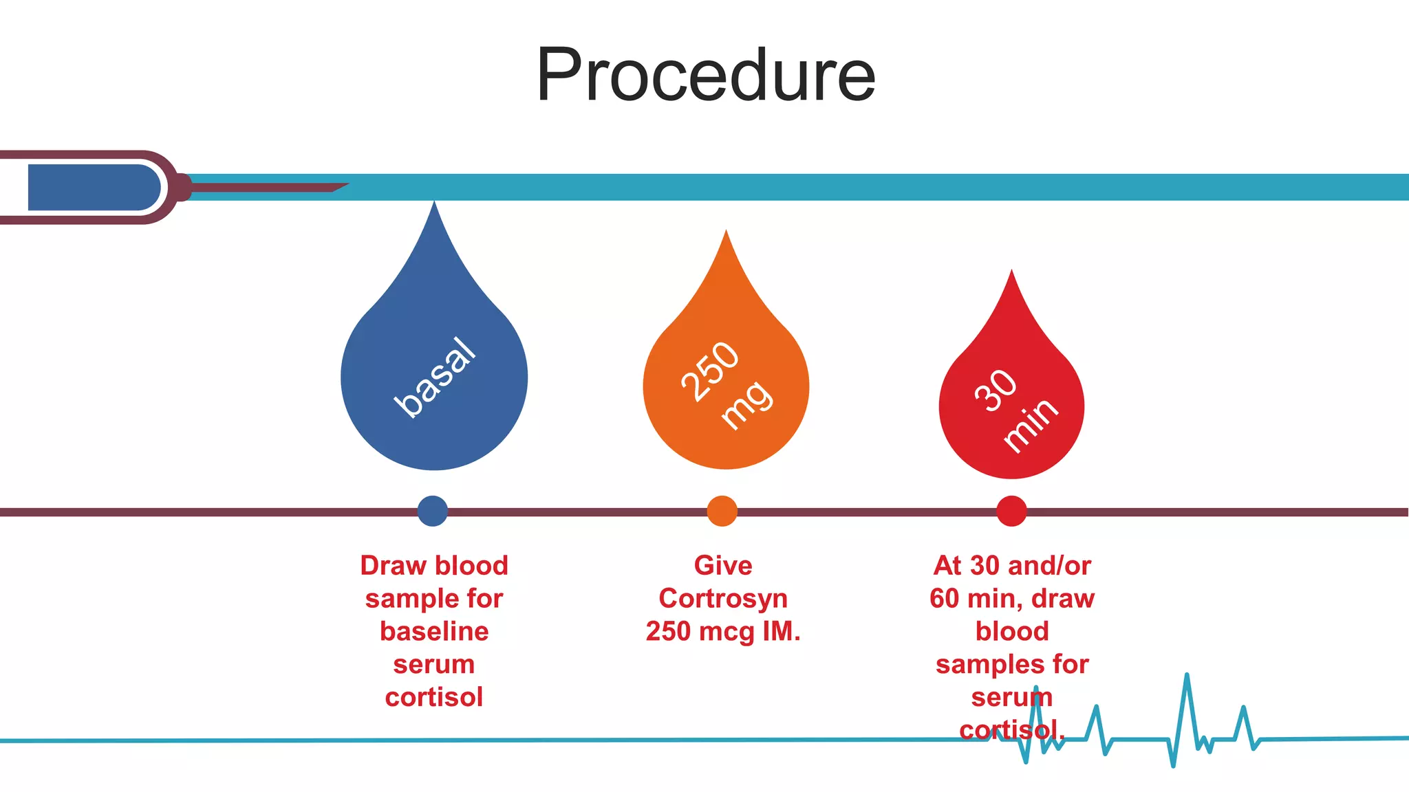
![ACTH Stimulation Test for Adrenal
Insufficiency with Total Cortisol Levels
Indication: to determine whether the adrenal glands can
respond normally to ACTH by producing cortisol.
Preparation: Patients should be off glucocorticoids that
potentially interfere with the cortisol assay
(hydrocortisone, prednisone) for 24 h pretesting.
Dexamethasone may be used.
Precautions: Cosyntropin is category C for pregnancy.
Caveats: • Taking oral estrogen may result in elevation of
the total cortisol level due to increased corticosteroid
binding globulin [3].
Patients with albumin < 2.5 gr/dL may have a low cortisol
level [4]. •
Sensitivity for the test is limited in secondary adrenal
insufficiency. Specificity is > 95 %, thus a positive
cosyntropin test result substantially increases the
likelihood that the patient has secondary adrenal
insufficiency.
Normal response:
Peak stimulated
cortisol value > 18
mcg/dl at 30 min](https://image.slidesharecdn.com/dynamicendocrinetests-190415205146/75/Dynamic-endocrine-tests-15-2048.jpg)
![ACTH Stimulation Test for Adrenal
Insufficiency with Free Cortisol Levels
IndicationThis test is particularly helpful in patient with
albumin < 2.5 mg/dL or low CBG.
Caveats: This cut-off value may not apply to ICU
patients. Further studies are needed to establish
appropriate levels in such patients.
Benefit from glucocorticoid therapy may be beyond
adrenal function status in patients with septic shock
Free cortisol index (FCI) may be used as an alternative
to this dynamic test, if free cortisol assay is not available.
•
FCI: Total cortisol (nmol/L)/CBG (mg/dl) < 12 suggests
adrenal insufficiency [5]. Total cortisol should be
measured in the morning between 8–10 am.
Normal response:
Peak stimulated
cortisol value >
1.2 mcg/dl at 30 or
60 min](https://image.slidesharecdn.com/dynamicendocrinetests-190415205146/75/Dynamic-endocrine-tests-17-2048.jpg)
![Overnight Low Dose Dexamethasone
Suppression Test—1 mg
Indication: To evaluate for Cushing’s syndrome,
to evaluate adrenal incidentaloma for subclinical
Cushing’s syndrome [1]. Preparation: None.
Caveats: • At the 1.8 µg/dL cutoff, the sensitivity
is high (> 95 %) with specificity rates of 80 %.
Specificity increases to greater than 95 % if the
diagnostic threshold is raised to 5 µg/dL [3]. •
Do not use this test if patient is on estrogens as
they increase CBG resulting in falsely elevated
cortisol levels [4]. •
Drugs, such as phenytoin, phenobarbital,
carbamazepine, rifampicin, and alcohol, induce
hepatic enzymatic clearance of dexamethasone,
mediated through CYP 3A4, thereby reducing the
plasma dexamethasone concentrations and may
be associated with a false positive result
Interpretation: Normal
response: Early
morning cortisol < 1.8
µg/dL [2]. Normal
response in adrenal
incidentaloma can be
set at < 3–5 µg/dL if
clinical significance and
increased specificity
are to be pursued](https://image.slidesharecdn.com/dynamicendocrinetests-190415205146/75/Dynamic-endocrine-tests-21-2048.jpg)
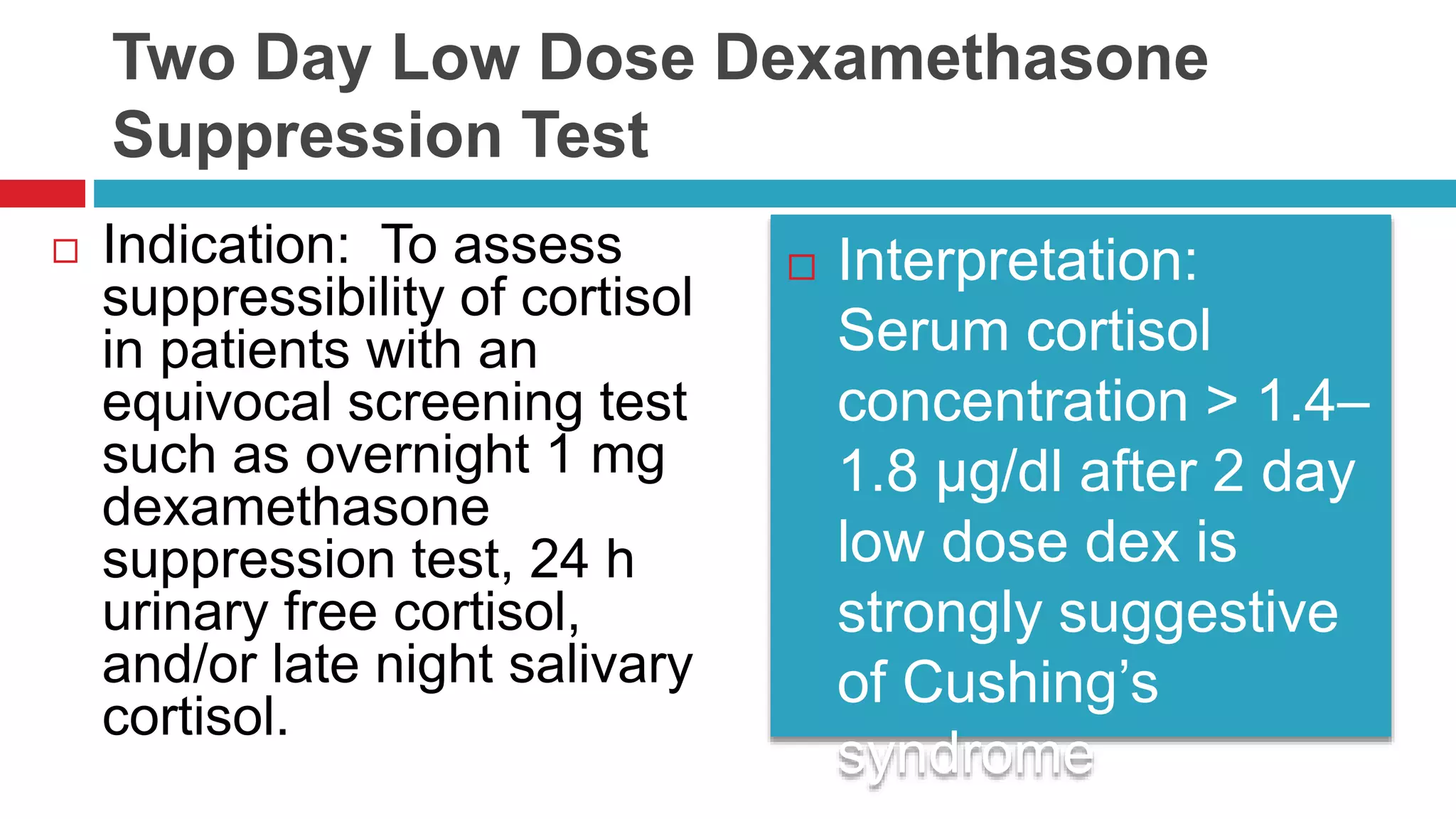
![ Use of the 2 mg 2-day test has greater specificity
at high sensitivity compared to the 1 mg overnight
test. However, it requires more patience on the
part of the patient [2, 3]. • We do not recommend
24 h urine cortisol measurement during 2 mg
dexamethasone suppression test (DST) because
measurement of serum cortisol concentration
during the low dose dexamethasone test is
simpler and more reliable than measurements of
urinary steroids [3]. • Do not use this test if the
patient is on estrogens which increase cortisol
binding globulin (CBG) and falsely elevate cortisol
levels [4]. • Drugs such as phenytoin,
phenobarbital, phenobarbitone, carbamazepine,
rifampicin, and alcohol induce hepatic enzymatic
clearance of dexamethasone, mediated through
CYP 3A4, thereby reducing the plasma
dexamethasone concentrations and may be
associated with a false positive result [5].
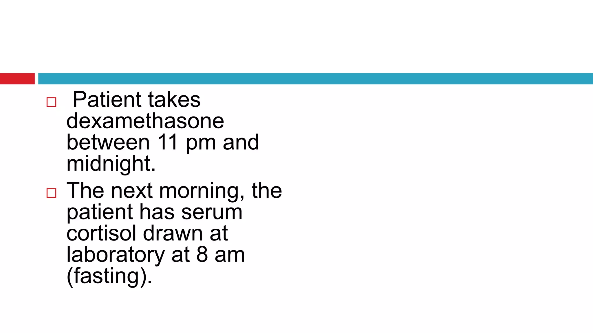
Instruct patient to begin
dexamethasone tablets.
Patient takes one tablet every
6 h for a total of 8 doses (8
am, 2 pm, 8 pm, and 2 am).
Some clinicians prefer a
different schedule such as 6
am, 12 pm, 6 pm, and 12 am
as a more convenient
alternative. Studies were
performed in the former
schedule. 2. 6 h after the last
dose, draw blood for cortisol
(8 am).](https://image.slidesharecdn.com/dynamicendocrinetests-190415205146/75/Dynamic-endocrine-tests-24-2048.jpg)

![Adrenal Venous Sampling
Indication: In order to distinguish between unilateral and
bilateral adrenal disease in patients with primary
aldosteronism.
Preparation: Patient should be in fasting state. The test
should be done in the morning, with the patient in the
supine position for at least 1 h before sampling . Patient
should be normokalemic prior to procedure. .
Precautions: At centers with experience with AVS, the
complication rate is 2.5 % or less
Caveats:
We do not recommend AVS testing without cosyntropin
stimulation due to high false positive rates .
• Medications that may increase renin secretion (e.g.,
mineralocorticoid receptor (MR) antagonists
[spironolactone and eplerenone], high-dose amiloride
[i.e., > 5 mg/day], ACE inhibitors, ARBs, and renin
inhibitors [e.g., aliskiren]) should be discontinued for at
least 4 weeks before AVS until more data on the
]. It is plausible that keeping a patient
with a history of severe uncontrolled
hypertension and/or hypokalemia on
these agents may be allowed in the
presence of low renin (< 1 ng/ml.hr)
levels. However, wherever possible,
MR antagonists should be avoided
because they have the potential to
allow a rise in renin secretion, which
can stimulate aldosterone secretion
from the unaffected side, thus
minimizing the lateralization. Such an
approach is in line with our institutional
experience. • Preferably, extended
release verapamil, peripheral alpha
adrenergic receptor antagonists (e.g.
doxazosin, terazosin and prazosin), and
hydralazine should be used for blood
pressure control prior to AVS.](https://image.slidesharecdn.com/dynamicendocrinetests-190415205146/75/Dynamic-endocrine-tests-32-2048.jpg)
![ Interpretation: 1. Confirm
successful catheterization With
cosyntropin infusion, the adrenal
vein (right and left) to IVC cortisol
ratio is typically more than 10:1;
a ratio of at least 3:1 is required
to be confident that the adrenal
veins were successfully
catheterized [4]. When
cosyntropin infusion is not used,
an adrenal vein to IVC cortisol
gradient of more than 2:1 is
recommended [5, 6]. Most
institutions perform AVS with
cosyntropin stimulation. 2.
Correct aldosterone levels for cortisol
and interpret A/C ratios in order to
localize the tumor side. Dividing the
right and left adrenal vein plasma
aldosterone concentrations by same-
sided cortisol concentrations (A/C
ratio) corrects for the dilutional effect
of the inferior phrenic vein flow into
the left adrenal vein; these are
termed cortisol-corrected ratios (A/C
ratio). When cosyntropin infusion
used criteria for localization is a
cutoff for the cortisol–corrected
aldosterone ratio (A/C) from high side
to low side of more than 4:1 to
indicate unilateral aldosterone
excess [2], the true positive rate is 88
% [6].](https://image.slidesharecdn.com/dynamicendocrinetests-190415205146/75/Dynamic-endocrine-tests-33-2048.jpg)
![ at An A/C ratio less than 3.0 is
consistent with bilateral
aldosterone hypersecretion. •
Ratios between 3 and 4.0
represent a zone of overlap. If
the A/C ratio of low side is less
than the IVC A/C ratio, the tumor
will be on the contralateral site in
93 % of patients with surgically
confirmed APA [4]. This criterion
may be particularly helpful in
patients with only unilateral
successful adrenal vein
catheterization.](https://image.slidesharecdn.com/dynamicendocrinetests-190415205146/75/Dynamic-endocrine-tests-34-2048.jpg)

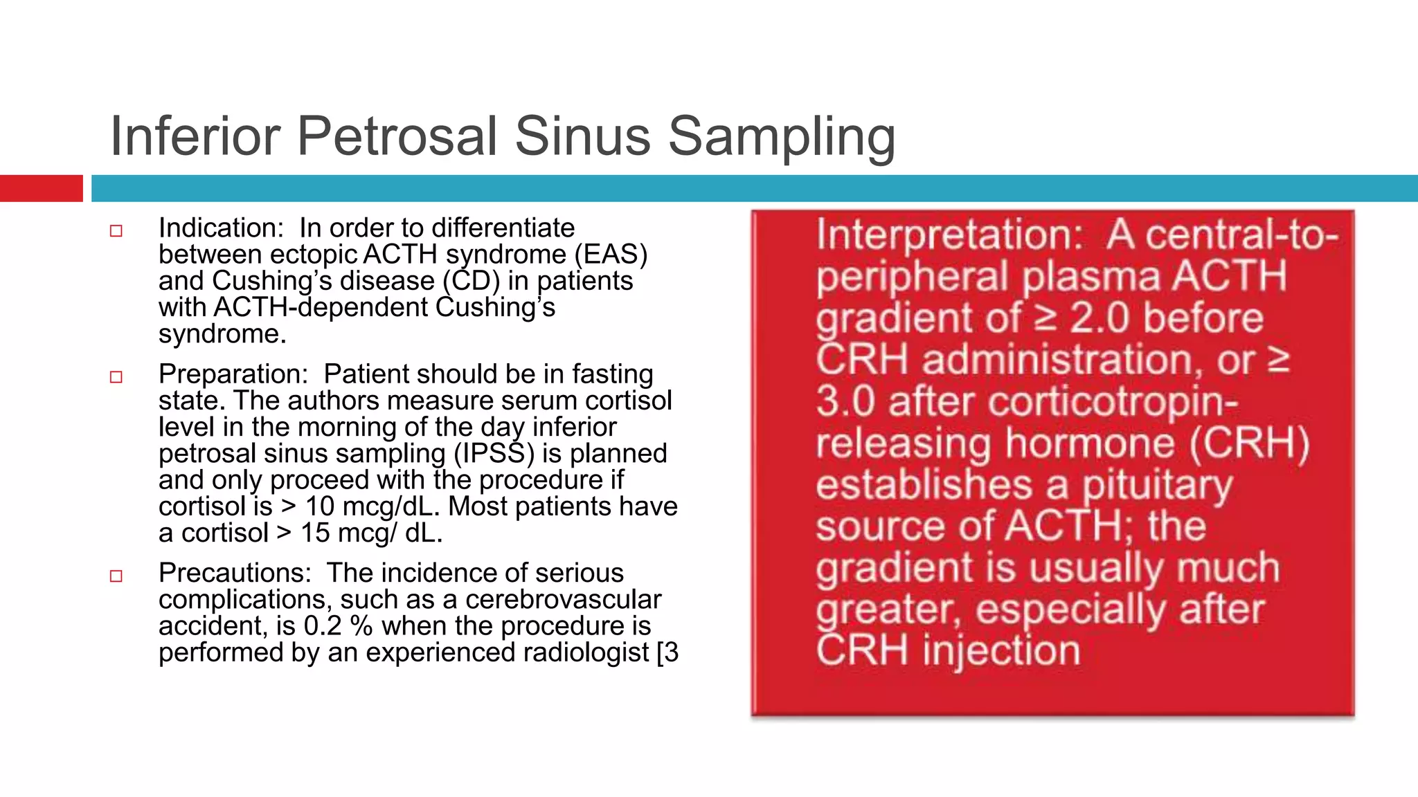
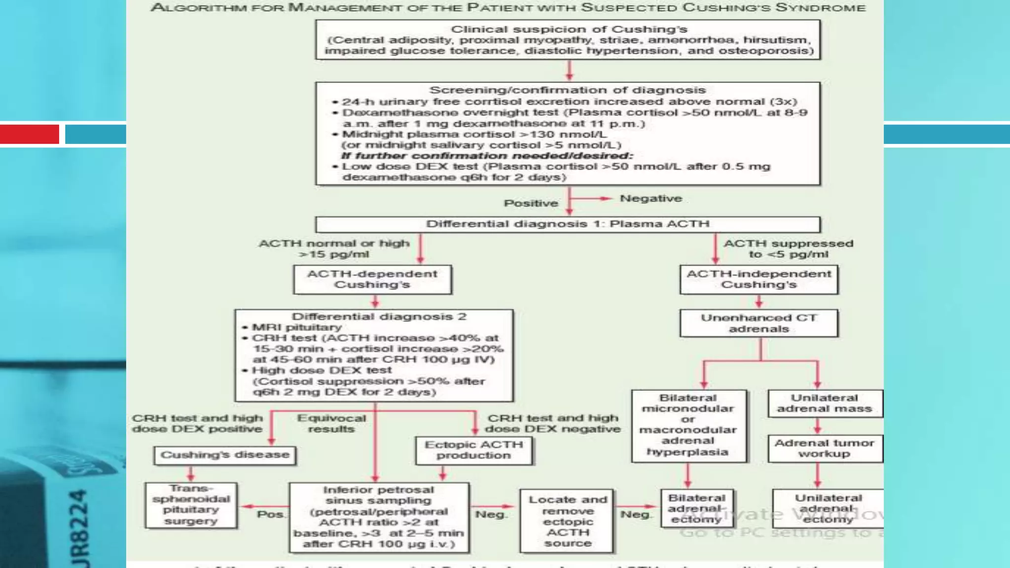
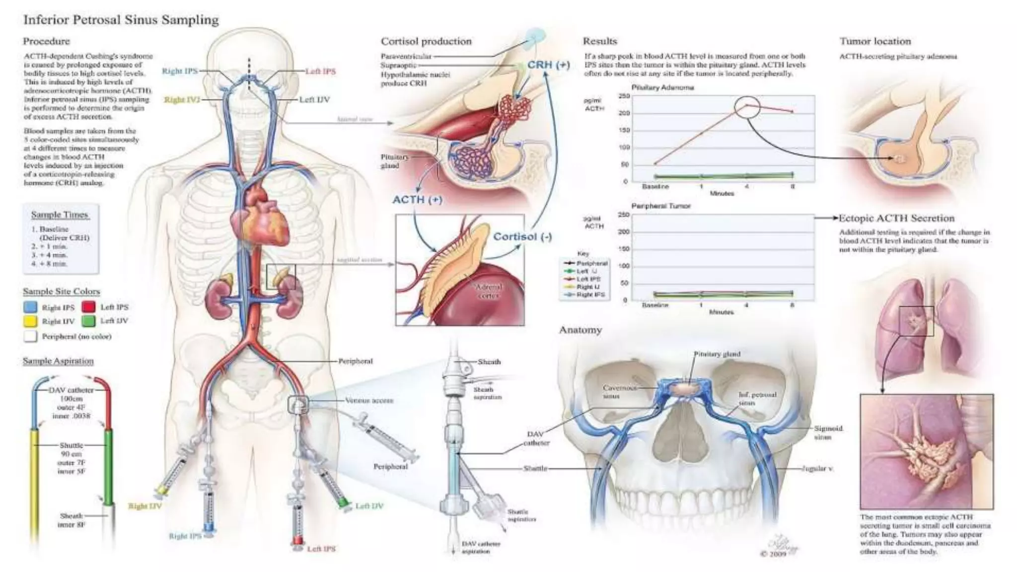
![ Caveats: • Sensitivity and specificity of
IPSS is > 88 % and close to 100 % after
CRH administration respectively [1]. • In
cases where the IPS to peripheral (IPS:P)
ACTH gradient is not consistent with a
pituitary source, peripheral ACTH
response (> 35 %) to CRH administration
suggests central etiology rather than
ectopic source [5]. • Radiologic
confirmation of placement of catheter tip in
IPS may not be reliable [4]. • IPS:P
prolactin ratios greater than 1.8, confirm
accurate catheterization [2]. Most patients
with appropriate IPS catheterization have a
gradient > 1.3 [5]. Prolactin may be
measured routinely during IPSS, or in
order to cut back on the cost, be stored
and measured later on if IPS:P ACTH ratio
is not consistent with a pituitary source. •
In the absence of appropriate bilateral IPS
catheterization, which may be confirmed
by measurement of IPS:P prolactin ratio, a
lack of significant IPS:P ACTH gradient
does not rule a pituitary source as the
underlying etiology for Cushing’s
syndrome. In addition, confirmation of
accurate venous sampling in only one IPS
may not rule out a pituitary source in the
contralateral side of the pituitary gland due
to variable venous drainage [4]. • Petrosal
sinus sampling is of limited value in
distinguishing between patients with
Cushing’s syndrome and normal
individuals or those with pseudo-Cushing’s
states. Therefore, a diagnosis of ACTH-
dependent Cushing’s syndrome should be
established before referring a patient for
IPSS [6].](https://image.slidesharecdn.com/dynamicendocrinetests-190415205146/75/Dynamic-endocrine-tests-37-2048.jpg)
![Indication: Assessment of beta cell reserve.
Preparation [1:] NPO except water after midnight
and during the test. Ten hours after the last dose of
short-acting or intermediate-acting insulin, metformin
or thiazolidinedione. At least 24 h after a dose of
sulfonylurea or long-acting insulin (glargine or
detemir).
Glucagon Stimulation
Interpretation: Beta Cell
functional reserve was defined
as preserved if the peak C-
peptide response to glucagon is
at least 1.5 ng/ dL (0.5 nmol/L)
or fasting C-peptide
concentration of at least 1 ng/dL
(0.33 nmol/L). Beta cell
functional reserve is defined as
absent if the glucagon
stimulated or fasting C-peptide
concentrations do not meet
these criteria [1]..](https://image.slidesharecdn.com/dynamicendocrinetests-190415205146/75/Dynamic-endocrine-tests-42-2048.jpg)


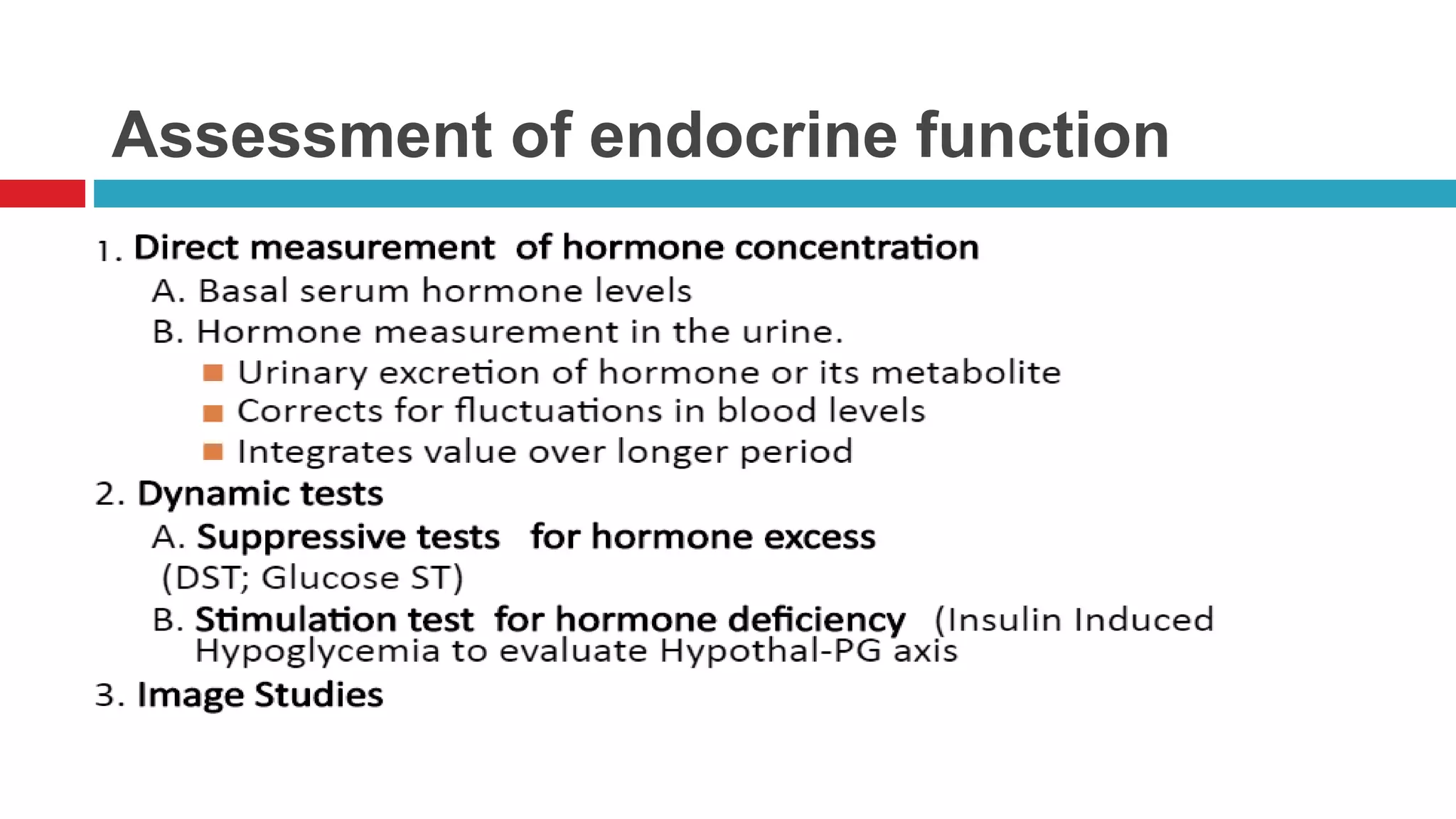
This document summarizes various endocrine tests used to assess different endocrine functions and disorders. It describes dynamic tests such as the insulin tolerance test used to evaluate the hypothalamic-pituitary-adrenal axis and growth hormone axis. It also summarizes tests used to evaluate disorders of the adrenal glands such as the ACTH stimulation test, dexamethasone suppression tests, and tests used to diagnose Cushing's syndrome and hyperaldosteronism. Precautions, interpretations and procedures are provided for many of the major endocrine dynamic tests.


