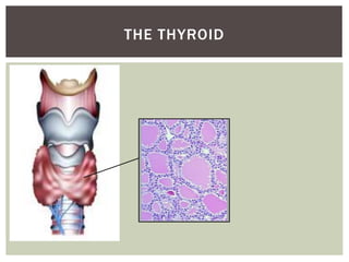
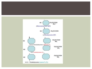
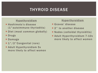
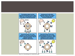
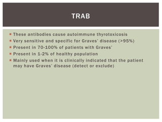
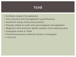
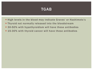
Jack Leitch
Clinical
Immunology
THYROID
AUTOANTIBODIES
 Briefly describe the thyroid and production of thyroid
hormones
 Understand some examples of thyroid disease and how they
are categorised
 Identify the main thyroid autoantibodies and how these are
investigated in the laboratory
 Understand how the main thyroid autoantibodies cause
disease of the thyroid
AIMS
THE THYROID
Hypothyroidism
 Hashimoto’s disease
(1°autoimmune thyrioditis)
 Diet (most common globally)
 Drugs
 Damage
 1°/2°Congenital (rare)
 Adult Hypothyroidism 5x
more likely to affect women
THYROID DISEASE
Hyperthyroidism
 Graves’ disease
 2° to another disease
 Nodes (colloidal thyroiditis)
 Adult Hyperthyroidism 7-10x
more likely to affect women
TPO - Thyroid peroxidase antibody
Autoimmune thyroid disease (Hashimoto’s, 1°Myxoedema 90%)
TRAb – Thyroid stimulating hormone receptor antibody
Graves’ disease (Goitre)
TgAb - Thyroglobulin antibody
Autoimmune thyroid disease + monitoring thyroid cancer
treatment
AUTOANTIBODY TESTING
 Antibody to glycoprotein (thyroperoxidase) responsible for
catalysing iodine and performing iodination of tyrosine to
produce thyroid hormones via colloid
 Patient sample added to wells containing the thyroperoxidase
protein
 If there are antibodies present these will bind to the protein in
the well
 Labelled mouse anti-TPO is displaced
 The light generated is inversely proportional to the amount of
patient TPO antibody present and bound
 Measured with a luminometer (0-60 U/mL)
TPO
 Hashimoto’s ≈ 90%
 Graves’ ≈ 60-80%
 10-12% of the healthy population may have detectable anti-
TPO antibodies
 Mainly used to help diagnose autoimmune thyroiditis
 Antibody titres can be done to assess disease activity
TPO
 Antibody to TSH receptors in the thyroid. Defined by their
action on signalling
 Activating = stimulates (hyperthyroidism  Graves’)
 Blocking = blocks receptor (Hypothyroidism  Hashimoto’s
thyroiditis)
 Tubes coated with TSH receptor
 Patient sample incubated in tubes
 If antibody present the TSHr will be blocked
 Labelled anti-TSHr is then added to the tubes, which will bind
to any free TSHr in the tube
 The light emitted from these labelled antibodies is measured
via a luminometer
 >1.5 IU/L = positive
TRAB
 These antibodies cause autoimmune thyrotoxicosis
 Very sensitive and specific for Graves’ disease (>95%)
 Present in 70-100% of patients with Graves’
 Present in 1-2% of healthy population
 Mainly used when it is clinically indicated that the patient
may have Graves’ disease (detect or exclude)
TRAB
TRAB
 Assay to become automated
 Works in a similar way
 ELISA platform
 Current method very time consuming
 Antibody targets thyroglobulin
 Can interfere with thyroglobulin quantification
 Sandwich assay (Immunoenzymatic)
 Sample added to wells with paramagnetic thyroglobulin
 Magnetic field prevents Ag:Ab complex from washing away
 Conjugate binds to TGAb
 Chemiluminescent substrate binds to Conjugate
 <4 IU/mL
TGAB
 High levels in the blood may indicate Graves’ or Hashimoto’s
 Thyroid not normally released into the bloodstream
 30-50% with hypothyroidism will have these antibodies
 15-30% with thyroid cancer will have these antibodies
TGAB
 Generally, higher levels of antibody are indicative of an
autoimmune thyroid disorder
 Treatment often involves inhibiting an overactive thyroid, or
replacing what is not being produced in an underactive thyroid
 If severe inflammation occurs surgery may be needed
 Antibodies are organ specific, rather than systemic
 Laboratory testing key in diagnosis
CONCLUSION
Thank you
 Tortora GJ, Derrickson B. The Endocrine System In: Tortora GJ,
Derrickson B editors. Principles of Anatomy and Physiology
[13th Ed]. John Wiley & Sons; 2011. Pg:696-700.
 Pyne D, Isenberg DA. Autoimmune Thyroid Disease in
Systemic Lupus Erythematosus. Annals of the Rheumatic
Diseases. 2001;61:70-72.
 Murphy K, Travers P, Walport M. Autoimmunity and
Transplantation In: Murphy K, Travers P, Walportm editors.
Immunobiology [7th Ed]. Garland Science; 2008. Pg:620-21.
 Lab Tests Online[Internet]. London: The Association for
Clinical Biochemistry and Laboratory Medicine; c2008 Oct.
Thyroid Autoantibodies; 2008 [cited 2014 July 14]; [about 2
screens]. Available from
http://labtestsonline.org.uk/understanding/analytes/thyroid-
antibodies/tab/sample/
REFERENCES

Thyroid autoantibodies

  • 1.
  • 2.
     Briefly describethe thyroid and production of thyroid hormones  Understand some examples of thyroid disease and how they are categorised  Identify the main thyroid autoantibodies and how these are investigated in the laboratory  Understand how the main thyroid autoantibodies cause disease of the thyroid AIMS
  • 3.
  • 5.
    Hypothyroidism  Hashimoto’s disease (1°autoimmunethyrioditis)  Diet (most common globally)  Drugs  Damage  1°/2°Congenital (rare)  Adult Hypothyroidism 5x more likely to affect women THYROID DISEASE Hyperthyroidism  Graves’ disease  2° to another disease  Nodes (colloidal thyroiditis)  Adult Hyperthyroidism 7-10x more likely to affect women
  • 7.
    TPO - Thyroidperoxidase antibody Autoimmune thyroid disease (Hashimoto’s, 1°Myxoedema 90%) TRAb – Thyroid stimulating hormone receptor antibody Graves’ disease (Goitre) TgAb - Thyroglobulin antibody Autoimmune thyroid disease + monitoring thyroid cancer treatment AUTOANTIBODY TESTING
  • 8.
     Antibody toglycoprotein (thyroperoxidase) responsible for catalysing iodine and performing iodination of tyrosine to produce thyroid hormones via colloid  Patient sample added to wells containing the thyroperoxidase protein  If there are antibodies present these will bind to the protein in the well  Labelled mouse anti-TPO is displaced  The light generated is inversely proportional to the amount of patient TPO antibody present and bound  Measured with a luminometer (0-60 U/mL) TPO
  • 9.
     Hashimoto’s ≈90%  Graves’ ≈ 60-80%  10-12% of the healthy population may have detectable anti- TPO antibodies  Mainly used to help diagnose autoimmune thyroiditis  Antibody titres can be done to assess disease activity TPO
  • 10.
     Antibody toTSH receptors in the thyroid. Defined by their action on signalling  Activating = stimulates (hyperthyroidism  Graves’)  Blocking = blocks receptor (Hypothyroidism  Hashimoto’s thyroiditis)  Tubes coated with TSH receptor  Patient sample incubated in tubes  If antibody present the TSHr will be blocked  Labelled anti-TSHr is then added to the tubes, which will bind to any free TSHr in the tube  The light emitted from these labelled antibodies is measured via a luminometer  >1.5 IU/L = positive TRAB
  • 11.
     These antibodiescause autoimmune thyrotoxicosis  Very sensitive and specific for Graves’ disease (>95%)  Present in 70-100% of patients with Graves’  Present in 1-2% of healthy population  Mainly used when it is clinically indicated that the patient may have Graves’ disease (detect or exclude) TRAB
  • 12.
    TRAB  Assay tobecome automated  Works in a similar way  ELISA platform  Current method very time consuming
  • 13.
     Antibody targetsthyroglobulin  Can interfere with thyroglobulin quantification  Sandwich assay (Immunoenzymatic)  Sample added to wells with paramagnetic thyroglobulin  Magnetic field prevents Ag:Ab complex from washing away  Conjugate binds to TGAb  Chemiluminescent substrate binds to Conjugate  <4 IU/mL TGAB
  • 14.
     High levelsin the blood may indicate Graves’ or Hashimoto’s  Thyroid not normally released into the bloodstream  30-50% with hypothyroidism will have these antibodies  15-30% with thyroid cancer will have these antibodies TGAB
  • 15.
     Generally, higherlevels of antibody are indicative of an autoimmune thyroid disorder  Treatment often involves inhibiting an overactive thyroid, or replacing what is not being produced in an underactive thyroid  If severe inflammation occurs surgery may be needed  Antibodies are organ specific, rather than systemic  Laboratory testing key in diagnosis CONCLUSION
  • 16.
  • 17.
     Tortora GJ,Derrickson B. The Endocrine System In: Tortora GJ, Derrickson B editors. Principles of Anatomy and Physiology [13th Ed]. John Wiley & Sons; 2011. Pg:696-700.  Pyne D, Isenberg DA. Autoimmune Thyroid Disease in Systemic Lupus Erythematosus. Annals of the Rheumatic Diseases. 2001;61:70-72.  Murphy K, Travers P, Walport M. Autoimmunity and Transplantation In: Murphy K, Travers P, Walportm editors. Immunobiology [7th Ed]. Garland Science; 2008. Pg:620-21.  Lab Tests Online[Internet]. London: The Association for Clinical Biochemistry and Laboratory Medicine; c2008 Oct. Thyroid Autoantibodies; 2008 [cited 2014 July 14]; [about 2 screens]. Available from http://labtestsonline.org.uk/understanding/analytes/thyroid- antibodies/tab/sample/ REFERENCES

Editor's Notes

  • #4 Lobes give distinct butterfly shape to thyroid. Thyroglobulin precursor to thyroid hormones. Follicular cells trap iodide ions to synth thyroglobulin. Located anterior to trachea Right and left lateral lobes Thyroid follicle: -Follicular cell, surround large spaces -Thyroglobulin (colloid)  large spaces
  • #5 Tyrosine iodination. One = monoiodotyrosine (t1) two = diiodotyrosine (t2). Tyrosine found on thyroglobulin. Tyrosine plus iodine = colloid. Lysosomes break down TGB, releasing t3 and t4 molecules. ^ basal metabolic rate, body temp via forcing cells to use more ATP, skeletal + nervous system development
  • #6 Thyroiditis means the inflammation of the thyroid gland. Hashimoto’s  primary is low t3/4 and high TSH. Secondary is low t3/4 and low or normal TSH. Primary is failure of thyroid function, secondary is failure of thyroid stimulation. TPO, autoantibodies attack the thyroid. Hypo  Myxoedema (swelling due to subcutaneous separation of connective tissues e.g. mucopolysaccarides) , lowered renal function, anaemia, low heart rate (bradycardia). Diet low iodine Secondary congenital  impaired pituitary signalling to stimulate the thyroid (TSH). Primary congenital  Need to treat asap to prevent developmental issues in child  mental development impaired, short stature, deaf/ mute. Thyroid not developed correctly. Damage  neck irradiation, radioiodine (form of cancer treatment as thyroid cells preferentially take up the radioiodine to produce T3 +T4)/ surgery for thyrotoxicosis (partial or total removal of thyroid gland  only if treatments don’t work. DeQuervain’s Thyroiditis  Colloidal regions break down releasing thyroid hormones into circulation  hyperthyroidism. The hormones trigger a response from the pituitary and lots of TSH is released, leading to a hypothyroidism. Usually self limiting but can look like Graves’. Clinical information important as it is usually caused by viral infections (mumps, flu) Hyper Goitre (swollen thyroid + neck) weight loss, heart palpitations, severe = thyroid storm or thyrotoxic crisis (rare, life threatening  fever, vomiting, heart failure) Nodes  abnormal thyroid tissue develops in the thyroid (nodes), these produce too much of the thyroid hormones. Other diseases  SLE, RA, pernicious anaemia (although it is still debated as to whether this is a true correlation or a by product of the fact that diseases such as SLE and thyroiditis are most prevalent in the same groups  young to middle aged women)
  • #7 Graves’ disease  autoantibodies to thyroid stimulating hormone receptor mimic the actions of TSH. Antibodies continually stimulate the thyroid into making more and more thyroid hormones, without the negative feedback from the pituitary preventing this from happening. Both environmental and genetic factors are involved and the cause of Graves’ is not known Usually patients have an enlarged thyroid and have exophthalmos, which is a distinct oedema behind the eyes associated with the disease. (Can cause blindness as the swelling puts pressure on the optical nerve) Pounding heart rate, weight loss, sweating are all symptoms. Treated with surgery to remove thyroid tissue, or with radioactive iodine to destroy thyroid tissue or with anti-thyroid drugs to block thyroid hormone synthesis.
  • #8 TPO – Hypo TRAb – Hyper Lab testing provides the clinical team with information direct treatment and refine differential diagnoses. Although clinical presentation will immediately make it apparent if the patient has hyper or hypothyroidism, it is important to know which antibodies are involved as they each have a different affect on the patient and how the disease is likely to develop. Thyroiditis (inflammation) can damage the thyroid, resulting in exposure of further antigens to the immune system
  • #9 TPO is a membrane associated glycoprotein found only in thyocytes . TPO catalyses the oxidation of iodine on tyrosine used to synthesize T3 and T4 Assay is a competitive immunoassay The patient antibodies need to compete with acridinium-ester labelled mouse anti-TPO antibodies that are bound to the human TPO in the assay. In the second stage of the assay mouse anti-TPO coupled to paramagnetic particles are added, which bind to the TPO. The magnetic particles prevent the TPO (and what is bound to the TPO, be it patient antibodies or the original labelled mouse antibodies) from being washed away, but any unbound antibodies will be washed away. If the patient antibodies do not bind, the labelled mouse antibodies will remain and the patient sample will be washed away. The mouse anti-TPO is acridinium-ester labelled, which emits light when stimulated. If the patient antibodies have bound, they will displace the labelled mouse antibody, resulting in a lower amount of light emitted. Inverse relationship  as antibodies in sample increase, amount of light detected decreases as the thyroperoxidase is being bound Samples with a very high TPO result (>1300) should be diluted to obtain a more accurate result. According to the chemistry SOP the assay can react with heterophiles.
  • #10 If clinical presentation and presence of anti-TPO then thyroiditis likely (Hashimoto’s) Easier and more accurate to measure than TRAB antibodies, so often used instead to determine if the patient has Graves’
  • #11 Can also be used as a differential diagnostic tool for thyrotoxicosis Known as long acting thyroid stimulating antibodies Blocks receptor to prevent stimulation from TSH Activating TRAB antibodies are characteristic of Graves’ Signal emitted inversely proportional to patient antibody concentration
  • #12 Method used in Immunology uses human recombinant TSH receptor, rather than porcine (pig) or bovine (cow) derivatives.
  • #13 Colour change in ELISA is inversely proportional to levels of antibody.
  • #14 Interference is a problem when testing patients who are being treated for thyroid cancer because the cancer cells can produce thyroglobulin, so to ensure the treatment is working correctly the true thyroglobulin level must be know. This is especially relevant if the patient has had their thyroid gland removed because there should be no thyroglobulin in the blood unless the cancerous cells have returned. Measured with a Luminometer
  • #15 Thyroiditis can cause damage to the follicular cells, which causes thyroglobulin to be released. Antibodies to thyroglobulin in the blood can then form. Most commonly associated with Hashimoto’s hypothyroidism  TPO is a more sensitive and specific test.
  • #16 Levothyroxine replaces thyroxine hormone (hypo treatment) Thionamide inhibits thyroid peroxidase (hyper treatment) Organ specific = only that organ affected. Systemic = several tissues can be affected.