Downloaded 55 times

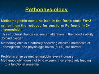
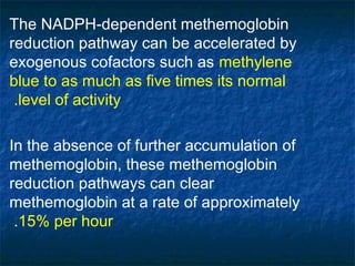

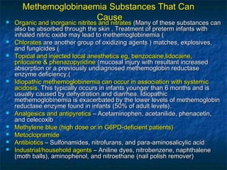
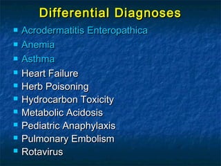
![The four types are as followsThe four types are as follows
Type IType I – This is the most common variant, and the enzyme– This is the most common variant, and the enzyme
deficiency isdeficiency is limited to the erythrocyteslimited to the erythrocytes causing cyanosis;causing cyanosis;
cyanosis usually, but not always, developscyanosis usually, but not always, develops during infancyduring infancy
Type IIType II – Widespread deficiency of the enzyme occurs in– Widespread deficiency of the enzyme occurs in
various tissues, includingvarious tissues, including erythrocytes, liver, fibroblasts, anderythrocytes, liver, fibroblasts, and
brainbrain; it is associated with severe CNS symptoms, including; it is associated with severe CNS symptoms, including
encephalopathy, microcephaly, hypertonia, athetosis,encephalopathy, microcephaly, hypertonia, athetosis,
opisthotonos, strabismus, mental retardation, and growthopisthotonos, strabismus, mental retardation, and growth
retardationretardation; cyanosis is evident at an early age; cyanosis is evident at an early age
Type IIIType III – Although the– Although the hematopoietic systemhematopoietic system ((platelets, RBCs,platelets, RBCs,
and white blood cellsand white blood cells [[WBCsWBCs]])) is involved, the only clinicalis involved, the only clinical
consequence is cyanosisconsequence is cyanosis
Type IVType IV –– Like type ILike type I, this type has isolated involvement of the, this type has isolated involvement of the
erythrocytes but results inerythrocytes but results in chronic cyanosischronic cyanosis](https://image.slidesharecdn.com/methemoglobinaemia-190222121310/85/Methemoglobinaemia-12-320.jpg)
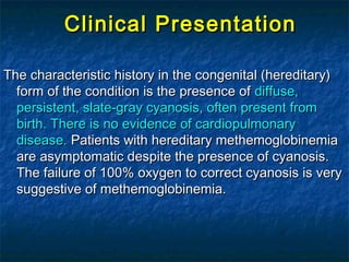
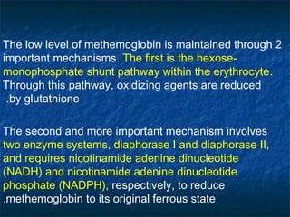
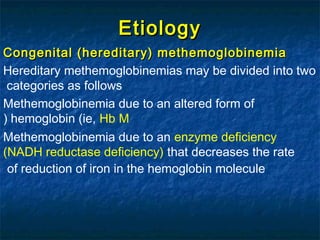
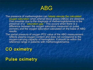
![WorkupWorkup
Investigations to rule out hemolysisInvestigations to rule out hemolysis ((complete bloodcomplete blood
countcount [[CBCCBC]], reticulocyte count, peripheral smear review, lactate, reticulocyte count, peripheral smear review, lactate
dehydrogenasedehydrogenase [[LDHLDH]], bilirubin, haptoglobin & Heinz body preparation, bilirubin, haptoglobin & Heinz body preparation))
EndEnd--organ dysfunction or failureorgan dysfunction or failure ((liver function tests,liver function tests,
electrolytes, renal function testselectrolytes, renal function tests))
Investigations to evaluate a hereditary causeInvestigations to evaluate a hereditary cause forfor
methemoglobinemia should be ordered when appropriatemethemoglobinemia should be ordered when appropriate..
Hemoglobin electrophoresis and DNA sequencing of theHemoglobin electrophoresis and DNA sequencing of the
globin chain gene can be used to identify hemoglobin Mglobin chain gene can be used to identify hemoglobin M..
Specific enzyme assaysSpecific enzyme assays ((nicotinamide adeninenicotinamide adenine
dinucleotidedinucleotide [[NADHNADH]]–dependent reductase, cytochrome b5 reductase–dependent reductase, cytochrome b5 reductase))
may be determined, often in multiple cell linesmay be determined, often in multiple cell lines ((ie, platelets,ie, platelets,
granulocytes, and fibroblastsgranulocytes, and fibroblasts)), to diagnose inherited cases, to diagnose inherited cases..](https://image.slidesharecdn.com/methemoglobinaemia-190222121310/85/Methemoglobinaemia-20-320.jpg)


This document provides information on methemoglobinemia, including: - Definition as an abnormal increase in methemoglobin levels in red blood cells above 1% - Symptoms vary based on methemoglobin levels and include cyanosis, headaches, and loss of consciousness - Causes include ingestion of drugs/toxins like local anesthetics, nitrates, sulfonamides, and certain antibiotics - Workup involves blood tests to check for anemia and enzyme levels to identify potential hereditary causes


![Approach to Cyanosis [Paediatrics presentation for medical (MBBS) students]](https://cdn.slidesharecdn.com/ss_thumbnails/approachtocyanosis71to77final-210927113642-thumbnail.jpg?width=640&height=640&fit=bounds)


![CASE_PRESENTATION_ON_subdural_hematoma(SDH)[1 FINAL PPT]-1.pptx](https://cdn.slidesharecdn.com/ss_thumbnails/casepresentationonsubduralhematomasdh1finalppt-1-260129172522-d405d375-thumbnail.jpg?width=640&height=640&fit=bounds)


