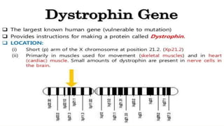
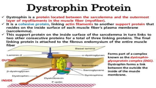
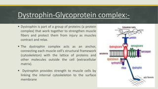
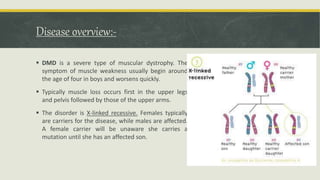
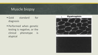
INTERNATIONAL SCHOOL
OF MEDICINE
Research studies on Duchenne Muscular Dystrophy.
Presented By:-
Mohammed Abdul Aziz -4th Group
Dinesh Pal Singh- 3rd Group
Tanwee Fatima - 3rd Group
Sana Momin - 3rd Group
Definition
Ducchenne muscular dystrophy is most common of
several childhood muscular dystrophies and it is an
inherited disorder ( X-linked recessive ) Characterized
by progressive degeneration of muscle.
Dystrophin-Glycoprotein complex:-
 Dystrophin is part of a group of proteins (a protein
complex) that work together to strengthen muscle
fibers and protect them from injury as muscles
contract and relax.
 The dystrophin complex acts as an anchor,
connecting each muscle cell's structural framework
(cytoskeleton) with the lattice of proteins and
other molecules outside the cell (extracellular
matrix).
 Dystrophin provides strength to muscle cells by
linking the internal cytoskeleton to the surface
membrane
Its Effect on cell:-
 Dystrophin provides strength to muscle cells by linking the internal cytoskeleton
to the surface membrane. Without this structural support, the cell membrane
becomes permeable.
 Mitochondrial dysfunction gives rise to an amplification of stress-induced
cytosolic calcium signals and an amplification of stress-induced reactive-oxygen
species production. In a complex cascading process that involves several
pathways and increased oxidative stress within the cell damages
the sarcolemma and eventually results in the death of the cell.
 As components from outside the cell are allowed to enter the internal pressure
of the cell increases until the cell bursts and dies.
 Muscle fibers undergo necrosis and are ultimately replaced
with adipose and connective tissue.
How Gene Defects:-
 Mutations of dystrophin gene lead to an absence of or defect in the protein
dystrophin, which results in progressive muscle degeneration.
 Mutation of the DMD gene is deletion of 1 or more exons, Duplications ,
nonsense or splice site mutations
 The most common mutation are repeats of the CAG nucleotides.
 Although there is no clear correlation found between the extent of the deletion
and the severity of the disorder, DMD deletions usually result in frameshift.
 Mutations within the dystrophin gene can either be inherited or occur
spontaneously during germline transmission.
Disease overview:-
 DMD is a severe type of muscular dystrophy. The
symptom of muscle weakness usually begin around
the age of four in boys and worsens quickly.
 Typically muscle loss occurs first in the upper legs
and pelvis followed by those of the upper arms.
 The disorder is X-linked recessive. Females typically
are carriers for the disease, while males are affected.
A female carrier will be unaware she carries a
mutation until she has an affected son.
Symptoms :-
 Delayed Onset
 Walking ,
 Difficulty in performing a standing jump
 Waddling when walking
 Difficulty standing up
 Enlarged Calves ( Pseudo hypertrophy due to fat infiltration )
 Muscle contractures of Achilles tendon and hamstrings impair functionality because the muscle fibers
shorten and fibrose in connective tissue
 Difficulty with motor skills.
 Skeletal deformities (including scoliosis in some cases)
 Higher risk of neurobehavioral disorders
 learning disorders (dyslexia)
 non-progressive weaknesses in specific cognitive skills (in particular short-term verbal memory), which
are believed to be the result of absent or dysfunctional dystrophin in the brain.
Signs:-
Gower’s
sign
Signs:-
Diagnosis:-
 The diagnosis of a dystrophinopathy is suspected based upon:
 Characteristic age and sex
 Presence of symptoms and signs suggestive of a myopathic
process
 Markedly increased serum creatine kinase values
 Myopathic changes on electromyography and muscle
biopsy
 A positive family history suggesting X-linked recessive
Serum muscle enzymes:-
 Markedly raised serum CK level, 10-20 times the upper limit of
normal
 – Levels peak at 2-3 years of age and then decline with
increasing age, due to progressive loss of dystrophic muscle
fibres
 Elevated serum ALT, AST, aldolase and LDH
Electromyography
 Needle electromyography
 Short duration, low amplitude polyphasic motor unit
potentials in proximal muscles
 Over time, some of these areas become electrically silent
Muscle biopsy
 Gold standard for
diagnosis
 Performed when genetic
testing is negative, or the
clinical phenotype is
atypical
Molecular Genetic Testing
 Multiplex polymerase chain reaction (PCR),
 Multiplex ligation-dependent probe amplification (MLPA) has
provided a more sensitive technique for detecting deletions.
 If MLPA testing is negative, the DMD gene can be tested for
point mutations.
Genetic Analysis
 Molecular genetic testing is indicated for patients with an
elevated serum CK level and clinical findings suggestive of a
dystrophinopathy.
 The diagnosis is established if a disease-causing mutation of the
dystrophin gene (DMD) is identified
 Deletion of one or more exons of DMD gene
 Duplication of one or more exons of DMD
 Small insertions/deletions/point mutations/splicing
mutations of DMD gene
Treatment:-
 There is no cure yet for DMD, however case and symptom management is
currently “successful”.
 Treat respiratory problems.
 Treat cardiac problems.
 Glucocorticoids therapy.
 Drugs used to improve muscle strength.
 Utrophin (sometimes can be substituted for dystrophin)
Complications:-
 Cardiomyopathy
 Congestive heart failure (rare)
 Deformities
 Heart arrhythmias (rare)
 Mental impairment (varies, usually minimal)
 Permanent, progressive disability
 Decreased mobility
 Decreased ability to care for self
 Pneumonia or other respiratory infections
 Respiratory failure .
Duchenne Muscular Dystophy ppt

Duchenne Muscular Dystophy ppt

  • 1.
    INTERNATIONAL SCHOOL OF MEDICINE Researchstudies on Duchenne Muscular Dystrophy. Presented By:- Mohammed Abdul Aziz -4th Group Dinesh Pal Singh- 3rd Group Tanwee Fatima - 3rd Group Sana Momin - 3rd Group
  • 2.
    Definition Ducchenne muscular dystrophyis most common of several childhood muscular dystrophies and it is an inherited disorder ( X-linked recessive ) Characterized by progressive degeneration of muscle.
  • 5.
    Dystrophin-Glycoprotein complex:-  Dystrophinis part of a group of proteins (a protein complex) that work together to strengthen muscle fibers and protect them from injury as muscles contract and relax.  The dystrophin complex acts as an anchor, connecting each muscle cell's structural framework (cytoskeleton) with the lattice of proteins and other molecules outside the cell (extracellular matrix).  Dystrophin provides strength to muscle cells by linking the internal cytoskeleton to the surface membrane
  • 6.
    Its Effect oncell:-  Dystrophin provides strength to muscle cells by linking the internal cytoskeleton to the surface membrane. Without this structural support, the cell membrane becomes permeable.  Mitochondrial dysfunction gives rise to an amplification of stress-induced cytosolic calcium signals and an amplification of stress-induced reactive-oxygen species production. In a complex cascading process that involves several pathways and increased oxidative stress within the cell damages the sarcolemma and eventually results in the death of the cell.  As components from outside the cell are allowed to enter the internal pressure of the cell increases until the cell bursts and dies.  Muscle fibers undergo necrosis and are ultimately replaced with adipose and connective tissue.
  • 7.
    How Gene Defects:- Mutations of dystrophin gene lead to an absence of or defect in the protein dystrophin, which results in progressive muscle degeneration.  Mutation of the DMD gene is deletion of 1 or more exons, Duplications , nonsense or splice site mutations  The most common mutation are repeats of the CAG nucleotides.  Although there is no clear correlation found between the extent of the deletion and the severity of the disorder, DMD deletions usually result in frameshift.  Mutations within the dystrophin gene can either be inherited or occur spontaneously during germline transmission.
  • 8.
    Disease overview:-  DMDis a severe type of muscular dystrophy. The symptom of muscle weakness usually begin around the age of four in boys and worsens quickly.  Typically muscle loss occurs first in the upper legs and pelvis followed by those of the upper arms.  The disorder is X-linked recessive. Females typically are carriers for the disease, while males are affected. A female carrier will be unaware she carries a mutation until she has an affected son.
  • 9.
    Symptoms :-  DelayedOnset  Walking ,  Difficulty in performing a standing jump  Waddling when walking  Difficulty standing up  Enlarged Calves ( Pseudo hypertrophy due to fat infiltration )  Muscle contractures of Achilles tendon and hamstrings impair functionality because the muscle fibers shorten and fibrose in connective tissue  Difficulty with motor skills.  Skeletal deformities (including scoliosis in some cases)  Higher risk of neurobehavioral disorders  learning disorders (dyslexia)  non-progressive weaknesses in specific cognitive skills (in particular short-term verbal memory), which are believed to be the result of absent or dysfunctional dystrophin in the brain.
  • 10.
  • 11.
  • 12.
    Diagnosis:-  The diagnosisof a dystrophinopathy is suspected based upon:  Characteristic age and sex  Presence of symptoms and signs suggestive of a myopathic process  Markedly increased serum creatine kinase values  Myopathic changes on electromyography and muscle biopsy  A positive family history suggesting X-linked recessive
  • 13.
    Serum muscle enzymes:- Markedly raised serum CK level, 10-20 times the upper limit of normal  – Levels peak at 2-3 years of age and then decline with increasing age, due to progressive loss of dystrophic muscle fibres  Elevated serum ALT, AST, aldolase and LDH
  • 14.
    Electromyography  Needle electromyography Short duration, low amplitude polyphasic motor unit potentials in proximal muscles  Over time, some of these areas become electrically silent
  • 15.
    Muscle biopsy  Goldstandard for diagnosis  Performed when genetic testing is negative, or the clinical phenotype is atypical
  • 16.
    Molecular Genetic Testing Multiplex polymerase chain reaction (PCR),  Multiplex ligation-dependent probe amplification (MLPA) has provided a more sensitive technique for detecting deletions.  If MLPA testing is negative, the DMD gene can be tested for point mutations.
  • 17.
    Genetic Analysis  Moleculargenetic testing is indicated for patients with an elevated serum CK level and clinical findings suggestive of a dystrophinopathy.  The diagnosis is established if a disease-causing mutation of the dystrophin gene (DMD) is identified  Deletion of one or more exons of DMD gene  Duplication of one or more exons of DMD  Small insertions/deletions/point mutations/splicing mutations of DMD gene
  • 18.
    Treatment:-  There isno cure yet for DMD, however case and symptom management is currently “successful”.  Treat respiratory problems.  Treat cardiac problems.  Glucocorticoids therapy.  Drugs used to improve muscle strength.  Utrophin (sometimes can be substituted for dystrophin)
  • 19.
    Complications:-  Cardiomyopathy  Congestiveheart failure (rare)  Deformities  Heart arrhythmias (rare)  Mental impairment (varies, usually minimal)  Permanent, progressive disability  Decreased mobility  Decreased ability to care for self  Pneumonia or other respiratory infections  Respiratory failure .