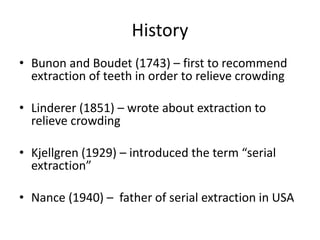
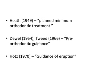
Downloaded 512 times
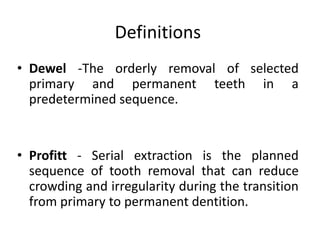
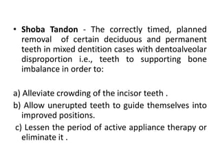
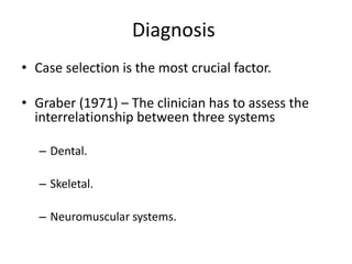
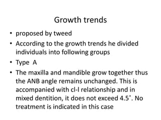
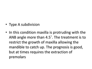
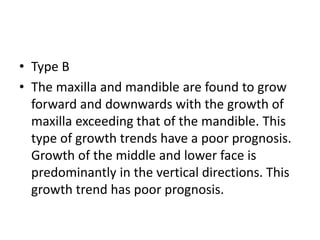
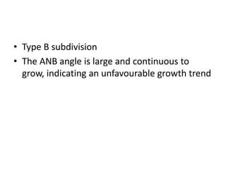

This document provides an overview of serial extraction in orthodontic treatment. It discusses the history, definitions, diagnosis, indications, contraindications, advantages, disadvantages, treatment sequence, appliances used, and conclusions regarding serial extraction. Key points covered include using serial extraction to relieve crowding by removing primary and permanent teeth in a planned sequence, the importance of understanding facial growth and development, and balancing the dental, skeletal, and muscular systems when determining case selection and treatment objectives.


![Hypothalamus short ppt by Dr. Neha [PT].pptx](https://cdn.slidesharecdn.com/ss_thumbnails/hypothalamusbydr-260124145759-b9f94a93-thumbnail.jpg?width=640&height=640&fit=bounds)


