Abstract
Objective(s):
Zinc oxide nanoparticles (ZNP) are increasingly used in sunscreens, biosensors, food additives and pigments. In this study the effects of ZNP on liver of rats was investigated.
Materials and Methods:
Experimental groups received 5, 50 and 300 mg/kg ZNP respectively for 14 days. Control group received only distilled water. ALT, AST and ALP were considered as biomarkers to indicate hepatotoxicity. Lipid peroxidation (MDA), SOD and GPx were detected for assessment of oxidative stress in liver tissue. Histological studies and TUNEL assay were also done.
Results:
Plasma concentration of zinc (Zn) was significantly increased in 5 mg/kg ZNP-treated rats. Liver concentration of Zn was significantly increased in the 300 mg/kg ZNP-treated animals. Weight of liver was markedly increased in both 5 and 300 mg/kg doses of ZNP. ZNP at the doses of 5 mg/kg induced a significant increase in oxidative stress through the increase in MDA content and a significant decrease in SOD and GPx enzymes activity in the liver tissue. Administration of ZNP at 5 mg/kg induced a significant elevation in plasma AST, ALT and ALP. Histological studies showed that treatment with 5 mg/kg of ZNP caused hepatocytes swelling, which was accompanied by congestion of RBC and accumulation of inflammatory cells. Apoptotic index was also significantly increased in this group. ZNP at the dose of 300 mg/kg had poor hepatotoxicity effect.
Conclusion:
It is concluded that lower doses of ZNP has more hepatotoxic effects on rats, and recommended to use it with caution if there is a hepatological problem.
Histopathological effects of nanosilver (Ag-NPs) in liver after dermal exposu...Nanomedicine Journal (NMJ)
Objective(s):
With the advent of nanotechnology, significant progress has been made in the area of nanoscale materials such as nanosilver (Ag-Nps). These nanoparticles have a wide range of applications and been used for antimicrobial purposes for more than a century. However, little
attention has been paid to the toxicity of nanosilver wound dressing. This study was designed to investigate the possible histopathological toxicity of Ag-NPs in liver of mice during wound healing.
Materials and Methods:
A group of 50 female BALB/c mice of about 8 weeks were randomly divided into two groups: Ag-NPs and control groups (n=25). After creating similar wound on the backs of all animals, the wound bed was treated in Ag-NPs group, with a volume of 50 microliters of the nanosilver solution (10ppm) ,and in control group, with the same amount of distilled water. The experiment lasted for 14 days. Histopathaological samplings of liver were conducted on days 2, 7 and 14 of the experiment.
Results:
Histopathological studies demonstrated time-dependent changes in mice liver treated with Ag-NPs compared to control group. Some changes include dilation in central venous, hyperemia, cell swelling, increase of Kupffer and inflammatory cells.
Conclusion:
This study suggests that use of nanosilver for wound healing may cause a mild toxicity, as indicated by time-dependent toxic responses in liver tissue. However, this issue will have to be considered more extensively in further studies
Enterocin 55 produced by non rabbit-derived strain Enterococcus faecium EF55 ...Agriculture Journal IJOEAR
— Ent55 is produced by poultry strain Enterococcus faecium EF55. It is substance which can be allotted to Class II enterocins; thermo-stable, small peptide. Because producer strain has shown beneficial effect in poultry and broiler rabbits as well, we decided to apply Ent55 in broiler rabbit husbandry. Ent55 showed antimicrobial activity in broiler rabbits by reduction of staphylococci, Clostridiae, pseudomonads and coliforms. Its beneficial effect was demonstrated by stimulation of phagocytic activity as well as by reduction of Eimeria spp. oocysts. GPx values were lower; it means, no oxidative stress was evoked. Moreover, it has not negative influence on growth performance and biochemical parameters. Our results indicated that enterocin produced by not-autochtonous strain can also have protective and beneficial effect in broiler rabbits.
Abstract
Objective(s):
Gold nanoparticles (GNPs) command a great deal of attention for biomedical applications nowadays. The data about the degree of toxicity and the accumulation of gold nanoparticles in-vivo is not enough to judge.
Materials and Methods:
A total of 32 healthy male Wistar rats were randomly divided into 4 including: three GNP-treated and one control group. Groups 1, 2 and 3 received 0.5 cc of a solution containing 5, 10, and 100 ppm Au daily via intraperitoneal (IP) injection for 7 days, respectively. The control group was treated with 0.5 cc normal saline with same procedure. Then, several biochemical parameters such as serum glutamate oxaloacetat transaminase (SGOT) and serum glutamate pyrvate transaminase (SGPT) were evaluated at 2, 7 and 14 days after the last injection. After 14 days, all the rats were sacrificed and liver, lung tissues were separated and evaluated.
Results:
SGOT two days after intervention was significantly greater in the group 2 than the control group. In liver histological assessment, in group 1, basophils were observed around the central veins, in group 2 fading and no observation of central veins was seen, and in group 3 hepatic damage was noticed. The lung histological results showed severe vascular hyperemia in group 1, air sacs damage in group 2, and complete air sacs destruction in group 3.
Conclusion:
The results showed extreme changes in the histopathology of lung and liver tissues caused by spherical nanogold with 5-10 nm size in all of three treatment groups.
Subacute dermal toxicity investigation of nanosilver on serum chemical biomar...Nanomedicine Journal (NMJ)
Abstract
Objective(s):
Nanosilver is one of the most widely used nanomaterials due to its strong antimicrobial activity. Thus, because of increasing potential for exposure of human to nanosilver, there is an increasing concern about possible side effects of these nanoparticles. In this study, we tested the potential dermal toxicity of nanosilver bandage on serum chemical biomarkers in mice.
Materials and Methods:
In this study, 20 male BALB/c mice were randomly allocated into the treatment and control groups (n=10). After general anesthesia and shaving the back of all animals in near the vertebral column, in the nanosilver group, a volume of 50μl of 10 μg/ml of nanosilver solution (40 nm), and in the control group the same amount of distilled water was added to the sterile bandage of mice, then the bandages were fixed on the skin surface with cloth glue. After 3 and 7 days, the bandages were opened and serum levels of blood urea
nitrogen (BUN), creatinine (Cr), alanine aminotransferase (ALT) and aspartate aminotransferase (AST) were measured by using standard kits for two groups of mice.
Results:
In treatment group, a significant increase in ALT, AST and BUN levels were observed compared with control group during experiment periods (p<0.05),>0.05).
Conclusion:
The present results indicated that the dermal absorption of 10 μg/ml nanosilver (40 nm) can lead to hepatotoxicity and renal toxicity in mice.
Genotoxicity of Goji Berry (Lyciumbarbarum) In Vivo Mammalian Cellsinventionjournals
Lyciumbarbarum (Gojji berry) belongs to family Salonaceae which is found in China and Himalayan. This herb is used to prevent various diseases and in medical treatments as an alternative medicine being widely used for its antioxidant and revitalizing potential effects. In recent years, Gojji has become increasingly popular in Europe and North America as a "superfruit" and dietary supplement. The belief that herbal products do not bring any risk to health, is part of popular culture. However the term "natural" assigned to many products cannot assure no health risk. The aim of this study was to evaluate the possible genotoxic effects of aqueous extract of Lyciumbarbarum (Gojji berry) by micronucleus test and comet assay. Thirty Rattus norvegicus were divided into three equal groups: 1) experimental group, submitted to Gojji berry (200mg/kg orally); 2) positive control group (cyclophosphamide), and; 3) negative control group (distilled water). Micronucleus Tests were done by smear method of bone marrow cells performed after 48h for acute, and 72h for chronic exposure. The comet assay was performed on peripheral blood taken from the tail of each animal 4h, and 24h after intervention. Cytotoxicity was assessed by observing the DNA damage measuring the percentage of DNA in the tail (% DNA- measurement of the proportion of the total DNA present in the tail) and the tail moment (TM-tail length times the percentage of DNA in the tail), calculated by 100 nucleoids per animal and the presence of micronuclei in 2,000 polychromatic erythrocytes per animal. Analysis of variance (ANOVA) followed by Tukey test at 5% significance was used comparing the results. The data showed no significant difference in the frequency of DNA damage and the number of micronuclei between the experimental group and the negative control group. The results also suggest that the aqueous extract of Lyciumbarbarum (Gojji berry) at the dose of 200 mg/kg showed no genotoxic effect, which could, to a certain point, justifies its use.
Hepatoprotective Effect of Cestrum parqui L. aerial parts and Phytochemical ...Jing Zang
This study deals with the investigation of hepatoprotective effect of 70% methanolic extract from Cestrum parqui aerial parts and determination of the bioactive components of the plant. The hepatoprotective effect of Cestrum parqui methanol extract (100, 500, 1000 mg/kg) was analysed on carbon tetrachloride (CCl4)-induced acute liver injury. The administration of a single dose of 40% CCl4 (1ml/kg b.w.) causes an increase in the activities of serum alanine aminotransferase (ALT) and aspirate aminotransferase (AST) enzymes and so pretreated orally of a dose from Cestrum parqui methanol extract (100, 500, 1000 mg/kg) and silymarin (200 mg/kg) for three consecutive days prior to The administration of a single dose of CCl4 significantly prevented the increase in the activities of these enzymes. Histological analysis showed that Cestrum parqui methanol extract at doses of 500 and 1000 mg/kg and silymarin reduced the incidence of liver lesions including vacuole formation, neutrophil infiltration and necrosis of hepatocytes induced by CCl4. The extract cause a negative result on the antioxidative enzymes, superoxide dismutase (SOD), glutathione peroxidase (GPx), glutathione reductase (GRd) and decreased malondialdehyde (MDA) level in liver, as compared to those in the CCl4-treated group and this suggests that the hepatoprotective activity of the extract is due to the antioxidant effect of the extract. Phytochemical analysis of the methanol extract from Cestrum parqui aerial parts showed that it contained different phytoconstituents, flavonoids, tannins, saponins, alkaloids, terpenes and carbohydrates.
IOSR Journal of Pharmacy (IOSRPHR), www.iosrphr.org, call for paper, research...iosrphr_editor
IOSR Journal of Pharmacy (IOSRPHR), www.iosrphr.org, call for paper, research paper publishing, where to publish research paper, journal publishing, how to publish research paper, Call for research paper, international journal, publishing a paper, call for paper 2012, journal of pharmacy, how to get a research paper published, publishing a paper, publishing of journal, research and review articles, Pharmacy journal, International Journal of Pharmacy, hard copy of journal, hard copy of certificates, online Submission, where to publish research paper, journal publishing, international journal, publishing a paper
Objective: To investigate the protective effect of lo- sartan, an angiotensin II type 1 receptor blocker with antioxidative effect on intestinal ischemia-reperfusion (I/R) injury in rats, against inflammation and apoptotic development.
Study Design: Forty male Wistar albino rats with a mean weight of 200–250 g each were divided into 4 groups: (1) Sham operation (laparotomy only, sham surgical preparation including isolation of the superior mesenteric artery [SMA] without occlusion), (2) Ischemia model with SMA closure for 2 hours, (3) I/R group (2 hours of ischemia followed by 3-hour reperfusion (SMA occlusion for 120 minutes followed by 240 minutes reperfusion), and (4) Losartan group (2 hours of ischemia, 40 mg/kg losartan was administered to the animals; losartan was dissolved in 1 mL distilled water and administered intraperitoneally after 2 hours of ischemia). Malondialdehyde (MDA), superoxide dismutase (SOD), catalase (CAT), and glutathione (GSH) levels were examined in jejunum tissue.
Results: Losartan treatment reduced the I/R-induced increase in MDA levels in the gut. Statistically, while SOD, CAT, and GSH activities decreased significantly in the I/R group, they increased in the I/R+Losartan group. Villus loss and increase in inflammation after ischemia persisted after reperfusion. Losartan treatment played a role in the reduction of inflammation and apoptosis and in the regulation of TNF-α and caspase-9 activity.
Conclusion: It has been thought that losartan in I/R may reduce mucosal damage and cell apoptosis in the direction of inflammation and may stabilize caspase-9 activity by inhibiting TNF-α stimulus.
Keywords: caspase-9, ischemia, ischemia/reperfusion, rat, reperfusion injury, TNF-α, tumor necrosis factor-alpha
Protective Effect of β-Carotene Extracted From the Cyanobacterium Oscillatori...iosrphr_editor
The IOSR Journal of Pharmacy (IOSRPHR) is an open access online & offline peer reviewed international journal, which publishes innovative research papers, reviews, mini-reviews, short communications and notes dealing with Pharmaceutical Sciences( Pharmaceutical Technology, Pharmaceutics, Biopharmaceutics, Pharmacokinetics, Pharmaceutical/Medicinal Chemistry, Computational Chemistry and Molecular Drug Design, Pharmacognosy & Phytochemistry, Pharmacology, Pharmaceutical Analysis, Pharmacy Practice, Clinical and Hospital Pharmacy, Cell Biology, Genomics and Proteomics, Pharmacogenomics, Bioinformatics and Biotechnology of Pharmaceutical Interest........more details on Aim & Scope).
Objective: To identify interstitial cells of Cajal (ICC) in the common bile duct of Kunming mice.
Study Design: Common bile ducts obtained from the Kunming mice were prepared for immunohistochemical investigations using the c-kit antibody. Immunoelectron microscopy was used to detect the expression of c-kit in the ICC of the common bile duct. Transmission electron microscopy showed ultrastructure of ICC in the murine bile duct. Reverse transcription–polymerase chain reaction (RT-PCR) and western blot were used to confirm the expression of mRNA specific for the c-kit gene and production of c-kit protein in the Kunming mice common bile duct.
Results: Immunohistochemistry revealed that ICC in the murine common bile duct are c-kit positive and the ICC are located in the tela submucosa and the tunica muscularis of the murine common bile duct and do not connect with each other. Immunoelectron microscopy confirmed the expression of Kit by ICC in the murine common bile duct. Transmission electron microscopy showed that ICC in the murine common bile duct have long processes, abundant mitochondria, plenty of smooth endoplasmic reticulum (sER), a lot of lysosomes, and dense bodies. The caveolae of ICC are distinctive. At the same time, RT-PCR indicated that the Kunming mice common bile duct expressed mRNA specific for the c-kit gene, and western blot analysis showed the evidence of production of c-kit protein in the Kunming mice common bile duct.
Conclusion: ICC are found in the Kunming mice common bile duct, which is likely to lead to the development of motility study of the common bile duct.
Keywords: common bile duct; electron microscopy; immuno-electron microscopy; interstitial cells of Cajal; intestines; smooth muscle; tyrosine kinase receptor (c-kit)
Acute toxicity and anti-ulcerogenic activity of an aqueous extract from the s...Jing Zang
Terminalia superba is a plant used in traditional medicine to treat many illnesses particularly gastro-intestinal disorders. This study was aimed to evaluate the acute toxicity and gastric anti-ulcer activity of an aqueous extract of Terminalia superba (AETs). The LD50 was determined by the graphic method of Miller and Tainter (1944) and the calculation method of Dragsted and Lang (1957) in mice. The preventive anti-ulcerogenic action of the extract was assessed using four models of gastric ulcer induction namely HCl/Ethanol solution, indomethacin solution, pylorus ligation and cold restraint stress in rats. The LD50 obtained by the oral administration of AETs was 12.2 ± 0.21 g/kg b.w. and 12.33 ± 0.87 g/kg b.w. by the graphic method and the calculation method respectively. The administration of AETs intraperitoneally gave 1.97 ± 0.29 g/kg b.w. (graphic method) and 1.93 ± 0.21 g/kg b.w. (calculation method) as LD50s. The preventive gastric anti-ulcer study revealed that for doses ranging from 125 to 500 mg/kg body weight, EATs significantly (P<0.05)>< 0.05) increased mucus production and reduced gastric acid secretion. Phytochemical screening of the aqueous extract of the stem bark of Terminalia superba showed the presence of polyphenols, tannins, flavonoids, quinones, coumarines, saponins, reduced sugar, sterols and polyterpenes. These results suggested that the preventive anti-ulcer activity of AETs may be due to a cytoprotective effect. The LD50s found indicated that the extract was not toxic and that the phytochemical coumpounds present in EATs could be responsible for its effects. In conclusion, the preventive gastric anti-ulcer and the non toxic effects of the aqueous extract of Terminalia superba could justify the use of this plant in traditional medicine to treat abdominal disorder and pains.
Effect of Antioxidant status on liver following Atrazine exposure and its pro...IOSR Journals
The efficacy of Andrographis paniculata (AP) extract was studied on atrazine induced hepatic damage in rats. Ethanolic extract of AP (150mg/kg body weight) was found to protect the male wistar rats from hepato toxic action of atrazine as evidence by significant reduction in the level of lipid peroxidation and increased the antioxidant defense system activity in the atrazine intoxicated rats. However, AP treatment ameliorated the effects of atrazine suggesting it as potential antioxidant against atrazine induced oxidative stress.
Renal Toxicity Studies of Albino Rats Exposed to Common Emulsion Paint SolutionPremier Publishers
Emulsion paint solutions (EPS) are constantly used to decorate and protect different materials in the environment. The occupational exposure of professional painters and paint factory workers to EPS may predispose them to systemic toxicity. This work investigated the effects of EPS exposure on renal function indices in albino rats to assess potential renal toxicity. Twenty male albino rats weighing between 120-140g were collected and divided into four groups. Two groups were exposed to periodic intraperitoneal administration of 2.5mg/kg body weight (bw) of the EPS for 14days while the other two groups received the same dose of distilled water and served as control. The rats were sacrificed after 7and14days respectively, and their blood collected and analyzed for heavy metals, urea, creatinine and electrolytes, using standard methods. The rat kidneys were also excised, weighed and analyzed for total protein and histopathology. Results showed that bw, kidney weight, serum creatinine, urea and electrolytes decreased significantly(p<0.05) in the exposed groups compared to control. However, urea and Na+ increased significantly(p<0.05) after the initial 7days exposure. Kidney total protein increased by 20.83% and Pb, Ni, and Cd were detected in the exposed rats. Results of kidney histopathology showed intact renal tubules and glomeruli. These results suggest that exposure of albino rats to EPS caused significant alterations in the steady-state levels of renal function biomarkers. Therefore, prolonged exposure may exert deleterious effects on renal function.
Objective: To investigate the changes in the retina due to deltamethrin toxicity and the process in cell inflammation and apoptosis.
Study Design: Sixteen Wistar albino rats were randomly divided into two groups as control (n=8) and deltamethrin (n=8) groups. Saline was given to the control group, and 0.5 mL of 5 mg/kg deltamethrin was given to the deltamethrin group for 14 days each. Blood was collected for biochemical analysis. Retinal tissue was processed for histological examination.
Results: Compared to the control group, MDA levels were high while GSH and CAT levels were low in the deltamethrin group. Histopathological analysis showed spaces between the pigment epithelium, irregularity in the delimiting membrane, degenerated ganglion, cone and bacillus cell, pyknotic nuclei, thinned inner limitation membrane, and thickened vascular wall. The control group showed FAS expression in the pigment layer limiting membranes, in the nuclei of many cone and bacillus cells, and ganglion cells in the control group sections. In the deltamethrin group, FAS expression was observed in the inner and outer limiting membranes of the pigment epithelium, cone and bacillus cells, and ganglion cell nuclei. In the control group, negative NOS expression in the pigment epithelium and outer limiting membranes, internal limitation membrane, and ganglion cells in the cone and bacillus cell nuclei were observed. In the deltamethrin group, NOS expression was positive in the pigment epithelium, cone and bacillus, and ganglion cell nuclei.
Conclusion: We suggest that deltamethrin toxicity induced apoptotic process due to increased inflammation in the retina and may cause visual impairment as a result of neural damage.
Keywords: deltamethrin, FAS, insecticides, NOS, nitric oxide synthase, retina
Oral toxic exposure of titanium dioxide nanoparticles on serum biochemical ch...Nanomedicine Journal (NMJ)
Abstract
Objective(s):
Titanium dioxide (TiO2) nanoparticles (NPs) are widely used in commercial food additives and cosmetics worldwide. Uptake of these nanoparticulate into humans by different routes and may exhibit potential side effects, lags behind the rapid development of nanotechnology. Thus, the present study designed to evaluate the toxic effect of mixed rutile and anatase TiO2 NPs on serum biochemical changes in rats.
Materials and Methods:
In this study, adult male Wistar rats were randomly allotted into the experimental and control groups (n=6), which were orally administered with 50 and 100 mg/kg body weight of TiO2 NPs. Toxic effects were assessed by the changes of serum biochemical parameters such as glucose, total protein, albumin, globulin, cholesterol, triglyceride, high density lipoprotein, alanine transaminase, aspartate transaminase, alkaline phosphatase, total bilirubin, blood urea nitrogen, uric acid and creatinine. All the serum biochemical markers were experimented in rats, after 14-days of post exposure.
Results:
Changes of the serum specific parameters indicated that liver and kidney were significantly affected in both experimental groups. The changes between the levels of total protein, glucose, aspartate transaminase, alanine transaminase and alkaline phosphatase indicate that TiO2 NPs induces liver damage. Significant increase in the blood urea nitrogen and uric acid indicates the renal damage in the TiO2 NPs treated rats.
Conclusion:
The data shows that the oral administration of TiO2 NPs (<100nm) may lead to hepatic and renal toxicity in experimental rats.
Objective(s):
There is a rising use of gold nanoparticles (AuNPs) in goods and in the medical fields but there is concern about the toxicity of them. So in this study spherical AuNPs with 3 different concentrations were applied for investigating their effects in vivo.
Materials and Methods:
40 male albino mice were randomly divided into sham, control, 25 ppm, 50 ppm, 100 ppm groups and were treated by intraperitoneal injection for period of 14 days. Blood was taken for measuring of glutamate oxaloacetate transaminase and glutamate pyruvate transaminase (SGOT and SGPT) enzyme levels and Complete Blood Count (CBC).
Results:
After the treatment and comparing groups with sham group, in 50 ppm group significant increases on RBC, HCT, HGB, MCHC and in 25 ppm group significant increase on MCHC and significant decrease on MCV and in 100 ppm group significant increase on MCHC were observed. Also in 50 ppm group an increase on SGOT enzyme level was observed. However, it was nonsignificant.
Conclusion:
By observing the abnormality on the RBC count and SGOT enzyme level in the 50 ppm group, we concluded a slight toxicity effect for AuNPs and the threat potential of their use in human.
The Protective Role Of High Dietary Protein On Arsenic Induced Hepatotoxicity...IOSR Journals
The objective of the present investigation was to study the protective role of High dietary protein on arsenic induced hepatotoxicity model in adult male albino rats. Hepatotoxicity in rats was caused by arsenic tri oxide at a dose of 3mg- /ml/kg body weight. Hepamerz, a drug used as standard hepatoprotective agent, was administered orally as standard hepatoprotective agent for 14 consecutive days prior to arsenic treatment at a dose of 10mg- /ml/kg body weight. This drug has many side effects. These side effects have prompted the scientific world for the search of alternative natural remedies of liver damage. The High dietary protein was administered orally to rats along with arsenic. The biochemical parameters were investigated. The results indicated that biochemical changes produced by arsenic were restored to almost normal by High protein diet. The High protein diet produced hepatoprotective effect through the modulation of antioxidant - mediated mechanism by altering serum glutamate oxaloacetate transaminase (SGOT), serum glutamate pyruvate transaminase (SGPT), alkaline phosphatase (ALP), superoxide dismutase (SOD) and catalase (CAT) activities and reduced glutathione (GSH) and lipid peroxidation (LPO) levels - against arsenic induced hepatotoxicity model in rats.
Objective(s):
This study aimed to address the gold nanoparticle(GNP)-dose and exposure duration effect on the kidney function of rats: in vivo.
Materials and Methods:
A total of 32 healthy male Wistar rats were used in this study. Animals were randomly divided into groups, three GNP-treated groups and control group. Group 1, 2 and 3 received. /5 cc of solution containing 5, 10,100 ppm Au via IP injection for 7 successive days, respectively. The control group was treated with 0.5% normal saline. Several biochemical parameters such as BUN (blood urea nitrogen), creatine and uric acid were evaluated at various time points (7 and 14 days). After 14 days, the tissue of kidney was collected and investigated.
Results:
There was no significant difference between the control and the intervention group regarding the amount of creatine-BUN and uric acid. The amount of creatine-BUN and uric acid showed increase in all the groups [except group1 (creatine) and group 2 (uric acid)] in the 7 and 14 days after intervention compared to the control group, but this difference was not significant. Results of histopatological tissue kidney showed: in group 1 and 3, complete destruction of the proximal tubules and distal cortical, in group 2, almost complete destruction of proximal tubules and distal.
Conclusions:
The induced histological alterations might be an indication of injured renal tubules due to GNPs toxicity that become unable to deal with the accumulated residues resulting from metabolic and structural disturbances caused by these NPs
Objective(s):
Nanotechnology and nanoparticles are increasingly recognized for their potential applications in aerospace engineering, nanoelectronics, and environmental remediation, medicine and consumer products. More importantly is the potential for the application of silver nanoparticles (Ag NPs) in the treatment of diseases that require maintenance of circulating drug concentration or targeting of specific cells or organs the aim of this study was to investigate the possible protective role of Ag NP antioxidative biomarkers in rats. Ag NPs are used to investigate the potential risks for the environment and health.
Materials and Methods:
Rats received Ag NP, 5, 50, 250 and 500 mg/kg/day IP. After two week of treatment, the activity of enzymatic scavengers such as glutathione peroxidase (GPx), superoxide dismutase (SOD) and total antioxidant capacity (TAC) of blood samples were measured.
Results:
Ag NP in 5, 50, 250 and 500 mg/kg reduced activities of CAT, SOD and increased TAC in plasma.
Conclusion:
In this study, Ag NP with 500mg/kg induced activities of CAT, SOD and decreased TAC. It is concluded that antioxidative properties of Ag NP is dose dependent.
Hepatoprotective Activity of Cinnamon Zeylanicum Leaves against Alcohol Induc...IJERA Editor
Plants play an important role in the life of human, as the major source of food, as well as for the maintenance and improvement of health and for the elimination of the enemies since ages. Plants are the basic source of knowledge of modern medicine. The present study was conducted to evaluate the hepatoprotective activity of aqueos extract of aerial parts of Cinnamon zeylanicum are evaluated in alcohol induced hepatotoxicity in albino rats. Silymarin (100mg/kg) was given as reference standard. The aqueos extract of aerial parts of Cinnamon zeylanicum have shown very significant hepatoprotection against alcohol induced hepatotoxicity in albino rats in reducing SGOT, SGPT, Alkaline phosphatase (ALP) and GGT and levels of total bilirubin and total protein were investigated and showed an increase in alcohol induced rats when compared to control. The extracts of the test plant exhibited significant (p < 0.05) hepatoprotective activity against the alcohol induced liver models by improving liver function which was indicated by reduction in the levels of SGOT, SGPT, ALP, GGT, total bilirubin and total protein.
Protective effects of commelina benghalensis linn (root) extract on ethanol i...IJSIT Editor
The present study was undertaken to investigate the protective effect and possible mechanism of
alcoholic (AlE) and aqueous extract (AqE) from Commelina benghalensis root (CB) on EtOH-induced hepatic
injury in Wistar rat. Hepatotoxic parameters studied in vivo include serum transaminases (AST, and ALT),
ALP, bilirubin, protein, lipid profile (Cholesterol, triglyceride, VLDL and HDL) and level of antioxidants
together with histopathological examination. Liv 52® was used as a reference hepatoprotective agent
(5ml/kg-1b.w.). AlE and AqE (200 mg/kg-1b.w.) on oral administration decreased the level of AST, ALP, ALT,
bilirubin, cholesterol, triglyceride, VLDL, MDA and increased the level of protein, HDL and antioxidants (SOD,
GSH and CAT) in rats being treated with ethanol (EtOH). Pentobarbitone -induced sleeping time study was
carried out to verify the effect on microsomal enzymes Histopathological observations confirmed the
beneficial roles of MF against EtOH-induced liver injury in rats. Possible mechanism may involve their
antioxidant activity
Protective Effects of Alpha Lipoic Acid (α -LA) Against Lead Neuro-Toxicity i...inventionjournals
Aim of the work: The present study was conducted to elucidate the possible protective effect of alpha lipoic acid (α-LA) against the deleterious effect perturbation induced in rat brain exposed to lead acetate. Methods: 32 Wistar male rats (weighing 130 ± 10 g) were divided into four groups (n=8): (1) normal control group (C); (2) Initiation group (Pb as lead acetate 20 mg/kg.b.wt, i.p. for 2 wks); (3) treatment group (α-LA 20 mg/kg.b.wt, i.p. for 3 wks); (4) post-initiation treatment group (Pb for 2 wks then followed by α-LA for 3 wks). Levels of monoamines (norepinephrine NE and dopamine DA), the level of Ache activity and finally adenosine triphosphate (ATP), were estimated in the hippocampus and cerebral cortex, in addition, a Morris water maze and the histological study were performed after completion of the experiments. Results: The results of the present work demonstrated that Pb inhibited neurotransmitters releases and decrease the level of Ache activity, as well as it inhibited energy production ATP. Pb impaired performance on Morris Water Maze of rats and histological degeneration. However, treatment with α-LA significantly attenuated the behavioral impairment and biochemical parameters in rat treated with Pb. And amelioration of histological changes. Conclusion: As a conclusion, treatment with α-LA can improve the Pb-induced toxicity via antioxidant activity.
Protective Effects of Alpha Lipoic Acid (Α-LA) Against Lead Neuro-Toxicity in...inventionjournals
Aim of the work: The present study was conducted to elucidate the possible protective effect of alpha lipoic acid (α-LA) against the deleterious effect perturbation induced in rat brain exposed to lead acetate. Methods: 32 Wistar male rats (weighing 130 ± 10 g) were divided into four groups (n=8): (1) normal control group (C); (2) Initiation group (Pb as lead acetate 20 mg/kg.b.wt, i.p. for 2 wks); (3) treatment group (α-LA 20 mg/kg.b.wt, i.p. for 3 wks); (4) post-initiation treatment group (Pb for 2 wks then followed by α-LA for 3 wks). Levels of monoamines (norepinephrine NE and dopamine DA), the level of Ache activity and finally adenosine triphosphate (ATP), were estimated in the hippocampus and cerebral cortex, in addition, a Morris water maze and the histological study were performed after completion of the experiments. Results: The results of the present work demonstrated that Pb inhibited neurotransmitters releases and decrease the level of Ache activity, as well as it inhibited energy production ATP. Pb impaired performance on Morris Water Maze of rats and histological degeneration. However, treatment with α-LA significantly attenuated the behavioral impairment and biochemical parameters in rat treated with Pb. And amelioration of histological changes. Conclusion: As a conclusion, treatment with α-LA can improve the Pb-induced toxicity via antioxidant activity.
Objective: Ischemia-reperfusion (I/R) leads to reactive oxygen species formation and cell death in kidney tissue with injury and organ transplantation. Simvastatin (SIM) is an antioxidant, anti-inflammatory, and anticoagulant agent. Alterations in I/R-induced acute kidney injury model with SIM treatment were analyzed.
Study Design: Wistar rats (n=28) were grouped into Sham, Ischemia, I/R, and I/R+SIM treated. Left rat kidney renal vessels were clamped for 60 minutes for ischemia, and the I/R group had 6 hours of reperfusion. 10 mg/kg SIM was given orally for 28 days. MDA, GSH, and MPO were analyzed. Kidney tissues were paraffin embedded, and primary antibodies TNF-α and caspase-3 were applied for immunohistochemistry.
Results: In the I/R group, intense inflammatory cell infiltration around the vessels and necrosis in the glomerular structures were observed. In the treated group, proximal and distal tubular cells were found to be close to normal. Immunoexpression of caspase-3 in the ischemia group was positive in degenerative glomeruli. In the treated group, TNF-α expression was negative in the glomerular structures. MDA and MPO levels were significantly increased in ischemia and I/R.
Conclusion: We suggest that SIM treatment improved kidney tissue structure and function in a model of I/R injury.
Keywords: caspase-3; immunohistochemistry; ischemia/reperfusion; kidney; MPO; simvastatin
Hepatoprotective Activity of Methanolic Extract of Whole Plant of Pulicaria W...IOSRJPBS
Natural remedies from medicinal plants are considered to be effective and safe alternative treatment for liver injury. The present study was conducted to evaluate the hepatoprotective activity of methanolic extract of whole plant of Pulicaria wightiana in wistar rats. The studies were conducted using the two popular inducing agents Paracetamol (2 g/kg, p.o.) in 1% NaCMC and Carbon tetrachloride (1 ml/kg). Silymarin (100 mg/kg, p.o.) was used as reference drug in the respective models. The effect was estimated by measuring the enzymatic levels and histo- pathological studies. The methanolic extract of whole plant of Pulicaria wightiana has shown very significant hepatoprotection against both Paracetamol and CCl4 - induced hepatotoxicity study models in wistar rats. This was evidenced by marked reduction in marker enzymes in serum. Histopathological studies also confirmed the hepatoprotective nature of the extract
Evaluation of the effect of crocetin on antitumor activity of doxorubicin enc...Nanomedicine Journal (NMJ)
Objective(s): The current study reports investigation of codelivery by PLGA nanoparticles (NPs) loaded with crocetin (Cro), a natural carotenoid dicarboxylicHYPERLINK “http://en.wikipedia.org/wiki/Carboxylic_acid” acid that is found in the crocus flower, and Doxorubicin (DOX).
Materials and Methods: Double emulsion/solvent evaporation method was used for preparation of PLGA nanoparticles containing Dox and Cro. Characterizations of prepared NPs were investigated by atomic force microscopy (AFM) and dynamic light scattering analysis. In vitro Cytotoxicity of DOX and Cro loaded PLGA NPs (PLGA-DOX-Cro) on MCF-7 cell line was evaluated using MTT test. Flow cytometry experiments were implemented to distinguish cells undergoing apoptosis from those undergoing necrosis. Furthermore the expression of caspase 3 was examined by western blot analysis.
Results: The prepared formulations had size of 150- 300 nm. Furthermore, PLGA-DOX-Cro nanoparticles inhibited MCF-7 tumor cells growth more efficiently than either DOX or Cro alone at the same concentrations, as quantified by MTT assay and flow cytometry. Studies on cellular uptake of DOX-Cro-NPs demonstrated that NPs were effectively taken up by MCF-7 tumor cells.
Conclusion: This study suggested that DOX-Cro-NPs may have promising applications in breast cancer therapy.
Effects of combination of magnesium and zinc oxide nanoparticles and heat on ...Nanomedicine Journal (NMJ)
Objective: The objective of this study was to investigate the antibacterial activities of combination of MgO and ZnO nanoparticles in the presence of heat against Escherichia coli and Staphylococcus aureus.
Materials and Methods:Bacteria were grown on either agar or broth media followed by the addition of ZnO and MgO nanoparticles. Then the combined effect of ZnO and MgO nanoparticles was investigated. Furthermore, the media containing nanoparticles were treated with mild heat and their synergistic antibacterial activity was investigated against E. coli and S. aureus in milk.
Results: The data showed that the nanoparticles used in this study had no effect on the bacteria in the agar medium. However, the results showed that ZnO and MgO nanoparticles resulted in a significant decrease in the number of E. coli (P<0.000) and S. aureus (Pd”0.05) in the broth medium. The combination of nanoparticles and mild heat exhibited a significant decrease in the number of E. coli and S. aureus indicating the synergistic effects of nanoparticles and heat.
Conclusion: Using a combination of mild heat, ZnO and MgO nanoparticles, E. coli and S. aureus can be controlled successfully in the milk. Mild heating plus ZnO and MgO nanoparticles has a synergistic effect which would reduce the need for high temperature and also the concentrations of ZnO and MgO nanoparticles required for pathogen control in minimally processed milk during maintaining.
Preparation and evaluation of electrospun nanofibers containing pectin and ti...Nanomedicine Journal (NMJ)
Objective(s):The aim of this study was to prepare electrospun nanofibers of celecoxib using combination of time-dependent polymers with pectin to achieve a colon-specific drug delivery system for celecoxib.
Materials and Methods:Formulations were produced based on two multilevel 22 full factorial designs. The independent variables were the ratio of drug:time-dependent polymer (X1) and the amount of pectin in formulations (X2). Electrospinning process was used for preparation of nanofibers. The spinning solutions were loaded in 5 mL syringes. The feeding rate was fixed by a syringe pump at 2.0 mL/h and a high voltage supply at range 10-18 kV was applied for electrospinning. Electrospun nanofibers were collected and evaluated by scanning electron microscopy and drug release in the acid and buffer with pH 6.8 with and without pectinase.
Results:Electrospun nanofibers of celecoxib with appropriate morphological properties were produced via electrospinning process. Drug release from electrospun nanofibers was very low in the acidic media; while, drug release in the simulated colonic media was the highest from formulations containing pectin.
Conclusion: Formulation F2 (containing drug:ERS with the ratio of 1:2 and 10% pectin) exhibited acceptable morphological characteristics and protection of drug in the upper GI tract and could be a good candidate as a colonic drug delivery system for celecoxib.
The combined effects of Aloe vera gel and silver nanoparticles on wound heali...Nanomedicine Journal (NMJ)
Objective(s): This study was aimed at investigating the synergy effects of Aloe vera gel and silver nanoparticles on the healing rate of the cutting wounds.
Materials and Methods: In order to determine the concentration of silver nanoparticles in Aloe vera gel, the MBC methods were applied on the most common bacteria infecting wounds, Staphylococcus aureus, Escherichia coli, Pseudomonas aeruginosa. The cutting wounds with Full-thickness skin were dorsally created on rats; then the rats were divided into 4 groups. The treatments groups included: mixture of Aloe vera gel and silver nanoparticles, Aloe vera gel alone and silver nanoparticles alone in addition to control groups. The treatment was carried out for 2 weeks and the size of the wound closures were measured by an image software analysis.
Results:There was no significant difference (p<0.05) in healing rate between the control and mixture group. However, there were significant differences between the silver nanoparticles and Aloe vera groups using Tukey’s analysis on the 6th, 8th and 10th days.
Conclusion:The Aloe vera gel increased the rate of wound healing whereas the silver nanoparticles had a delay effect; and when they were mixed, it was similar to the average effect of both Aloe vera gel and silver nanoparticles.
Simultaneous loading of 5-florouracil and SPIONs in HSA nanoparticles: Optimi...Nanomedicine Journal (NMJ)
Objective(s): Over the past two decades, considerable interest has been focused on utilizing biocompatible magnetic nanoparticles (MNPs) for biomedical applications. In this study, production of human serum albumin (HSA) nanoparticles using desolvation technique that were simultaneous loaded with high amounts of superparamagnetic iron oxide nanoparticles (SPIONs) and 5-flourouracil (5-FU) was investigated.
Materials and Methods: 5-FU loading (%) and SPIONs entrapment efficiency (%) were optimized using response surface methodology (RSM). The design expert software used to analyse the interactive effects of pH, 5-FU and SPIONs concentrations.
Results:The optimum conditions found to be pH of 8.2, drug concentration of 1.5 mg/ml and SPIONs concentration of 2.79 mg/ml. Under the mentioned optimum conditions, particles with the size of 111.8 nm, zeta potential of -37.1 mV, 5-FU loading of 15.8% and SPIONs entrapment efficiency of 41.1% were obtained. In vitro cumulative release of 5-FU from the nanoparticles was evaluated in phosphate buffer saline (pH 7.4, 37 °C). Results indicated that 85% of the 5-FU released during 95 h, which revealed a sustained release profile. In addition, Vibrating Sample Magnetometer (VSM) analyses confirmed the superparamagnetic properties of magnetic albumin nanoparticles manufactured under the optimum conditions.
Conclusion: According to the findings,SPIONs and 5-FU loaded HAS nanoparticles arepromising for use as novel targeted delivery system due to proper magnetic and drug release behaviours.
Antimicrobial and cytotoxicity effect of silver nanoparticle synthesized by C...Nanomedicine Journal (NMJ)
Objective(s): For the development of reliable, ecofriendly, less expensive process for the synthesis of silver nanoparticles and to evaluate the bactericidal, and cytotoxicity properties of silver nanoparticles synthesized from root extract of Croton bonplandianum, Baill.
Materials and Methods: The synthesis of silver nanoparticles by plant part of Croton bonplandianum was carried out. The formation of nanoparticles was confirmed by Transmission Electron Microscopy (TEM), Scanning Electron Microscopy (SEM), XRD and UV-Vis spectrophotometric analysis. The biochemical properties were assayed by antibacterial study, cytotoxicity assay using cancer cell line.
Results: The formation of silver nanoparticles was confirmed by UV-VIS spectroscopic analysis which showed absorbance peak at 425 nm. X-ray diffraction photograph indicated the face centered cubic structure of the synthesized AgNPs. TEM has displayed the different dimensional images of biogenic silver nanoparticles with particle size distribution ranging from 15-40 nm with an average size of 32 nm. Silver particles are spherical in shape, clustered. The EDX analysis was used to identify the elemental composition of synthesized AgNPs. Antibacterial activity of the synthesized AgNPs against three Gram positive and Gram negative bacteria strains like Staphylococcus aureus, Escherichia coli and Pseudomonas aeruginosa carried out showed significant zones of inhibition. The cytotoxicity study by AgNPS also showed cytotoxicity on ovarian cancer cell line PA-1 and lung epithelial cancer cell line A549.
Conclusion: The present study confirms that the AgNPs have great promise as antibacterial, and anticancer agent.
Investigation of the effect of different parameters on the phase inversion te...Nanomedicine Journal (NMJ)
Objective(s): Nanoemulsions are a kind of emulsions that can be transparent, translucent (size range 50-200 nm) or “milky” (up to 500 nm). Nanoemulsions are adequatly effective for transfer of active component through skin which facilitate the entrance of the active component . The transparent nature of the system and lack of the thickener and fluidity are among advantages of nanoemulsion.
Materials and Methods: In this study, a nanoemulsion of lemon oil in water was prepared by the phase inversion temperature (PIT) emulsification method in which the tween 40 was used as surfactant. The effect of concentration of NaCl in aqueous phase, pH and weight percent of surfactant and aqueous on the PIT and droplet size were investigated. Results: The results showed that with increasing of concentration of NaCl from 0.05 M to 1 M, PIT decrease from 72 to 50. The average droplet sizes, for 0.1, 0.5 and 1 M of NaCl in 25 ºC are 497.3, 308.1 and 189.9 nm, respectively and the polydispersity indexes are 0.348, 0.334 and 0.307, respectively.
Conclusion: Considering the characteristics of nanoemulsions such as being transparent, endurance of solution and droplet size can provide suitable reaction environment for polymerization process used in making hygienic and medical materials.
Mechanism of oxidative stress involved in the toxicity of ZnO nanoparticles a...Nanomedicine Journal (NMJ)
ZnO NPs (zinc oxide nanoparticles) has generated significant scientific interest as a novel antibacterial and anticancer agent. Since oxidative stress is a critical determinant of ZnO NPs-induced damage, it is necessary to characterize their underlying mode of action. Different structural and physicochemical properties of ZnO NPs such as particle surface, size, shape, crystal structure, chemical position, and presence of metals can lead to changes in biological activities including ROS (reactive oxygen species) production. However, there are some inconsistencies in the literature on the relation between the physicochemical features of ZnO NPs and their plausible oxidative stress mechanism. Herein, the possible oxidative stress mechanism of ZnO NPs was reviewed. This is worthy of further detailed evaluations in order to improve our understanding of vital NPs characteristics governing their toxicity. Therefore, this study focuses on the different reported oxidative stress paradigms induced by ZnO NPs including ROS generated by NPs, oxidative stress due to the NPs-cell interaction, and role of the particle dissolution in the oxidative damage. Also, this study tries to characterize and understand the multiple pathways involved in oxidative stress induced by ZnO NPs. Knowledge about different cellular signaling cascades stimulated by ZnO NPs lead to the better interpretation of the toxic influences induced by the cellular and acellular parameters. Regarding the potential benefits of toxic effects of ZnO NPs, in-depth evaluation of their toxicity mechanism and various effects of these nanoparticles would facilitate their implementation for biomedical applications.
Combined effects of PEGylation and particle size on uptake of PLGA particles ...Nanomedicine Journal (NMJ)
Abstract
Objective:
At the present study, relationship between phagocytosis of PLGA particles and combined effects of particle size and surface PEGylation was investigated.
Materials and Methods:
Microspheres and nanospheres (3500 nm and 700 nm) were prepared from three types of PLGA polymers (non-PEGylated and PEGylation percents of 9% and 15%). These particles were prepared by solvent evaporation method. All particles were labeled with FITC-Albumin. Interaction of particles with J744.A.1 mouse macrophage cells, was evaluated in the absence or presence of 7% of the serum by flowcytometry method.
Results:
The study revealed more phagocytosis of nanospheres. In the presence of the serum, PEGylated particles were phagocytosed less than non-PEGylated particles. For nanospheres, this difference was significant (P<0/05) and their uptake was affected by PEGylation degree. In the case of microsphere formulation, PEGylation did not affect the cell uptake. In the serum-free medium, the bigger particles had more cell uptake rate than smaller ones but the cell uptake rate was not influenced by PEGylation.
Conclusion:
The results indicated that in nanosized particles both size and PEgylation degree could affect the phagocytosis, but in micron sized particles just size, and not the PEGylation degree, could affect this.
Synthesis of silver nanoparticles and its synergistic effects in combination ...Nanomedicine Journal (NMJ)
Abstract
Objectives:
Biofilms are communities of bacteria attached to surfaces through an external polymeric substances matrix. In the meantime, Acinetobacterbaumannii is the predominant species related to nosocomial infections. In the present study, the effect of silver nanoparticles alone and in combination with biocides and imipenem against planktonic and biofilms of A. baumannii was assessed.
Materials and Methods:
Minimum inhibitory concentrations (MICs) of 75 planktonic isolates of A. baumannii were determined by using the microdilution method as described via clinical and laboratory standards institute (CLSI). Among all strains, 10 isolates which formed strong biofilms were selected and exposed to silver nanoparticles alone and in combination with imipenem, bismuth ethandithiol (BisEDT) and bismuth propanedithiol (BisPDT) to determine minimum biofilm inhibitory concentrations (MBIC). Subsequently, minimum biofilm eradication concentrations (MBECs) of silver nanoparticles alone and in combination with imipenem against mature biofilm of the isolates were evaluated.
Results:
Results showed that 29.3% of isolates were susceptible to silver nanoparticles and could inhibit the growth and eradicate biofilms produced by the isolates. For this reason, ∑FIC, ∑FBIC and ∑FBEC ≤ 0.05 were reported which shows synergism between silver nanoparticles and imipenem against not only planktonic cells but also inhibition and eradication of biofilms. The results of ∑FBIC >2 indicated to antagonistic impacts between silver nanoparticles and BisEDT/BisPDT against biofilms.
Conclusion:
It can be concluded that silver nanoparticles alone can inhibit biofilm formation but in combination with imipenem are more effective against A. baumannii in planktonic and biofilm forms.
Synthesis of graphene oxide-TiO2 nanocomposite as an adsorbent for the enrich...Nanomedicine Journal (NMJ)
Abstract
Objective(s):
In our study, graphene oxide-TiO2 nanocomposite (GO/TiO2) was prepared and used for the enrichment of rutin from real samples for the first time.
Materials and Methods:
The synthesized GO/TiO2 was characterized by X-ray diffraction, scanning electron microscopy, and FT-IR spectra. The enrichment process is fast and highly efficient. The factors including contact time, pH, and amount of GO/TiO2 affecting the adsorption process were studied.
Results:
The maximum adsorption capacity for ciprofloxacin was calculated to be 59.5 mg/g according to the Langmuir adsorption isotherm. The method yielded a linear calibration curve in the concentration ranges from 15 to 200 μg/L for the rutin with regression coefficients (r2) of 0.9990. The limits of detection (LODs, S/N=3) and limits of quantification (LOQs, S/N=10) were found to be 8 μg/Land 28 μg/L, respectively. Both the intra-day and inter-day precisions (RSDs) were < 10% .
Conclusion:
The developed approach offered wide linear range, and good reproducibility. Owing to the diverse structures and unique characteristic, GO/TiO2 possesses great potential in the enrichment and analysis of trace rutin in real aqueous samples.
Preparation and evaluation of vitamin A nanosuspension as a novel ocular drug...Nanomedicine Journal (NMJ)
Abstract
Objective(s):
The aim of this study was to prepare a nanosuspension formulation as a new vehicle for the improvement of the ocular delivery of vitamin A.
Material and Methods:
Formulations were designed based on full factorial design. A high pressure homogenization technique was used to produce nanosuspensions. Fifteen formulations were prepared by the use of different combinations of surfactants Tween 80, benzalkonium chloride and Pluronic and evaluated for pH, particle size, entrapment efficiency, differential scanning calorimetry (DSC), stability and drug release. Also, Draize test was used to evaluate the irritation of rabbit eye by formulations.
Results:
All formulations showed a small mean size that is well suited for ocular application. Also it was observed that the particle size decreased with increase in the amount of surfactant. Drug entrapment increased with increasing amount of surfactant. It was shown that initial and final drug release can be controlled by the ratio and the total amount of surfactants, respectively.
Conclusion:
It was concluded that the use of Tween 80 and Pluronic in the formualtions with a proper ratio does not show eye irritation and could be useful to achieve a suitable nanosuspension of vitamin A as a novel ocular delivery system.
A comparative study about toxicity of CdSe quantum dots on reproductive syste...Nanomedicine Journal (NMJ)
Abstract
Objective(s):
Medicinal benefits of quantum dots have been proved in recent years but there is little known about their toxicity especially in vivo toxicity. In order to use quantum dots in medical applications, studies ontheir in vivo toxicity is important.
Materials and Methods:
CdSe:ZnS quantum dots were injected in 10, 20, and 40 mg/kg doses to male mice10 days later, mice were sacrificed and five micron slides were prepared structural and optical properties of quantum dots were evaluated using XRD.
Results:
Histological studies of testis tissue showed high toxic effect of CdSe:ZnS in 40 mg/kg group. Histological studies of epididymis did not show any effect of quantum dots in terms of morphology and tube structure. Mean concentration of LH and testosterone and testis weight showed considerable changes in mice injected with 40 mg/kg dose of CdSe:ZnS compared to control group. However, FSH and body weight did not show any difference with control group.
Conclusion:
Although it has been reported that CdSe is highly protected from the environment by its shell, but this study showed high toxicity for CdSe:ZnS when it is used in vivo which could be suggested that shell could contribute to increased toxicity of quantum dots. Considering lack of any previous study on this subject, our study could potentially be used as an basis for further extensive studies investigating the effects of quantum dots toxicity on development of male sexual system.
Functionalization of carbon nanotubes and its application in nanomedicine: A ...Nanomedicine Journal (NMJ)
Abstract
This review focuses on the latest developments in applications of carbon nanotubes (CNTs) in medicine. A brief history of CNTs and a general introduction to the field are presented.
Then, surface modification of CNTs that makes them ideal for use in medical applications is highlighted. Examples of common applications, including cell penetration, drug delivery, gene delivery and imaging, are given. At the same time, there are concerns about their possible adverse effects on human health, since there is evidence that exposure to CNTs induces toxic effects in experimental models. However, CNTs are not a single substance but a growing family of different materials possibly eliciting different biological responses. As a consequence, the hazards associated with the exposure of humans to the different forms of CNTs may be different. Understanding the structure–toxicity relationships would help towards the assessment of the risk related to these materials. Finally, toxicity of CNTs, are discussed. This review article overviews the most recent applications of CNTs in Nanomedicine, covering the period from 1991 to early 2015.
The role of surface charge of ISCOMATRIX nanoparticles on the type of immune ...Nanomedicine Journal (NMJ)
Abstract
Objective(s):
ISCOMATRIX vaccines have now been shown to induce strong antigen-specific cellular or humoral immune responses to a broad range of antigens of viral, bacterial, parasite or tumor. In the present study, we investigated the role of ISCOMATRIX charge in induction of a Th1 type of immune response and protection against Leishmania major infection in BALB/c mice.
Materials and Methods:
Positively and negatively charged ISCOMATRIX were prepared. BALB/C mice were immunized subcutaneously, three times with 2-week intervals, with different ISCOMATRIX formulations. Soluble Leishmania antigens (SLA) were mixed with ISCOMATRIX right before injection. The extent of protection and type of immune response were studied in different groups of mice.
Results:
The group of mice immunized with negatively charged ISCOMATRIX showed smaller footpad swelling upon challenge with L. major and the highest IgG2a production compared with positively charged one. The mice immunized with positively charged ISCOMATRIX showed the lowest splenic parasite burden compared to the other groups. Cytokine assay results indicated that the highest level of IFN- γ and IL-4 secretion was observed in the splenocytes of mice immunized with negatively charged ISCOMATRIX as compared to other groups.
Conclusion:
The results indicated that ISCOMATRIX formulations generate an immune response with mixed Th1/Th2 response that was not protective against challenge against L. major.
Abstract
In the last decade, developments in nanotechnology have provided a new field in medicine called “Nanomedicine”. Nanomedicine has provided new tools for photodynamic therapy. Quantum dots (QDs) are approximately spherical nanoparticles that have attracted broad attention and have been used in nanomedicine applications. QDs have high molar extinction coefficients and photoluminescence quantum yield, narrow emission spectra, broad absorption, large effective stokes shifts. QDs are more photostable and resistant to metabolic degradation. These photosensitizing properties can be used as photosensitizers for Photodynamic Therapy (PDT). PDT has been recommended for its unique characteristic, such as low side effect and more efficiency. Therefore, nanomedicine leads a promising future for targeted therapy in cancer tumor. Furthermore, QDs have recently been applied in PDT, which will be addressed in this review letter. Also this review letter evaluates key aspects of nano-particulate design and engineering, including the advantage of the nanometer scale size range, biological behavior, and safety profile.
Preparation of protein-loaded PLGA-PVP blend nanoparticles by nanoprecipitati...Nanomedicine Journal (NMJ)
Abstract
Objective(s):
Despite of wide range applications of polymeric nanoparticles in protein delivery, there are some problems for the field of protein entrapment, initial burst and controlled release profile.
Materials and Methods:
In this study, we investigated the influence of some changes in PLGA nanoparticles formulation to improve the initial and controlled release profile. Selected parameters were: pluronic F127, polysorbate 80 as surfactant, pH of inner aqueous phase, L/G ratio of PLGA polymer, volume of inner aqueous phase and addition of polyvinylpyrrolidone as an excipient. FITC-HSA was used as a model hydrophilic drug. The nanoparticles were prepared by nanoprecipitation.
Results:
Initial release of FITC-HSA from PLGA-tween 80 nanoparticles (opt-4, 61%) was faster than control (PLGA-pluronic) after 2.30 h of incubation. Results showed that decrease in pH of inner aqueous phase to pI of protein can decrease IBR but the release profile of protein is the same as control. Release profile with three phases including a) initial burst b) plateau and c) final release phase was observed when we changed volume of inner aqueous phase and L/G ratio in formulation. Co-entrapment of HSA with PVP and pluronic reduced the IBR and controlled release profile in opt-19. Encapsulation efficiency was more than 97% and nanoparticles size and zeta potentials were mono-modal and -18.99 mV, respectively.
Conclusion:
In this research, we optimized a process for preparation of PLGA-PVP-pluronic nanoparticles of diameter less than 300 nm using nanoprecipitation method. This formulation showed a decreased initial burst and long lasting controlled release profile for FITC-HSA as a model drug for proteins.
Abstract
Objective(s):
Abdominal adhesions are one of the most important problems, occurring after intra-abdominal surgery in more than 90% of cases. This condition is the leading cause of bowel obstruction, infertility, and abdominal/pelvic pain. Gold nanoparticles (GNPs) have been shown to be non-toxic and exhibit anti-inflammatory, anti-angiogenic and antioxidant activities. The purpose of this study was to determine the effect of intraperitoneal lavage with GNP solutions on the development of postoperative peritoneal adhesion (PPA).
Materials and Methods:
In the current experimental study, thirty-five male Wistar rats were randomly assigned to seven groups of five rats. After a standardized peritoneal injury, GNP solutions in different concentrations (1, 2.5, 5, 10, 50 and 100 ng/ml) were locally administered through nebulization; normal saline (NS) was administered to the control group. Two weeks later, the rats were sacrificed and cecum and peritoneal samples were harvested for histopathological assessment. Blood samples were obtained to determine serum concentrations of inflammatory biomarkers including tumor necrosis factor alpha (TNF-α), interleukin-1 beta (IL-1β) and vascular endothelial growth factor (VEGF).
Results:
The rats treated with GNPs had significantly lower microscopic and macroscopic peritoneal adhesion scores, compared to the control group (P<0.05). Score 5 of macroscopic adhesions was reported in all the rats of the control group, unlike the GNP groups. Furthermore, microscopic adhesions were reported with all rats in the control group, unlike the GNP groups (reported in 0 out of 5 rats in all GNP groups). In addition, serum levels of IL-1β, TNF-α and VEGF underwent no significant changes.
Conclusion:
Compared to the control group, GNPs decreased the severity of peritoneal adhesions, although they did not alter TNF-α, IL-1β or VEGF serum levels.
Synthetic Fiber Construction in lab .pptxPavel ( NSTU)
Synthetic fiber production is a fascinating and complex field that blends chemistry, engineering, and environmental science. By understanding these aspects, students can gain a comprehensive view of synthetic fiber production, its impact on society and the environment, and the potential for future innovations. Synthetic fibers play a crucial role in modern society, impacting various aspects of daily life, industry, and the environment. ynthetic fibers are integral to modern life, offering a range of benefits from cost-effectiveness and versatility to innovative applications and performance characteristics. While they pose environmental challenges, ongoing research and development aim to create more sustainable and eco-friendly alternatives. Understanding the importance of synthetic fibers helps in appreciating their role in the economy, industry, and daily life, while also emphasizing the need for sustainable practices and innovation.
Welcome to TechSoup New Member Orientation and Q&A (May 2024).pdfTechSoup
In this webinar you will learn how your organization can access TechSoup's wide variety of product discount and donation programs. From hardware to software, we'll give you a tour of the tools available to help your nonprofit with productivity, collaboration, financial management, donor tracking, security, and more.
How to Make a Field invisible in Odoo 17Celine George
It is possible to hide or invisible some fields in odoo. Commonly using “invisible” attribute in the field definition to invisible the fields. This slide will show how to make a field invisible in odoo 17.
Model Attribute Check Company Auto PropertyCeline George
In Odoo, the multi-company feature allows you to manage multiple companies within a single Odoo database instance. Each company can have its own configurations while still sharing common resources such as products, customers, and suppliers.
Operation “Blue Star” is the only event in the history of Independent India where the state went into war with its own people. Even after about 40 years it is not clear if it was culmination of states anger over people of the region, a political game of power or start of dictatorial chapter in the democratic setup.
The people of Punjab felt alienated from main stream due to denial of their just demands during a long democratic struggle since independence. As it happen all over the word, it led to militant struggle with great loss of lives of military, police and civilian personnel. Killing of Indira Gandhi and massacre of innocent Sikhs in Delhi and other India cities was also associated with this movement.
Ethnobotany and Ethnopharmacology:
Ethnobotany in herbal drug evaluation,
Impact of Ethnobotany in traditional medicine,
New development in herbals,
Bio-prospecting tools for drug discovery,
Role of Ethnopharmacology in drug evaluation,
Reverse Pharmacology.
The Roman Empire A Historical Colossus.pdfkaushalkr1407
The Roman Empire, a vast and enduring power, stands as one of history's most remarkable civilizations, leaving an indelible imprint on the world. It emerged from the Roman Republic, transitioning into an imperial powerhouse under the leadership of Augustus Caesar in 27 BCE. This transformation marked the beginning of an era defined by unprecedented territorial expansion, architectural marvels, and profound cultural influence.
The empire's roots lie in the city of Rome, founded, according to legend, by Romulus in 753 BCE. Over centuries, Rome evolved from a small settlement to a formidable republic, characterized by a complex political system with elected officials and checks on power. However, internal strife, class conflicts, and military ambitions paved the way for the end of the Republic. Julius Caesar’s dictatorship and subsequent assassination in 44 BCE created a power vacuum, leading to a civil war. Octavian, later Augustus, emerged victorious, heralding the Roman Empire’s birth.
Under Augustus, the empire experienced the Pax Romana, a 200-year period of relative peace and stability. Augustus reformed the military, established efficient administrative systems, and initiated grand construction projects. The empire's borders expanded, encompassing territories from Britain to Egypt and from Spain to the Euphrates. Roman legions, renowned for their discipline and engineering prowess, secured and maintained these vast territories, building roads, fortifications, and cities that facilitated control and integration.
The Roman Empire’s society was hierarchical, with a rigid class system. At the top were the patricians, wealthy elites who held significant political power. Below them were the plebeians, free citizens with limited political influence, and the vast numbers of slaves who formed the backbone of the economy. The family unit was central, governed by the paterfamilias, the male head who held absolute authority.
Culturally, the Romans were eclectic, absorbing and adapting elements from the civilizations they encountered, particularly the Greeks. Roman art, literature, and philosophy reflected this synthesis, creating a rich cultural tapestry. Latin, the Roman language, became the lingua franca of the Western world, influencing numerous modern languages.
Roman architecture and engineering achievements were monumental. They perfected the arch, vault, and dome, constructing enduring structures like the Colosseum, Pantheon, and aqueducts. These engineering marvels not only showcased Roman ingenuity but also served practical purposes, from public entertainment to water supply.
2024.06.01 Introducing a competency framework for languag learning materials ...Sandy Millin
http://sandymillin.wordpress.com/iateflwebinar2024
Published classroom materials form the basis of syllabuses, drive teacher professional development, and have a potentially huge influence on learners, teachers and education systems. All teachers also create their own materials, whether a few sentences on a blackboard, a highly-structured fully-realised online course, or anything in between. Despite this, the knowledge and skills needed to create effective language learning materials are rarely part of teacher training, and are mostly learnt by trial and error.
Knowledge and skills frameworks, generally called competency frameworks, for ELT teachers, trainers and managers have existed for a few years now. However, until I created one for my MA dissertation, there wasn’t one drawing together what we need to know and do to be able to effectively produce language learning materials.
This webinar will introduce you to my framework, highlighting the key competencies I identified from my research. It will also show how anybody involved in language teaching (any language, not just English!), teacher training, managing schools or developing language learning materials can benefit from using the framework.
Unit 8 - Information and Communication Technology (Paper I).pdfThiyagu K
This slides describes the basic concepts of ICT, basics of Email, Emerging Technology and Digital Initiatives in Education. This presentations aligns with the UGC Paper I syllabus.
Dose-dependent hepatotoxicity effects of Zinc oxide nanoparticles
1. Nanomed. J., 2(4): 273-282, Autumn 2015
273
Dose-dependent hepatotoxicity effects of Zinc oxide nanoparticles
1, 2
E. Mansouri;1, 2*
L. Khorsandi; 1, 2
M. Orazizadeh; 2
Z. Jozi
1
Cell & Molecular Research Center, Faculty of Medicine, Ahvaz Jundishapur University of Medical
Sciences, Ahvaz, Iran
2
Department of Anatomical Sciences, Faculty of Medicine, Ahvaz Jundishapur University of
Medical Sciences, Ahvaz, Iran
ABSTRACT:
Objective(s): Zinc oxide nanoparticles (ZNP) are increasingly used in sunscreens, biosensors, food additives and
pigments. In this study the effects of ZNP on liver of rats was investigated.
Materials and Methods: Experimental groups received 5, 50 and 300 mg/kg ZNP respectively for 14 days. Control
group received only distilled water.ALT, AST and ALP were considered as biomarkers to indicate hepatotoxicity. Lipid
peroxidation (MDA), SOD and GPx were detected for assessment of oxidative stress in liver tissue. Histological studies
and TUNEL assay were also done.
Results: Plasma concentration of zinc (Zn) was significantly increased in 5 mg/kg ZNP-treated rats. Liver concentration
of Zn was significantly increased in the 300 mg/kg ZNP-treated animals. Weight of liver was markedly increased in both
5 and 300 mg/kg doses of ZNP. ZNP at the doses of 5 mg/kg induced a significant increase in oxidative stress through the
increase in MDAcontent and a significant decrease in SOD and GPx enzymes activity in the liver tissue.Administration
of ZNP at 5 mg/kg induced a significant elevation in plasma AST, ALT and ALP. Histological studies showed that
treatment with 5 mg/kg of ZNP caused hepatocytes swelling, which was accompanied by congestion of RBC and
accumulation of inflammatory cells. Apoptotic index was also significantly increased in this group. ZNP at the dose of
300 mg/kg had poor hepatotoxicity effect.
Conclusion: It is concluded that lower doses of ZNP has more hepatotoxic effects on rats, and recommended to use it
with caution if there is a hepatological problem.
Keywords: Apoptosis, Nanomaterials, Oxidative stress
Nanomed. J., 2(4): 273-282, Autumn 2015
DOI: 10.7508/nmj. 2015.04.005
*Corresponding Author Email: layasadat@yahoo.com
Tel: (+98) 611- 3720458
Note. This manuscript was submitted on May 10, 2015;
approved on July 20, 2015
Received; 10 May 2015 Accepted; 20 July 2015
INTRODUCTION
Metal nanoparticles (NPs) and their oxides have a
considerable number of present and future applications
in the medical and industrial fields. The smaller size
and unique properties of the NPs has substantially
improved the application of them (1-3). NPs are
considered to be more highly absorbed into the
respiratory, skin, and gastrointestinal systems than
micron-sized particles because of their unique
physicochemical properties, such as their size and
surface modifications (4). Previous studies revealed
that administration of NPs to rodents resulted in their
accumulation in the various tissues including liver,
brain and spleen (5-6).
One of the most popular industrial applications of NPs
is sunscreens (7). The major component of sunscreens
is zinc oxide nanoparticles (ZNP), which can effectively
absorb ultraviolet light (8). No published data is
available about the daily exposure doses of ZNP in
human. However, ZNP increasing use increases the
health risk of people exposed to these particles, either
occupationally or environmentally.An important factor
to be considered in toxicity tests is the diversity of the
exposure routes, which includes inhalation, dermal
uptake, ingestion and injection (9). Although most of
the exposure to ZNP is via epidermal contact, several
studies have reported that ZNP do not permeate the
skin (10, 11). However, small fractions of lip products
and sun protection products may accidentally ingest.
ZNP may used for its antimicrobial properties in food
packaging. ZNP can also be ingested directly when
2. Nanomed. J., 2(4): 273-282, Autumn 2015
274
E. Mansouri et al.
used in food packaging or drug delivery (12, 13).
Emamifar et al. studied the migration of silver and ZNP
in LDPEnano-composite packaging. The zinc (ZN) ions
showed an increase in their migration when compared
to silver (15). Workers involved in the synthesis of
ZNP can be exposed by unintentional hand-to-mouth
transfer of nanomaterials. When discharged
accidentally into the environment, ZNP may enter the
human body through the food chain (16). In addition,
some of the NPs can be swallowed into and reach the
gastrointestinal tract when they are expelled from the
mucociliary system of the lungs after inhalation (17).
Oberdorseter et al. demonstrated that following whole-
body inhalation of ultrafine carbon particles, the NPs
were translocated and accumulated in liver (18). It has
been reported that ZNP administered orally dissolve in
the stomach, with Zn ions then absorbed to enter into
systemic circulation (19).
De Louise has reported that ZNP are nontoxic to
cultured human dermal fibroblasts (20). Other reports
suggest that ZNP are toxic to dermal fibroblast,
neuroblastoma cells and vascular endothelial cells (21-
23).It has been reported that ZNP can induce oxidative
stress and apoptosis in HepG2 (24). However, published
toxicity data are still considered inadequate to earn a
full understanding of the potential toxicity of ZNP. In
present study toxic effects of ZNP on liver was
investigated.
MATERIALS AND METHODS
Animals
In this experimental study, 32 healthyand adult male
Wistar rats (8-10 weeks old, 180-200 g) were used. The
animals were obtained from Ahvaz Jundishapur
University of Medical Sciences, Experimental Research
Center, and this study was approved by the ethics
committee of Jundishapur University and carried out
in an ethically proper way by following the guidelines
provided. The animals were kept under standard
laboratory conditions (12 h-dark and 12 h- light cycle,
relative humidity of 50 ± 5% and 22 ± 3°C) for at least 1
week before the experiment and those conditions were
preserved until the end of the experiment.Animal cages
were kept clean, and commercial food (pellet) and water
were provided ad libitum.
Experimental design
The rats were randomly divided into 4 groups of 8
animals each. Control group received 0.2 ml normal
saline for 14 days. Experimental groups received 5, 50
and 300 mg/kg ZNP respectively (ZNP-1, 2, 3) for 14
days (25).
After characterization of ZNP, the stock solution (2
mg/ml) was prepared in Milli-Q water and dispersed for
10 min by using a sonicator. The stock solution of ZNP
was kept at 4°C and used within 1 week for the
experiments. Just before use, the stock solution diluted
in Milli-Q water and prepared by ultrasonication (Solid
State/Ultrasonic FS-14; Fisher Scientific) for 15 min to
prevent aggregation. To ensure non-aggregation of
ZNP before administration, the time interval from
preparation to oral gavage was strictly limited in less
than 20 min. In addition, 20 min after the preparation,
the particle size of ZNP was analyzed by atomic force
microscopy (AFM).
Changes in body weight and behavior, and possible
appearance of symptoms in the rats were carefully
recorded every day after administration of the ZNP.
One day after the last administration, after blood
sampling, the rats were sacrificed by cervical
dislocation under ether anesthesia and liver from each
animal were removed quickly and weighed. Small pieces
of liver were stored separately in a deep freezer (-80°C)
for the MDA, SOD and GPx assay, and measuring the
concentration of zinc (Zn) in liver. Other pieces were
fixed in 10% buffered formalin for histological
assessments.
Zn content analysis
Approximately 1 g of the liver tissue was taken and
digested in concentrated nitric acid overnight. Next
day, 5 ml of concentrated nitric acid and perchloric acid
mixture (6:1) was added to each sample and heated at
80-90 °C until the solutions were colourless and clear.
The concentrated solutions were diluted to 5 ml with
1% nitric acid. The Zn content was analyzed by an
atomic absorption spectrophotometer (ZEEnit 700 P,
Analytikjena, Germany). Blood samples were collected
via the tail vein to evaluate the plasma Zn
concentration. The blood samples were centrifuged at
3000 rpm for 15 minutes at 4°C to obtain the plasma
(26).
Biochemical tests
The blood sample were collected in heparinized
centrifuge tube and centrifuged. The plasma enzyme
levels including plasma alaine aminotransferase (ALT),
aspartate aminotransferase (AST) and alkaline
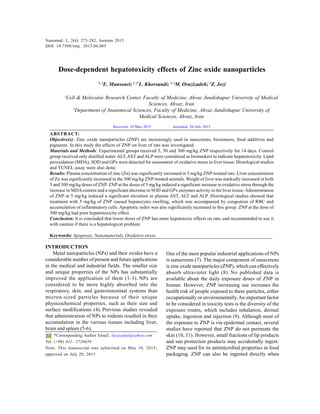
3. Nanomed. J., 2(4): 273-282, Autumn 2015
275
phosphatase (ALP) were determined spectro-
photometrically from plasma samples using
commercially available kits (Sigma).
Estimation of lipid peroxidation
Degree of lipid peroxidation in liver tissue
homogenate of all the experimental animals was
determined in terms of thiobarbituric acid reactive
substances (TBARS) formation as previously described
(27).Avolume of 500 μl of supernatant was mixed with
1.5 ml trichloroacetic acid (10%) and after centrifugation
(4,000× g for 10 min), 1.5 ml of supernatant was added
to 2 ml TBA (0.67%) and heated at 100°C for 30 min.
After cooling, the sample was extracted with 2 ml n-
butanol and after centrifugation at 4,000× g for 15 min,
the organic phase was collected. The absorbance was
read spectrophotometrically at 535 nm. Values were
expressed as nmol per milligram of tissue.
Superoxide dismutase (SOD) activity assay
The assay for SOD activity was made according to
the method of Suttle (28) using Ransod kit (Randox
Labs, Crumlin, UK). This method is based on the
formation of red formazan from the reaction of 2-(4-
iodophenyl)-3-(4-nitrophenol)-5-phenyltetrazolium
chloride and superoxide radical (produced in the
incubation medium from xanthine oxidase reaction),
which is assayed in a spectrophotometer at 505 nm.
The inhibition of the produced chromogen is
proportional to the activity of the SOD present in the
sample. A 50% inhibition is defined as 1 unit of SOD,
and specific activity is expressed as units per milligram
of tissue.
Glutathione peroxidase (GPx) activity assay
GPx activity was determined using the Ransel kit
(Randox Labs.,Crumlin, UK) according to Paglia and
Valentine (29). GPx catalyzes the oxidation of
glutathione by cumene hydroperoxide. In the presence
of glutathione reductase and NADPH, the oxidized
Glutathione (GSSG) was immediately converted to the
reduced form with a concomitant oxidation of NADPH
to NADP+. The decrease in absorbance was monitored
with a spectrophotometer at 340 nm. One GPx unit is
defined as 1 μ mol of NADPH consumed per minute,
and specific activity is reported as units per milligram
of tissue.
Histology changes
The formalin fixed samples were embedded in
paraffin, sectioned (5 μm) and stained with
haematoxylin and eosin (H&E). Six microscopy stained
slides per animal were examined forsigns of histological
features, such as inflammatory-cell infiltration,
hepatocyte vacuolization (fatty deposits) and
congestion of red blood cells (RBC).
TUNEL assay
For detection of apoptosis at a single cell level,
based on labeling of DNA strand breaks, the In Situ
Cell Death Detection Kit, POD (Roche, Germany) was
used. The paraffin sections were dewaxed and
rehydrated by standard methods. Proteases were added
and incubated with 5% of appropriate normal serum
for 30 min at 37°C. The slides were washed in phosphate
buffered saline. The sections were permeabilized (2 min,
on ice) and incubated with TUNELreaction mixture (60
min, 37°C). Anti-fluorescein-AP was added and
incubated (30 min, 37°C). Subsequently, the sections
were washed in PBS and incubated for 20 min with
substrate. They were analyzed by light microscopy. A
cell was considered TUNEL-positive when the nuclear
staining was intense, dark brown and homogenous.
Apoptotic index was calculated by dividing the number
of TUNEL-positive hepatocytes in a randomly focused
field by the total number of hepatocytes in that field,
and the result was multiplied by 100. The apoptotic
indexes of 10 randomly field for each slide were
evaluated and the mean apoptotic index of each case
was calculated (30). Four slides/ animal were used in
this study.
Statistical analysis
The data were analyzed using one-way ANOVA
followed by post hoc LSD test and were presented as
the mean ± SD. p < 0.05 was considered significant.
RESULTS
Nanoparticle characterizations
AFM revealed the size and morphology of the
synthesized particles. The complexes appear spherical
with a mean size that is inferior to 100 nm as can be
seen in Fig. 1.
Organ weight
No animal was found dead or in a moribund state
during the experiment. There were also no treatment
related clinical signs such as lethargy, ataxia, etc.
However, body weight in ZNP-1 group was
significantly lower than control group (p < 0.05). Liver
weights were significantly incresed in ZNP-1 group. In
ZNP-2 group, liver weight was slightly increased and
body weight was similar to control animals. ZNP-3
group showed a significant increase in liver weight (p
< 0.05). Body weight was not significantly changed in
this group. These results are shown in Table 1.
4. Nanomed. J., 2(4): 273-282, Autumn 2015
276
Hepatotoxicity effects of Zinc oxide nanoparticles
Fig. 1. AFM image of NPs showed distinct spherical particles
in size range between 20 and 30 nm
Table 1. Liver and body weight for control and experimental
groups
Values expressed as mean ± SD for 8 rats. *p < 0.05,
**p < 0.01, * indicate comparison to control group
Absorption of ZNP
In ZNP-1 group, concentration of Zn in blood was
significantly increased in compare to control group.
Zn contents of liver tissue was similar to control group
(p>0.05). In ZNP-2 group, concentration of Zn in blood
was similar to control group. Zn contents of liver tissue
was slightly increased in compare to control group
(p>0.05). In ZNP-3 group, concentration of Zn in blood
was slightly higher than control group (p>0.05). Zn
contents of liver tissue was significantly increased in
compare to control or ZNP-1 groups (p<0.01). These
results are shown in Fig. 2.
Group body weight (g) Liver weight (mg)
Control 241 ± 18.29 10.1± 1.2
ZNP-1 218 ± 21.3*
16.2 ± 3.3**
ZNP-2 238 ± 14.3 11.6 ± 0.8
ZNP-3 240 ± 16.7 15.2 ± 2.4**
Fig. 2. Plasma and liver concentrations of Zn in control and
experimental groups. Values are expressed as mean ± SD for
8 rats. *p < 0.05
Biochemical tests
ZNP-1 group showed a significant increase in
plasma levels of ALT, AST and ALP compared to
controlgroup(p<0.001). Plasma levels ofall biochemical
tests were significantly increased in ZNP-2 group (p <
0. 01). In ZNP-3 group, biochemical tests were not
significantly changed. These findings are depicted in
Fig. 3.
MDA level, SOD and GPx activities
ZNP-1 showed a significant increase in the hepatic
level of MDA when compared the control animals. GPx
and SOD activities were significantly decreased in ZNP-
1 group compared to the control (p < 0.001). These
effects were less in ZNP-2 group. There were not
significant changes in MDA, SOD and GPx in ZNP-3
group in compare to the control group. These results
are reported in Fig. 4.
Histological Analysis
Under light microscope, liver lobular structures in
control group was clear and regular, and single layer of
5. Nanomed. J., 2(4): 273-282, Autumn 2015
277
hepatocytes arranged around the central vein in a radial
pattern (Fig. 5-A). In ZNP-1 treated rats normal liver
lobular structures were damaged. The hepatocytes
showed swelling and vacuolization. Severe sinusoidal
congestion, hemorrhage and inflammatory cell
infiltration were also observed (Fig. 5-B). These
histological changes were considerably decreased in
ZNP-2 group (Fig. 5-C). In ZNP-3 group, no
considerable changes in histology of liver were
observed.
Fig. 3. Biochemical tests of control and experimental
groups. Values expressed as mean ± SD for 8 rats. *p < 0.01,
**p < 0.001
Fig. 4. MDA level, SOD and GPx activities of control and
experimental groups. Values are expressed as mean ± SD for
8 rats. *p < 0.01, **p < 0.001
6. Nanomed. J., 2(4): 273-282, Autumn 2015
278
E. Mansouri et al.
Fig. 5. Light microscopy of cross sections of H&E stained liver from control and experimental groups. A, Control groups; B,
ZNP-1 group; C, ZNP-2 group. D, ZNP-1 group. WBC: White blood cells, F: Fat deposit, C: Congestion of RBC.
Magnification: ×250
Fig. 6. Light microscopy of cross sections of TUNEL stained liver from control and experimental groups. A, Control
groups; B, ZNP-1 group; C, ZNP-2 group. D, ZNP-1 group. Magnification: ×250
7. Nanomed. J., 2(4): 273-282, Autumn 2015
279
TUNEL assay
Spontaneous apoptosis was mainly observed in
some hepatocytes of normal liver from the control
group (Fig. 6). In ZNP-1 group, apoptosis was observed
in all lobules and apoptotic index significantly increased
in compare to the control group (p < 0.001). In the ZNP-
2 group, apoptotic index was also significantly higher
than control group (p > 0.001). In ZNP-3 group, no
significant changes in apoptotic index were observed
in compare to control group. The results of TUNEL
assay are shown in Figs 6 and 7.
Fig. 7. Apoptotic index of control and experimental groups.
Values are expressed as mean ± SD for 8 rats. *p < 0.01,
**p < 0.001
DISCUSSION
Our results demonstrated that ZNP had
concentration-dependent toxic effects on liver
inducing stress oxidative, apoptosis induction and
histological changes. We found that lower doses had
more toxicity.
Despite intensive research efforts, reports of cellular
responses to nanomaterials are often inconsistent and
even contradictory. Pasupuleti et al. found that the
incidences of toxicity were higher in lower doses of
nano-size zinc oxide compared to higher dose (31).
However, Talebi et al. have demonstrated that ZNP at
the doses of 50 and 300 mg/kg body weight have toxic
effects on mouse spermatogenesis while no significant
changes in spermatogenesis was reported at the dose
of 5 mg/kg (25). Akhtar et al. showed that ZNP has no
impact on normal rat astrocytes and hepatocytes (32).
On the other hand, Guan et al. have reported that ZNP
has toxic effects on human hepatocyte (L02) and human
embryonic kidney (HEK293) cells (33). Interestingly,
Wang et al. have reported that ZNP induces oxidative
stress and apoptosis in cultured primary astrocytes
(34).
Adverse effects of NPs on human health depend on
individual factors such as genetics and existing disease,
as well as exposure, and NPs chemistry, size, shape,
agglomeration state, and electromagnetic properties
(35).
Song et al. suggested that the morphologies, ion release
rate ofNPs as well as the species-specific vulnerabilities
of cells should all be considered when explaining and
extrapolating toxicity test results among particles and
among species (36).
Administration of carbon nanotube (20-50 nm) within
the trachea causes acute inflammatory effects in the
lungs in mice but not in rats (37, 38). This suggests
that toxic effects of NPs are dependent on the species
used. in this study, ZNP significantly elevated the levels
ofALT,AST and ALP. The levels of these enzymes are
indicative of the functional efficiency of the liver, and
are very sensitive to any disease process of the liver
(39).
Due to damaged liver cells, such enzymes (which are
inside the hepatocytes) are released into the blood.
Therefore, a high amount of these enzymes indicates
destruction of liver cells. In the present study, the
decreased SOD and GPx activity and elevated MDA
levels in liver indicated that the presence of oxidative
stress and lipid peroxidation response were generated
by ZNP exposure. It has been reported that oxidative
stress mediated DNA damage and cytotoxicity induced
by ZNP in HepG2 cells (24). The role of oxidative stress
in the mechanism of NPs induced hepatotoxicity has
also been reported by Sha et al. (40).
Mechanism of the generation of the oxidative stress
after NPs treatment is not clear, but Singh et al. (41)
indicated that it is related to the large particle surface
area. as shown in results, liver weights were
significantly increased in ZNP-1 and ZNP-3groups. It
may have been the result of RBC congestion and WBC
infiltration as well as accumulation of ZNP in liver
tissue. Zn content of liver in ZNP-3 was significantly
higher than control while histological changes were
8. Nanomed. J., 2(4): 273-282, Autumn 2015
280
E. Mansouri et al.
not observed in this group. Thus, the increase in liver
weight of ZNP-3 group relates to accumulation of ZNP
in this organ.
The destruction of lobular structure, vacuolization of
hepatocytes (fat deposits), congestion of RBC and
infiltration of leukocytes indicate necrotic effects of
ZNP at lower doseson liver tissue. On the other hand,
these accumulations of fat, RBCs and leukocytes can
increase the weight of liver. The appearance of
inflammatory cells in hepatic tissue suggests that the
ZNP at lowdose can interact with proteins and enzymes
in the interstitial tissue of the liver, interfering with the
antioxidant defense mechanism and leading to
generation of reactive oxygen species, which in turn
may induce an inflammatory response (42).
Swelling of hepatocytes together with dilatation of the
central vein and blood sinusoids indicate that these
NPs may affect permeability of the cell membrane in
hepatocytes and the endothelial lining of blood
vessels. Hepatocytes fatty deposits might be due to
lipid peroxidation that leads to rough endoplasmic
damage and detachment of the cytoplasmic lipoprotein
which indicate abnormal fat metabolism (43).
The abnormal retention of lipids in hepatocytes might
indicate toxic injury to the liver in the form of
hepatocytes lipolysis by ZNP.
In addition to necrosis, apoptosis were also observed
in liver tissue of ZNP treated animals. Park et al. found
that, out of6 different NPs, ZNP were the most cytotoxic
to A549 cells, as assessed by DNA fragmentation and
apoptosis experiments (44). Both apoptosis and
necrosis has been reported to occur in cells treated
w i t h Z N P . W i l h e l m i e t a l . h a v e r e p o r t e d t h a t ZNP can
induce necrosis and apoptosis in macrophages (45).
Meyer et al. have reported that ZNP can induce
apoptosis in human dermal fibroblasts (21). Sharma et
al. have also demonstrated that ZNP even at low
concentrations possess a genotoxic potential in human
epidermal cells (46).
In addition, severe oxidative stress is associated with
lysosomal membrane permeabilization and subsequent
necrosis, which is controlled by complicated signaling
pathways (47, 48). It has also been reported that
apoptosis and necrosis are not completely independent
process (49),In particular, when a large number of cells
undergoing apoptosis (50). As mentioned above ZNP
at low dose showed more toxicity. The mechanism of
this finding is not clear. It has been demonstrated that
NPs at high concentrations tend to cluster, forming
aggregates often larger than 100 nm. Larger NPs (>100
nm) can be readily phagocytized by macrophages (51).
Results of studies involving inhalation or intra-tracheal
instillation of high concentrations of NPs (silver, iron,
India ink,ortitaniumdioxide)smallerthan100 nm, which
aggregate in larger particles, suggest that most NPs
are indeed stopped by alveolar macrophages (51, 52).
Hence, minute concentrations of NPs with size smaller
than 100 nm can have a higher probability of
translocation to the circulatory system and organs (and
produce damage) than high concentrations of the same
particles, which are likely to formaggregates, and which
will be stopped from translocation by macrophage.As
demonstrated in results, plasma levels of Zn were
significantly higher in lower dose of ZNP-treated rats.
Overall, our results highlight the need for caution
during the use of ZNP to prevent health impacts. We
demonstrated that ZNP exhibited toxicity at lower
concentrations. Thus future nanotoxicology research
needs to be focused on importance of dose metrics
rather following the conventional methods while
conducting in vivo experiments.
ACKNOWLEDGMENTS
This paper is part of M. Sc thesis for Zahra Jozi and
was supported by a Grant (CMRC-108) from the
research council of the Ahvaz Jundishapur University
of Medical Sciences in 2013.
REFERENCES
1. Adams LK, Lyon DY, Alvarez PJJ. Comparative eco-toxicity
of nanoscale TiO2, SiO2, and ZnO water suspensions. Water
Res. 2006; 40 (19): 3527-3532.
2. Mody VV, Siwale R, Singh A, Mody HR. Introduction to
metallic nanoparticles. J Pharm Bioallied Sci. 2010; 2(4):
282-289.
3. Warheit DB. Nanoparticles: Health impacts? Materials Today
2004; 7(2): 32-35.
4. Fubini B, Ghiazza M, Fenoglio I. Physico-chemical features
of engineered nanoparticles relevant to their toxicity.
Nanotoxicol. 2010; 4: 347-363.
5. Borm PJ, Kreyling W. Toxicological hazards of inhaled
nanoparticles potential implications for drug delivery. J
Nanosci Nanotechnol. 2004; 4(5): 521-531.
6. Chen Y, Xue Z, Zheng D, Xia K, Zhao Y, Liu T, et al. Sodium
chloride modified silica nanoparticles as a non-viral vector
with a high efficiency of DNA transfer into cells. Curr
Gene Ther. 2003; 3(3): 273-279.
7. Burnett ME, Wang SQ. Current sunscreen controversies: a
critical review. Photodermatol Photoimmunol Photomed.
2011; 27(2): 58-67.
9. Nanomed. J., 2(4): 273-282, Autumn 2015
281
8. Nohynek GJ, Lademann J, Ribaud C, Roberts MS. Grey goo
on the skin? Nanotechnology, cosmetic and sunscreen safety.
Crit Rev Toxicol. 2007; 37(3): 251-277.
9. Oberdörster G, Maynard A, Donaldson K, Castranova V,
Fitzpatrick J, Ausman K, et al. Principles for characterizing
the potential human health effects from exposure to
nanomaterials: elements of a screening strategy. Part Fibre
Toxicol. 2005; 2(8): 1-35.
10. Zvyagin AV, Zhao X, Gierden A, Sanchez W, Ross JA, Roberts
MS. Imaging of zinc oxide nanoparticle penetration in human
skin in vitro and in vivo. J Biomed Opt. 2008; 13(6): 064031.
11. Filipe P, Silva JN, Silva R, Cirne de Castro JL, Marques
Gomes M, Alves LC, et al. Stratum corneum is an effective
barrier to TiO2
and ZnO nanoparticle percutaneous
absorption. Skin Pharmacol Physiol. 2010; 22(5): 266-275.
12. John S, Marpu S, Li J, Omary M, Hu Z, Fujita Y, et al. Hybrid
zinc oxide nanoparticles for biophotonics. J Nanosci
Nanotechnol. 2010; 10(3):1707-12.
13. Tankhiwale R, Bajpai SK. Preparation, characterization and
antibacterial applications of ZnO-nanoparticles coated
polyethylene films for food packaging. Colloids Surf. B
Biointerfaces. 2012; 90: 16-20.
14. Emamifar A, Kadivar M, Shahedi M, Soleimanian-Zad S.
Effect of nanocomposite packaging containing Ag and ZnO
on inactivation of Lactobacillus plantarum in orange juice.
Food Control. 2011; 22(3-4): 408-413.
15. Ates M, Arslan Z, Demir V, Daniels J, Farah IO. Accumulation
and toxicity of CuO and ZnO nanoparticles through
waterborne and dietary exposure of goldfish (Carassius
auratus). Environ Toxicol. 2015;30(1):119-28
16. Hernandez-Viezcas JA, Castillo-Michel H, Andrews JC, Cotte
M, Rico C, Peralta-Videa JR, et al. In Situ synchrotron X-ray
fluorescence mapping and speciation of CeO2
and ZnO
nanoparticles in soil cultivated Soybean (Glycine max). ACS
Nano. 2013; 7(2): 1415-23.
17. Oberdorster G, Oberdorster E, OberdorsterJ.
Nanotoxicology: an emerging discipline evolving from studies
of ultrafine particles. Environ Health Perspect. 2005; 113(7):
823-39.
18. Oberdörster G, Sharp Z, Atudorei V, Elder A, Gelein R, Lunts
A, et al. Extrapulmonary translocation of ultrafine carbon
particles following whole-body inhalation exposure of rats. J
Toxicol Environ Health A. 2002; 65(20): 1531-43.
19. Cho WS, Kang BC, Lee JK, Jeong J, Che JH, Seok SH.
Comparative absorption, distribution, and excretion of
titanium dioxide and zinc oxide nanoparticles after repeated
oral administration. Part Fibre Toxicol. 2013; 10:9
20. De Louise LA. Applications of nanotechnology in
dermatology. J Invest Dermatol. 2012; 132 (3): 964-975.
21. Meyer K, Rajanahalli P, Ahamed M, Rowe JJ, Hong Y. ZnO
nanoparticles induce apoptosis in human dermal fibroblasts
via p53 and p38 pathways. Toxicol in Vitro. 2011; 25(8):
1721-6.
22. Jeng HA, Swanson J. Toxicity of metal oxide nanoparticles
in mammalian cells. J Environ Sci Health A Tox Hazard
Subst Environ Eng. 2006; 41(12): 2699-2711.
23. Gojova A, Guo B, Kota RS, Rutledge JC, Kennedy IM, Barakat
AI. Induction of inflammation in vascular endothelial cells
by metal oxide nanoparticles: Effect of particle composition.
Environ Health Perspect. 2007; 115(3): 403-409.
24. Sharma V, Anderson D, Dhawan A. Zinc oxide nanoparticles
induce oxidative stress and genotoxicity in human liver cells
(HepG2). J Biomed Nanotechnol. 2011; 7(1): 98-99.
25. Talebi AR, Khorsandi L, Moridian M. The effect of zinc
oxide nanoparticles on mouse spermatogenesis. J Assist
Reprod Genet. 2013; 30(9):1203-9.
26. Sharma V, Singh P, Pandey AK, Dhawan A. Induction of
oxidative stress, DNA damage and apoptosis in mouse liver
after sub-acute oral exposure to zinc oxide nanoparticles.
Mutat Res. 2012; 45(1-2): 84-91.
27. Mansouri E, Panahi M, Ghaffari MA, Ghorbani A. Effects
of grape seed proanthocyanidin extract on oxidative stress
induced by diabetes in rat kidney. Iran Biomed J. 2010;
15(3):100-6.
28. Suttle NF. Copper deficiency in ruminants recent
developments. Vet Rec. 1998; 119(21): 519-522.
29. Paglia DE, Valentine WN. Studies on the quantitative and
qualitative characterization of erythrocyte glutathione
peroxidase. J Lab Clin Med. 1967; 70(1): 158-69.
30. Orazizadeh M, Hashemitabar M, Khorsandi L. Protective
effect of minocycline on dexamethasone induced testicular
germ cell apoptosis in mice. Eur Rev Med Pharmacol Sci.
2009; 13(1):1-5.
31. Pasupuleti S, Alapati S, Ganapathy S, Anumolu G, Pully NR,
Prakhya BM. Toxicity of zinc oxide nanoparticles through
oral route. Toxicol Ind Health. 2012; 28(8): 675-86.
32. Akhtar MJ, Ahamed M, Kumar S, Khan MM, Ahmad J,
Alrokayan SA. Zinc oxide nanoparticles selectively induce
apoptosis in human cancer cells through reactive oxygen
species. Int J Nanomedicine. 2012; 7: 845-857.
33. Guan R, Kang T, Lu F, Zhang Z, Shen H, Liu M. Cytotoxicity,
oxidative stress, and genotoxicity in human hepatocyte and
embryonic kidneycells exposed to ZnO nanoparticles.
Nanoscale Res Lett. 2012; 7(1): 602.
34. Wang J, Deng X, Zhang F, Chen D, Ding W. ZnO
nanoparticle-induced oxidative stress triggers apoptosis by
activating JNK signaling pathway in cultured primary
astrocytes. Nanoscale Res Lett. 2014; 9(1): 117.
35. Buzea C, Pacheco II, Robbie K. Nanomaterials and
nanoparticles: Sources and toxicity. Biointerphases. 2007;
2(4): 17-71.
36. Song L, Connolly M, Fernández-Cruz ML, Vijver MG,
Fernández M, Conde E, et al. Species-specific toxicity of
copper nanoparticles among mammalian and piscine cell
lines. Nanotoxicology. 2014; 8(4): 383-93.
37. Shvedova AA, Kisin ER, Mercer R, Murray AR, Johnson VJ,
Potapovich AI, et al. Unusual inflammatory and fibrogenic
pulmonary responses to single-walled carbon nanotubes in
mice. Am J Physiol Lung Cell Mol Physiol. 2005; 289(5):
698-708.
38. Warheit DB, Laurence BR, Reed KL, Roach DH, Reynolds
GA, Webb TR. Comparative pulmonary toxicity assessment
of single-wall carbon nanotubes in rats. Toxicol Sci. 2004;
77(1): 117-125.
39. Aragon G, Younossi ZM. When and how to evaluate mildly
elevated liver enzymes in apparently healthy patients. Cleve
Clin J Med. 2010; 77(3): 195-204.
40. Sha B, Gao W, Wang S, Gou X, Li W, Liang X, et al. Oxidative
stress increased hepatotoxicity induced by nano-titanium
dioxide in BRL-3A cells and Sprague-Dawley rats. J Appl
Toxicol. 2014; 34(4): 345-356.
10. Nanomed. J., 2(4): 273-282, Autumn 2015
282
Hepatotoxicity effects of Zinc oxide nanoparticles
How to cite this article:
Mansouri E, Khorsandi L, Orazizadeh M, Jozi Z . Dose-dependent hepatotoxicity effects of Zinc oxide nanoparticles. Nanomed. J., 2015; 2(4):
273-282.
41. Singh S, Shi T, Duffin R, Albrecht C, van Berlo D, Höhr D,
et al. Endocytosis, oxidative stress and IL-8 expression in
human lung epithelial cells upon treatment with fine and
ultrafine TiO2: role of the specific surface area and of surface
methylation of the particles. Toxicol Appl Pharmacol. 2007;
222(2): 141-151.
42. Johar D, Roth JC, Bay GH, Walker JN, Kroczak TJ, Los M.
Inflammatory response, reactive oxygen species,
programmed (necrotic-like and apoptotic) cell death and
cancer. Rocz Akad Med Bialymst. 2004; 49: 31-39.
43. Ma L, Zhao J, Wang J, Liu J, Duan Y, Liu H, et al. The acute
liver injury in mice caused by nano-anatase TiO2
. Nanoscale
Res Lett. 4(11): 1275-1285.
44. Park S, Lee YK, Jung M, Kim KH, Chung N, Ahn EK, et al.
Cellular toxicity of various inhalable metal nanoparticles on
human alveolar epithelial cells. Inhal Toxicol. 2007; 1: 59-
65.
45. Wilhelmi V, Fischer U, Weighardt H, Schulze-Osthoff K,
Nickel C, Stahlmecke B, et al. Zinc oxide nanoparticles induce
necrosis and apoptosis in macrophages in a p47phox- and
Nrf2-independent manner. PLoS One. 2013; 8(6): e65704.
46. Sharma V, Shukla RK, Saxena N, Parmar D, Das M, Dhawan
A. DNA damaging potential of zinc oxide nanoparticles in
human epidermal cells. Toxicol Lett. 2009; 185(1-2): 211-
8.
47. Tang D, Kang R, Zeh HJ, Lotze MT. High-mobility group
box 1, oxidative stress, and disease. Antioxid Redox Signal.
2011; 14(7): 1315-1335.
48. Vandenabeele P, Galluzzi L, Vanden Berghe T, Kroemer G.
Molecular mechanisms of necroptosis: an ordered cellular
explosion. Nat Rev Mol Cell Biol. 2010; 11(10): 700-714.
49. Lemasters JJ. Necrapoptosis and the mitochondrial
permeability transition: Shared pathways to necrosis and
apoptosis. Am J Physiol. 1999; 276(1): 1-6.
50. Levin S, Bucci TJ, Cohen SM, Fix AS, Hardisty JF, LeGrand
EK, et al. The nomenclature of cell death: Recommendations
of an ad hoc Committee of the Society of Toxicologic
Pathologists. Toxicol Pathol. 1999; 27(4): 484-490.
51. Takenaka S, Karg E, Roth C, Schulz H, Ziesenis A, Heinzmann
U, et al. Pulmonary and systemic distribution of inhaled
ultrafine silver particles in rats. Environ Health Persp. 2001;
109(4): 547-55.
52. Oberdörster G. Pulmonary deposition, clearance and effects
of inhaled soluble and insoluble cadmium compounds. IARC
Sci Publ. 1992; 118:189-204.