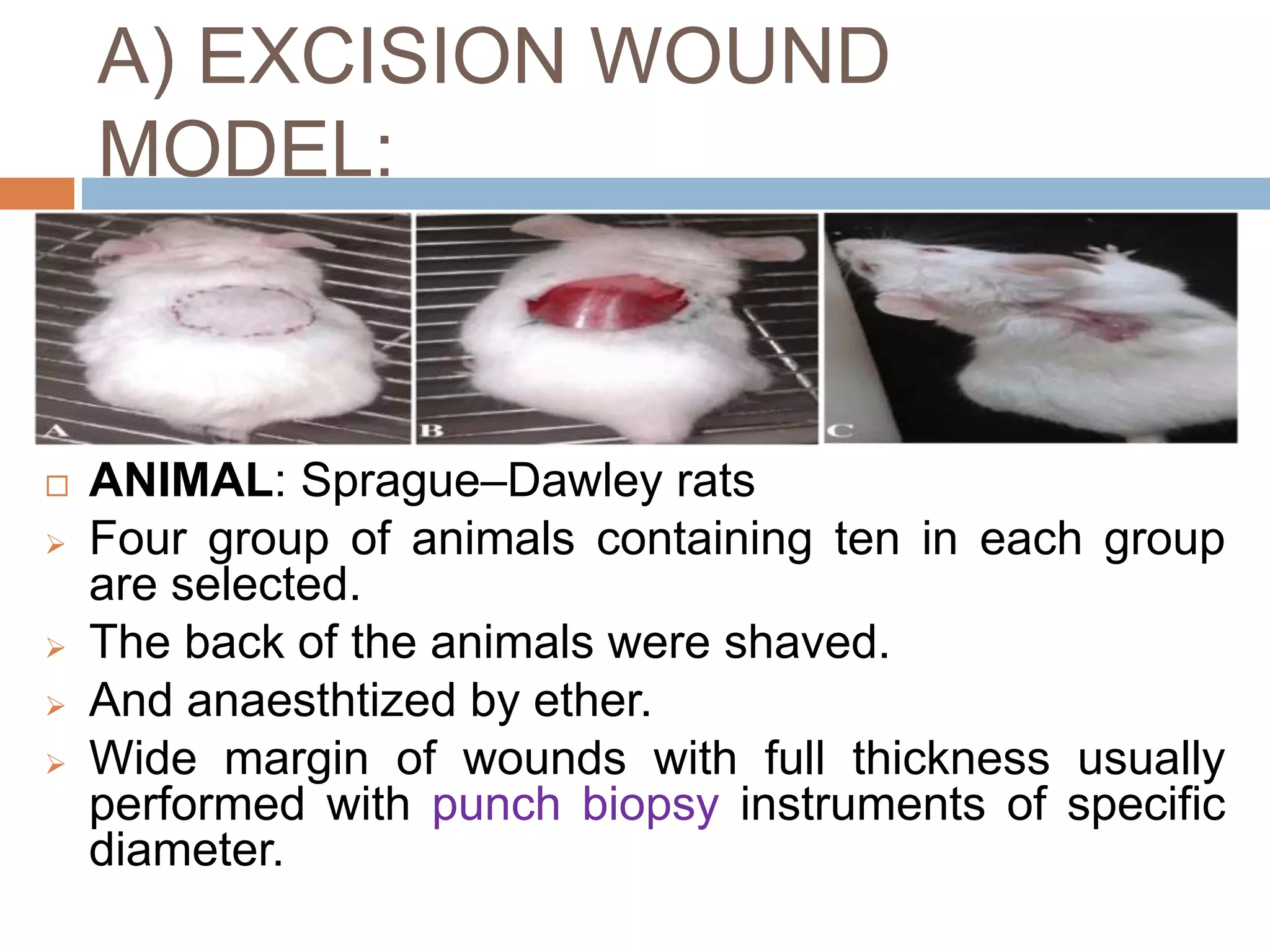
IN-VIVO EVALUATION TECHNIQUES FOR WOUND
HEALING ACTIVITY & HEPATOPROTECTIVE
ACTIVITY
 Adarsh Patil
1stM.Pharm(Pharmacognos
y),
National College of
Pharmacy,
Shivamogga.
 S M Prasanna
Dept. Of Pharmaconosy,
National College of
Pharmacy,
Shivamogga.
Submitted by : Submitted to :
Wound healing activity :
INTRODUCTION:
 Wound healing is a process in which
the skin repairs itself after injury.
 In undamaged skin,
the epidermis (surface layer)
and dermis (deeper layer) form a
protective barrier against the external
environment.
 When the barrier is broken, a
regulated sequence of biochemical
events is set into motion to repair the
damage.
 Wound healing process is
divided into 4 phases,
1)bloodclotting (hemostasis)
2)inflammation
3)tissue growth (proliferation)
4)tissue remodeling (maturation).
 Initially there is an inflammatory
response and the cells below
the dermis begin to increase
collagen production. Finally the
gap is filled up with the
regeneration of epithelial tissue.
IN-VIVO EVALUATION OF WOUND
HEALING PROPERTY
Selection of animals for wound-healing
study:
 The most important relevant animal model
which can be considered for wound-healing
research is the rat. Because there are many
similarities between the physiological nature
of the skin of rats and human beings.
Animal wound models :
 Wounds are made over the dorsal area of the
animal (rat).
 The region needs to be cleaned well with 70%
ethanol.
 Permissible anaesthetic agents such as
ketamine in a scheduled dose are injected
before the artificial production of a wound.
 The extracts obtained from plants are usually
made into different formulations, either as
ointments or lotion and applied to skin wound.
 It is used internally or even injected if required
depending on the nature of the constituents.
A) EXCISION WOUND
MODEL:
 ANIMAL: Sprague–Dawley rats
 Four group of animals containing ten in each group
are selected.
 The back of the animals were shaved.
 And anaesthtized by ether.
 Wide margin of wounds with full thickness usually
performed with punch biopsy instruments of specific
diameter.
 One excision wound is inflicted by cutting away
500mm2 skin of a predetermined area in circular
manner.
 Rats are left undressed to open environment and no
local or systemic antimicrobial agents were used.
 The drugs i.e, the reference standard(0.2% W/W
nitrofurazone ointment); sample ointment BP(conrol);
and test drug oinments or diffeernt forms are
administered till the wound is completely healed.
 This model is used to monitor wound contraction and
epithelisation time.
 Epithelisation time is noted as the number of days
after wounding required to fade off the scar leaving no
raw wound behind.
 Wound contraction is calculated as percentage
reduction in wound area.
 The progressive change in wound area are monitored
planimetrically by tracing wound margin on graph
paper every alternative day.
 For Histopathological examination, tissue are
collected from the completely healed wound when
scar is removed.
 A transverse section of tissues is prepared from each
groups of rat and stained with haematotoxilin and
eosin to reveal the tissue section clearly.
 Tissues are observed under microscope to study
different histopathological phenomenon.
B) INCISION WOUND MODEL:
 ANIMAL: Sprague–Dawley rats.
 Four group of animals containing
ten in each group are selected.
 Two linear-paravertebral long
incisions of 6 cm long were
created with a sterile surgical
blade at the distance of 1.5 cm
from the midline of each side of
the vertebral column.
 The parted skin is kept together
and stiched with black silk by 0.5
cm apart.
 The continuous threads on both wound edges
were tightened for good closure of the wound.
 After stitching, wound was left undressed then
ointment base, standard ointment and extracts
ointment were applied daily until 10 days.
 when wounds were cured thoroughly the sutures
were removed on the day 10.
 Tensile strength of cured wound skin was
measured using tensiometer.
 Tensile strenght indicates how much the repaired
tissue resists to breaking under tension and may
indicate in part the quality of repaired tissue.
C) BURN HEALING WOUND
MODEL:
 Animals :Male Wistar rats (200-250 g) of 2-3 months were used.
 Burn wound creation: Burn wounds were created on dorsal part
of shaved rats using a metal rod heated to 80-85°C and exposed
for 20s.
 After 24 Hours ,dead tissues were excised using sterile surgical
blade.
 Control rats were dressed with cold cream alone, while
experimental rats were dressed with the 10% (w/w) ointment
formulated.
 Experimental design: The animals were divided
randomly into 4 groups of nine each.
 Group-I was treated with standard drug. Groups-
II and –III are taken as positive and negetive
control,Group-IV was treated with test extract
(200 mg/kg/day).
 Burn healing: During the burn wound healing period
and at the certain period of intervals, the burn wound
area was traced manually and photographed.
 The burn wound area was calculated using Auto CAD
RL 14 (Autodesk Company) software.
 At days 6th,9th and 16th the experiment was
terminated and the wound area was removed from the
surviving animals for histological examination.
 Analysis of data: The relative burn wound area was
statistically analyzed as mean + S.D(standard
deviation) and statistically significance between treated
and control groups were analyzed by means of
Student’s t-test.
IN-VIVO EVALUATION OF
HEPATOPROTECTIVE AGENT:
INTRODUCTION:
 The various toxic dose or repeated doses of a
known hepatotoxin (such as carbon tetrachloride,
paracetamol, thioacetamide, alcohol, D-
galactosamine. Etc) are administrated, to induce
liver damage in experimental animals.
 The test substance is administered along with,
prior to and /or after the toxin treatment.
 If the hepatoxicity is prevented or reduced by the
pre-treatment then it is confirmed that the test
substance is effective.
 Liver damage and recovery from damage are
assessed by measuring the serum marker
enzymes, bilirubin, histopathological changes in
the liver, biochemical changes in liver and bile
flow.
 When the liver is damaged, the liver enzymes
such as glutamate pyruvate transaminase (GPT),
glutamate oxaloacetate transaminase (GOT), and
alkaline phosphatase enter into the circulation. An
increase in the levels of these enzymes in the
serum is an indication of liver damage.
A)CCl 4 induced hepatotoxicity:
 MECHANISM:
 Carbon tetrachloride is metabolized by cytochrome P-450
in endoplasmic reticulum and mitochondria with the
formation of CCl 3 O - , a reactive oxidative free radical,
which initiates lipid peroxidation.
 Administration of a single dose of CCl 4 to a rat produces,
within 24 hrs, a necrosis and fatty changes. The poison
reaches its maximum concentration in the liver within 3
hrs of administration. Thereafter, the level falls and by 24
hrs there is no CCl 4 left in the liver. The development of
necrosis is associated with leakage of hepatic enzymes
into serum
Procedure: Wistar Albino rats (100–150 g) are used.
 - Group I : Normal group.
 - Group II: CCl4 treated
 - Group III: CCl4 + standard drug ( Silymarin )
 - Group IV: CCl4 + extract.
 CCl4 is administerd orally daily for 5 days to induce
hepatotoxicity.
 From 6th day till 12th day (total 7 days) animals
received treatment of herbal formulations
suspended in vehicle at dose of 400 mg/kg orally.
The standard group received Standard drug
6mg/kg orally while the control group received
vehicle (1% carboxy methyl cellulose).
 On the 13th day blood was collected from each
animal for serum analysis. The rats were
sacrificed, liver removed and observed for weight,
volume and appearance and then fixed in 10%
formalin for histopathological studies of the liver to
determine the degree of hepatic damage
 A single dose or few repeated doses of D-galactosamine
cause’s acute hepatic necrosis in rats. Prolonged
administered administration leads to cirrhosis.
 Procedure:
 Animals; Albino rats
 Weight- 110-180gm is injected i.p three times weekly with
500mg/kg D-galactosamine over periods of one to 3
months. The test substances are administered orally with
food. The control group receives only vehicle or food
without drugs.
 The rats are sacrificed at various time intervals and liver
taken out and evaluated by light microscopy and
immunohistology using antibodies against macrophages,
lymphocytes and extracellular matrix component.
B)Galactosamine induced
hepatotoxicity:
C) Paracetamol induced
hepatotoxicity:
 Paracetamol, a widely used analgesic and antipyretic drug,
produces acute liver damage in high doses.
 Dose of Paracetamol: 1 gm/kg
 Procedure;
 Rats are fasted for 16h, and then divided into several groups of 8
each.
 Groups 1 –control, (receiving normal saline only (10ml/kg i.p)
 Group 2-Paracetamol
 Group3-reference (Silymarin) and
 Group4- test groups (at varying concentration depending upon on
the design of the experiment) are administered (different groups)
2hr before and 2 or 6hr after acetaminophen administration.
 All the animals are killed; whole blood is drawn from the carotid
artery. Liver tissues are removed, and then liver sections are taken
from each lobe of the liver and fixed in 10% neutral formalin.
 Assessment of liver function;
 In hepatotoxicity there is significant increased in
serum AST&ALT(ratio between the concentration of
enzyme transaminase and alanine transminase)
compared with the control group after
acetaminophen injection.
 The result of paracetamol induced hepatotoxicity
reveals that when the rats are treated with
paracetamol alone there is a significantly rise in
SGOT, SGPT, ALP and bilirubin values.
 Pretreatment with test drug with hepatoprotective
activity and standard drug result in significant
protection against the increase of SGOT, SGPT, ALP
 Histopathological observation;
 Histologically, paracetamol treated animals
showed central or submissive necrosis, where as
in the test and standard drug treated animals,
necrotic lesions should be absent.
In vivo studies of wound healing and hepatoprotective agents

In vivo studies of wound healing and hepatoprotective agents

  • 1.
    IN-VIVO EVALUATION TECHNIQUESFOR WOUND HEALING ACTIVITY & HEPATOPROTECTIVE ACTIVITY  Adarsh Patil 1stM.Pharm(Pharmacognos y), National College of Pharmacy, Shivamogga.  S M Prasanna Dept. Of Pharmaconosy, National College of Pharmacy, Shivamogga. Submitted by : Submitted to :
  • 2.
    Wound healing activity: INTRODUCTION:  Wound healing is a process in which the skin repairs itself after injury.  In undamaged skin, the epidermis (surface layer) and dermis (deeper layer) form a protective barrier against the external environment.  When the barrier is broken, a regulated sequence of biochemical events is set into motion to repair the damage.
  • 3.
     Wound healingprocess is divided into 4 phases, 1)bloodclotting (hemostasis) 2)inflammation 3)tissue growth (proliferation) 4)tissue remodeling (maturation).  Initially there is an inflammatory response and the cells below the dermis begin to increase collagen production. Finally the gap is filled up with the regeneration of epithelial tissue.
  • 4.
    IN-VIVO EVALUATION OFWOUND HEALING PROPERTY Selection of animals for wound-healing study:  The most important relevant animal model which can be considered for wound-healing research is the rat. Because there are many similarities between the physiological nature of the skin of rats and human beings.
  • 5.
    Animal wound models:  Wounds are made over the dorsal area of the animal (rat).  The region needs to be cleaned well with 70% ethanol.  Permissible anaesthetic agents such as ketamine in a scheduled dose are injected before the artificial production of a wound.
  • 6.
     The extractsobtained from plants are usually made into different formulations, either as ointments or lotion and applied to skin wound.  It is used internally or even injected if required depending on the nature of the constituents.
  • 7.
    A) EXCISION WOUND MODEL: ANIMAL: Sprague–Dawley rats  Four group of animals containing ten in each group are selected.  The back of the animals were shaved.  And anaesthtized by ether.  Wide margin of wounds with full thickness usually performed with punch biopsy instruments of specific diameter.
  • 8.
     One excisionwound is inflicted by cutting away 500mm2 skin of a predetermined area in circular manner.  Rats are left undressed to open environment and no local or systemic antimicrobial agents were used.  The drugs i.e, the reference standard(0.2% W/W nitrofurazone ointment); sample ointment BP(conrol); and test drug oinments or diffeernt forms are administered till the wound is completely healed.  This model is used to monitor wound contraction and epithelisation time.  Epithelisation time is noted as the number of days after wounding required to fade off the scar leaving no raw wound behind.
  • 9.
     Wound contractionis calculated as percentage reduction in wound area.  The progressive change in wound area are monitored planimetrically by tracing wound margin on graph paper every alternative day.  For Histopathological examination, tissue are collected from the completely healed wound when scar is removed.  A transverse section of tissues is prepared from each groups of rat and stained with haematotoxilin and eosin to reveal the tissue section clearly.  Tissues are observed under microscope to study different histopathological phenomenon.
  • 11.
    B) INCISION WOUNDMODEL:  ANIMAL: Sprague–Dawley rats.  Four group of animals containing ten in each group are selected.  Two linear-paravertebral long incisions of 6 cm long were created with a sterile surgical blade at the distance of 1.5 cm from the midline of each side of the vertebral column.  The parted skin is kept together and stiched with black silk by 0.5 cm apart.
  • 12.
     The continuousthreads on both wound edges were tightened for good closure of the wound.  After stitching, wound was left undressed then ointment base, standard ointment and extracts ointment were applied daily until 10 days.  when wounds were cured thoroughly the sutures were removed on the day 10.  Tensile strength of cured wound skin was measured using tensiometer.
  • 13.
     Tensile strenghtindicates how much the repaired tissue resists to breaking under tension and may indicate in part the quality of repaired tissue.
  • 14.
    C) BURN HEALINGWOUND MODEL:  Animals :Male Wistar rats (200-250 g) of 2-3 months were used.  Burn wound creation: Burn wounds were created on dorsal part of shaved rats using a metal rod heated to 80-85°C and exposed for 20s.  After 24 Hours ,dead tissues were excised using sterile surgical blade.  Control rats were dressed with cold cream alone, while experimental rats were dressed with the 10% (w/w) ointment formulated.
  • 15.
     Experimental design:The animals were divided randomly into 4 groups of nine each.  Group-I was treated with standard drug. Groups- II and –III are taken as positive and negetive control,Group-IV was treated with test extract (200 mg/kg/day).
  • 16.
     Burn healing:During the burn wound healing period and at the certain period of intervals, the burn wound area was traced manually and photographed.  The burn wound area was calculated using Auto CAD RL 14 (Autodesk Company) software.  At days 6th,9th and 16th the experiment was terminated and the wound area was removed from the surviving animals for histological examination.  Analysis of data: The relative burn wound area was statistically analyzed as mean + S.D(standard deviation) and statistically significance between treated and control groups were analyzed by means of Student’s t-test.
  • 17.
    IN-VIVO EVALUATION OF HEPATOPROTECTIVEAGENT: INTRODUCTION:  The various toxic dose or repeated doses of a known hepatotoxin (such as carbon tetrachloride, paracetamol, thioacetamide, alcohol, D- galactosamine. Etc) are administrated, to induce liver damage in experimental animals.  The test substance is administered along with, prior to and /or after the toxin treatment.  If the hepatoxicity is prevented or reduced by the pre-treatment then it is confirmed that the test substance is effective.
  • 18.
     Liver damageand recovery from damage are assessed by measuring the serum marker enzymes, bilirubin, histopathological changes in the liver, biochemical changes in liver and bile flow.  When the liver is damaged, the liver enzymes such as glutamate pyruvate transaminase (GPT), glutamate oxaloacetate transaminase (GOT), and alkaline phosphatase enter into the circulation. An increase in the levels of these enzymes in the serum is an indication of liver damage.
  • 19.
    A)CCl 4 inducedhepatotoxicity:  MECHANISM:  Carbon tetrachloride is metabolized by cytochrome P-450 in endoplasmic reticulum and mitochondria with the formation of CCl 3 O - , a reactive oxidative free radical, which initiates lipid peroxidation.  Administration of a single dose of CCl 4 to a rat produces, within 24 hrs, a necrosis and fatty changes. The poison reaches its maximum concentration in the liver within 3 hrs of administration. Thereafter, the level falls and by 24 hrs there is no CCl 4 left in the liver. The development of necrosis is associated with leakage of hepatic enzymes into serum
  • 20.
    Procedure: Wistar Albinorats (100–150 g) are used.  - Group I : Normal group.  - Group II: CCl4 treated  - Group III: CCl4 + standard drug ( Silymarin )  - Group IV: CCl4 + extract.  CCl4 is administerd orally daily for 5 days to induce hepatotoxicity.
  • 21.
     From 6thday till 12th day (total 7 days) animals received treatment of herbal formulations suspended in vehicle at dose of 400 mg/kg orally. The standard group received Standard drug 6mg/kg orally while the control group received vehicle (1% carboxy methyl cellulose).  On the 13th day blood was collected from each animal for serum analysis. The rats were sacrificed, liver removed and observed for weight, volume and appearance and then fixed in 10% formalin for histopathological studies of the liver to determine the degree of hepatic damage
  • 22.
     A singledose or few repeated doses of D-galactosamine cause’s acute hepatic necrosis in rats. Prolonged administered administration leads to cirrhosis.  Procedure:  Animals; Albino rats  Weight- 110-180gm is injected i.p three times weekly with 500mg/kg D-galactosamine over periods of one to 3 months. The test substances are administered orally with food. The control group receives only vehicle or food without drugs.  The rats are sacrificed at various time intervals and liver taken out and evaluated by light microscopy and immunohistology using antibodies against macrophages, lymphocytes and extracellular matrix component. B)Galactosamine induced hepatotoxicity:
  • 23.
    C) Paracetamol induced hepatotoxicity: Paracetamol, a widely used analgesic and antipyretic drug, produces acute liver damage in high doses.  Dose of Paracetamol: 1 gm/kg  Procedure;  Rats are fasted for 16h, and then divided into several groups of 8 each.  Groups 1 –control, (receiving normal saline only (10ml/kg i.p)  Group 2-Paracetamol  Group3-reference (Silymarin) and  Group4- test groups (at varying concentration depending upon on the design of the experiment) are administered (different groups) 2hr before and 2 or 6hr after acetaminophen administration.  All the animals are killed; whole blood is drawn from the carotid artery. Liver tissues are removed, and then liver sections are taken from each lobe of the liver and fixed in 10% neutral formalin.
  • 24.
     Assessment ofliver function;  In hepatotoxicity there is significant increased in serum AST&ALT(ratio between the concentration of enzyme transaminase and alanine transminase) compared with the control group after acetaminophen injection.  The result of paracetamol induced hepatotoxicity reveals that when the rats are treated with paracetamol alone there is a significantly rise in SGOT, SGPT, ALP and bilirubin values.  Pretreatment with test drug with hepatoprotective activity and standard drug result in significant protection against the increase of SGOT, SGPT, ALP
  • 25.
     Histopathological observation; Histologically, paracetamol treated animals showed central or submissive necrosis, where as in the test and standard drug treated animals, necrotic lesions should be absent.

Editor's Notes

  • #25 SGOT_Serum glutamic oxaloacetic transaminase SGPT- SERUM GLUTAMIC PYRUVIC TRANSAMINASE ALP-ALKALINE PHOSPHATASE.