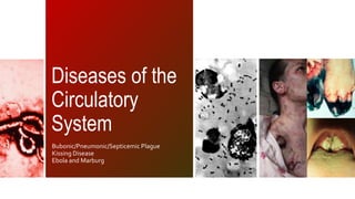
Diseases of the Circulatory System
•Download as PPTX, PDF•
3 likes•3,595 views

This document discusses several diseases of the circulatory system including plague, viral hemorrhagic fevers like Ebola and Marburg, and infectious mononucleosis. It provides details on the causative agents, symptoms, transmission, treatment and prevention of these diseases. Plague is caused by the bacterium Yersinia pestis and is transmitted via flea bites. Ebola and Marburg are viral hemorrhagic fevers transmitted through direct contact with body fluids. Infectious mononucleosis or "kissing disease" is caused by the Epstein-Barr virus and is most commonly spread through saliva.

Recommended
More Related Content
What's hot
What's hot (20)
Viewers also liked
Viewers also liked (20)
Similar to Diseases of the Circulatory System
Similar to Diseases of the Circulatory System (20)
Recently uploaded
Recently uploaded (20)
Diseases of the Circulatory System
- 1. Diseases of the Circulatory System Bubonic/Pneumonic/Septicemic Plague Kissing Disease Ebola and Marburg
- 3. an acute, often severe zoonosis infectious disease caused by Yersinia pestis symptoms depend on the concentrated areas of infection in each person Lymph nodes = BUBONIC PLAGUE Blood vessels = SEPTICEMIC PLAGUE Lungs = PULMONARY PLAGUE
- 4. YERSINIA PESTIs About the causative agent
- 5. Family Enterobacteriaceae Gram negative Facultative anaerobe Non-spore forming Pleomorphic coccobacillus Non-motile Obligate parasite One serotype 3 biovars Antiqua (1st pandemic) Medievalis (2nd pandemic) Orientalis (3rd pandemic) 11 species (3 pathogenic) Y. enterocolitis – enteropathogen Y. pseudotuberculosis – enteropathogen Y. pestis – systemic pathogen
- 6. Destroyed by •Sunlight •Desiccation Survival • 1 hour in air • Briefly in soil • 1 week in soft tissue • Years when frozen
- 7. Urban and domestic rats RESERVOI Ground squirrels Rock squirrels Prairie dogs RS Deer mice Field mice Gerbils Voles Chipmunks Marmots Guinea pigs Kangaroo rats …over 200 identified reservoirs
- 8. VECTOR XenoSpsylla cheopis (the oriental rat flea) Oropsylla montanus Nosopsyllus fasciatus (nearly worldwide in temperate climates) Xenopsylla brasiliensis (Africa, India, South America) Xenopsylla astia (Indonesia and Southeast Asia) Xenopsylla vexabilis (Pacific Islands) ~30 identified flea vectors
- 9. Incidental hosts Humans Domestic and feral Cats Dogs Lagomorphs (rabbits and hares) Coyotes Camels Goats Deer Antelope
- 10. TYPES OF PLAGUE PNEUMONIC PRIMARY SECONDARY BUBONIC SEPTICEMIC PRIMARY SECONDARY a ruptured inguinal lymph node, or bubo anteroposterior x-ray reveals a progressive plague infection affecting both lungs
- 11. July 22, 11 2012 Footer text here
- 12. bubonic septicemic pneumonic PORTAL OF ENTRY Any site of the body bitten by the vector (flea) PORTAL OF EXIT PORTAL OF ENTRY PORTAL OF EXIT PORTAL OF ENTRY PORTAL OF EXIT Eyes, nose, and mouth (Highly contagious) Eyes, nose, and mouth Eyes, nose, and mouth (Highly contagious) Breaks in the skin, nose, mouth Breaks in the skin, nose, mouth
- 14. bubonic septicemic pneumonic Bites from flea vectors Bites or scratches from infected animals, such as cats Direct contact with infected animal carcasses, such as rodents (especially marmots), rabbits, hares, carnivores (eg, wild cats, coyotes), and goats 1º Bites from flea vectors where Y pestis is inserted directly into bloodstream - no discernable bubo present 2º Develops as a complication of bubonic or 1º pneumonic plague - when Y pestis enters the bloodstream 1º Inhalation of respiratory droplets from infected animals such as cats Inhalation of respiratory droplets from a person with primary or secondary pneumonic plague Handling Y pestis cultures in the laboratory setting 2º Bubonic and 1° septicemic spread plague bacilli hematogenously to the lungs
- 16. bubonic Signs symptoms Incubation Period: 2-6 Days Fever Headache Abdominal pain Cough Pain/tenderness at regional lymph nodes enlarge to become “buboes” malaise Presence buboes in groin, axilla, cervical area Ulcer or skin lesions at site of flea bite Skin rash Rapid pulse Hypotension Vomiting Severe exhaustion Intestinal discomfort
- 17. septicemic Signs symptoms Incubation Period: 2-6 Days Fever Nausea Vomiting Diarrhea Patchy bilateral infiltrates (Chest x-ray) Prostration Hemorrhagic changes in skin called “purpuric lesions” Disseminated intravascular coagulation (DIC) Extremity gangrene Sepsis Altered mental status Abdominal pain
- 18. pneumonic Incubation Period: 1-3 Days Signs symptoms Hemoptysis Coughing Rapid, shallow breathing Cyanosis Vomiting Nausea Alveolar infiltrates on chest x-ray Chest pain dyspnea Cervical bubo Abdominal pain Fever Chills Malaise Myalgia
- 19. PATHOGENE SIS suppress and avoid normal immune system responses such as phagocytosis and antibody production
- 20. bubonic 1° Septicemic 1° pneumonic 2° SEPTICEMIC 2° PNEUMONIC
- 21. diagnosis Lymph node aspirate affected bubo should contain numerous organisms that can be evaluated microscopically and by culture Blood cultures smears taken from suspected plague patients early in the course of illness are usually negative in microscopic examination but may be positive by culture CSF (cerebrospinal fluid)a test to look at the fluid that surrounds the brain and spinal cord; gram stain of CSF may show plague bacilli; Limulus test of CSF demonstrates the presence of endotoxin Immunodiagnostic tests Observation of typical appearance (Bipolar-staining bacilli that resemble safety pins) in Gram-stained or Wright-Giemsa-stained sputum Gram stain of sputum often reveals Y pestis Biochemical Tests uses an antigen-antibody reaction as their primary means of detection
- 22. treatment In a contained casualty setting, parenteral antibiotic therapy, especially streptomycin or gentamycin, is suggested. In a mass casualty setting, intravenous or intramuscular therapy may not be possible, so oral therapy, preferably with doxycycline (or tetracycline) or ciprofloxacin, should be administered. Patients with pneumonic plague will suffer from complications and therefore require substantial advanced medical supportive care. Preferred Streptomycin Gentamycin alternative Alternative Doxycycline Ciprofloxacin Chloramphenicol
- 23. Prevention and control Reduce rodent habitat in homes, work places, and recreational areas remove brush, rock piles, junk, cluttered firewood, and possible rodent food supplies, such as pet and wild animal food. Make your home and buildings rodent-proof!!!
- 24. Prevention and control Wear gloves, protective masks, when handling or skinning potentially infected animals To prevent contact between skin and the plague bacteria
- 25. Prevention and control Use of Repellants during activities such as camping, hiking, or working outdoors It is suggested to use products containing DEET and permethrin
- 26. Prevention and control wash your hands regularly and avoid touching your eyes, nose, and mouth
- 27. Viral Hemorrhagic Fever Ebola and Marburg
- 28. Viruses of four distinct families ARENAVIRIDA E BUNYAVIRIDA E FILOVIRIDAE FLAVIVIRIDAE JUNIN CRIMEAN-CONGO H.F. EBOLA KYASANUR FOREST DISEASE MACHUPO HANTAVIRUS MARBURG OMSK H.F. SABIA RIGT VALLEY FEVER YELLOW FEVER GUANARITO DENGUE LASSA
- 29. Severe multi-system syndrome (multiple organs affected). Vascular system is damaged and body loses the ability to regulate itself Accompanied by hemorrhaging Many VHF viruses cause life threatening diseases Most have no established treatment or cure.
- 30. Features of these Viruses RNA Viruses, covered in lipid coating Humans are not natural reservoir, but can transmit virus Viruses are restricted to areas of their host species
- 31. mARBURG ZOONOTIC 1st recognize in 1967 when simultaneous outbreaks occurred in Marburg and Frankfurt, Germany, and in Belgrade, Yugoslavia. Bats have been implicated for this virus The area to which the virus is native is unknown , but it is believed to include parts of Uganda, Western Kenya, and Zimbabwe Original people who became ill had been exposed to the tissues of African green monkeys, which were imported from Uganda for research. Rousettus aegyptiacus Egyptian rousettes (bat) Bat, Human and Non-human primates destroyed by gamma and UV radiation, lipid solvents, and bleach
- 32. Portal of entry Portal of exit Respiratory Tract Eyes Skin Respiratory Tract Eyes Skin The investigation for the entry of exit of the causative agent is still under research. But some suggests that the entry of MAV is dependent on NPC1, a cholesterol transporter to enter and replicate.
- 33. transmission The animal host to the Marburg Virus is unknown, and so is the way that the animal transmits the disease to humans People who have been exposed to infected monkeys or their body fluids have become infected in the past. Disease is easily transmitted between humans. Direct contact with an infected person, or exposure to their body fluids, are both ways by which the disease is transmitted.
- 34. SIGNS SYMPTOM S Incubation period of 5-10 days Maculopapular rash Most prominent on the trunk (chest and back) Jaundice Inflammation of the pancreas Severe weight loss Delirium Shock Liver failure Massive hemorrhaging Multi-organ dysfunction. Fever Chills Headache Myalgia Nausea Vomiting Chest pain Abdominal pain Diarrhea
- 35. diagnosis Many of the signs and symptoms of Marburg hemorrhagic fever are similar to those of other infectious diseases, such as malaria or typhoid fever, diagnosis of the disease can be difficult, especially if only a single case is involved Readily diagnosed by: To confirm a case of Marburg hemorrhagic fever within a few days of the onset of symptoms: Antigen-capture enzyme-linked immunosorbent assay (ELISA) testing virus isolation IgM-capture ELISA polymerase chain reaction (PCR) Test appropriate for testing persons later in the course of disease or after recovery: The IgG-capture ELISA Immunohistochemistry virus isolation polymerase chain reaction (PCR)
- 36. treatment A specific treatment for this disease is unknown Isolation and quarantine Quick containment No standard treatment Use of heparin (which blocks clotting) to prevent the consumption of clotting factors Fresh-frozen plasma and other preparations to replace the blood proteins important in clotting Supportive therapy, which includes balancing the patient’s fluids and electrolytes, maintaining their oxygen status and blood pressure, and treating them for complicating infections.
- 37. Prevention and control Still no established preventive measures for the transmission of disease Use of barrier nursing techniques to prevent direct physical contact with the patient Use of protective clothing wearing of protective gowns, gloves, and masks Proper disposal of all needles, equipments, and patient excretions
- 38. ebola Has 4 types Ebola-Zaire Ebola-Sudan Ebola-Ivory Coast Ebola Reston zoonotic Named after the river in congo (zaire) – first outbreak - where 88% of the people died Sporadic appearance Fatal in humans and other non-human primates Natural reservoir remains unknown destroyed by gamma and Originated in africa UV radiation, lipid solvents, and bleach
- 39. Portal of entry and exit EYES AND MOUTH SKIN
- 40. transmission Intimate contact Direct contact with the blood or Aerosol transmission secretions of an infected person Nosicomial transmission • contact with objects, such as needles, that are contaminated with secretions or blood. • reuse of needles and syringes • exposure to infectious tissues, excretions, and hospital wastes Common among primates
- 41. SIGNS SYMPTOM S Incubation period of 2-21 days Arthritic pain and backache Chills Diarrhea Fatigue Fever Headache Malaise Nausea Sore throat Vomiting Bleeding from eyes, ears, and nose Gastrointestinal bleeding Eye inflammation (conjunctivitis) Genital swelling (labia and scrotum) Increased feeling of pain in skin Rash over the entire body that often contains blood (hemorrhagic) Roof of mouth looks red Seizures, coma, delirium Impaired kidney and liver Internal and external bleeding
- 43. diagnosis It is initially difficult to diagnose Ebola clinically because many of the symptoms are nonspecific RT-PCR and ELISA Polymerase Chain Reaction Enzyme-linked immunosorbent assays (ELISA) IgM response most useful in diagnosis of recent infections in surviving patients. somewhat delayed and expected only in the early convalescent sera IgG response
- 44. treatment there is no specific treatment or cure for Ebola HF Isolation and quarantine Quick containment No standard treatment Mechanical ventilation Renal dialysis Anti-seizure therapy Supportive therapy • balancing the patient’s fluids and electrolytes • maintaining oxygen status and blood pressure • Treatment of infection complications
- 46. Infectious mononucleosis disease “Kissing Disease”
- 47. Epstein- Barr Virus human herpesvirus 4 Herpesviridae family double stranded linear DNA core core surrounded by a nucleocapsid envelope contains glycoproteins affects B- lymphocytes
- 48. Portal of entry and Exit TONSILS transmission (bodily fluids, especially saliva, can also spread through blood and semen during sexual contact, blood transfusions, and organ transplantations)
- 49. July 22, 49 2012 Footer text here
- 50. SIGNS SYMPTOM S Incubation Period: 4-6 weeks swollen lymph nodes enlargement of liver or spleen skin rash Jaundice pharyngitis/ tonsilitis sore throat fever constant fatigue sore muscles abdominal pain loss of appetite nausea or vomiting headaches
- 51. diagnosis Heterophil Antibody/ Monospot Test - detects a type of antibody (heterophil antibody) that forms during certain infections - looks for antibodies that possess the unique ability to cause clumping of red cells - presence of heterophil antibodies indicates a mono infection. complete blood cell count EBV Antibody Test Blood sample is mixed with a substance that attaches to antibodies against EBV Davidson Differential Slide Test
- 52. diagnosis Mono- Plus Test Clumping of horse red blood cells by mono antibodies presumed to be in a person's serum
- 53. treatment Corticosteroids fever reducing medications may be prescribed in rare cases of airway obstruction, hemolytic anemia (an autoimmune process in which red blood cells are destroyed), severe thrombocytopenia (a decrease in platelets, which are clotting components in the blood), and complications involving the heart and nerves drinking fluids to stay hydrated medications to treat pain, and other symptoms bed rest
- 54. Prevention and control Avoid sharing drinks, food, or personal items, like toothbrushes, with people who have EBV infection. Avoid kissing with people who have EBV infection. Wash hands at all time
- 55. Diseases of the Circulatory System Casipe, Kimberly Chua, Charlean Lou Espinosa, Karl Elvis Sodusta, Patrick Jason
Editor's Notes
- There are 3 biovars of Y. pestis, each named for the pandemic that it is thought to have caused They are named based on their ability to convert nitrate to nitrite and ferment glycerol
- (survival in air increases its threat and aids in its dispersal as a potential bioterrorism weapon. Image: Wayson stain of blood shows the characteristic bipolar “safety pin” appearance of Yersinia pestis. From CDC.
- 1. Flea feeds on blood with y. pestis 2. enters the midgut and multiplies 3. clump of y. pestis blocks the foregut 4. because of the clump present in the foregut, during the fleas next meal, blood cannot enter the midgut thus flea gets very hungry 5. flea bites vigorously and injects the contents of its midgut into the next wound 6. only blocked fleas effectively transmit plague to mammals. 7.while growing inside the flea, the bacteria loses its antiphagocytic capsular layer (F1) and so many of the pathogenic organisms are phagocytosed and killed by mammalian leukocyte 8.. Howevernot all are killed, Those that are ingested by neutrophils appears to be readily killed, but cbacteria within macrophages are able to survive 9. macrophages provide protection. Giving time to the bacteria to resynthesize their protective F1 capsular layer and other irulence antigens. The ability of Y. pestis to survive and grow in macrophages is critical to the early pathogenesis of pague 10. The bacteria within the macrophages are then transported to the local draining lymph node. 11. the massive filtration of the phagocytic cells within the nodes cause them to become hot and swollen and hemorrhagic giving rise to buboes. 12. within the bubo, undergoing unknown mechanism , the bacteria escapes from the macrophages and adopt an extracellular lifestule where they further grow and replicate. 13. The newly formed protective capsular layer of the bacteria helps resist phagocytosis by the leukocytes. 14. Eventually, the infection can now sppill out into the bloodstream, leading to involvement of the liver, spleen, and the lungs (eading to second degree septicemic and pneumonic development). First degree septicemic plague 1, flea inserts directly into the bloodstream causing migration of Y. pestis to organs First degree pneumonic plague inhaled Y. pestis bacili would eneter into the lungs
- Infection Control The use of standard precautions is appropriate for managing plague patients who do not have respiratory infections. In past epidemics, patient-to-patient transmission only seemed to occur after close contact, often for prolonged periods, with a patient that coughed bloody sputum. It is extremely rare for patients in the early stage of pneumonic plague to transmit the infection to others. Bubonic: Standard and contact precautions if any open wounds. Pneumonic: Standard and respiratory droplet precautions. (Note: Available evidence indicates that person-to-person spread of pneumonic plague is via respiratory droplets, not fine aerosols or droplet nuclei.) Septicemic: Standard precautions. Pneumonic plague patients are no longer infective after 24 to 48 hours of antibiotic treatment, because the sputum no longer contains live bacilli. However, they may still be ill and continue demonstrating signs of pneumonia. Infection Control When individual isolation of suspected plague patients is not possible, they should be cohorted away from other hospital patients, and managed under respiratory droplet precautions until no longer considered to be contagious. Although quick diagnosis and appropriate antibiotic treatment is imperative in preventing the spread of disease throughout the community, isolation of contacts and/or quarantine may increase in importance for outbreak control. The bodies of persons who have died from plague should be handled with standard infection control precautions Occupational Exposures – Hospital and Laboratory Medical staff or laboratory workers accidentally exposed to infectious materials via needle sticks, cuts, or abrasions should immediately wash the area with a nonabrasive soap and water and follow the standard policy of their institution regarding workplace exposures. When eye exposure occurs, the eye should be flushed with copious amounts of water or eye wash solution for at least 15 minutes. In addition, postexposure antimicrobial prophylaxis with doxycycline or ciprofloxacin should be started immediately and continued for 7 days. Laboratory workers who handle cultures should be alerted to the possibility of Y. pestis and take precautions to avoid aerosolization of cultures or other infectious materials.
- Infection Control The use of standard precautions is appropriate for managing plague patients who do not have respiratory infections. In past epidemics, patient-to-patient transmission only seemed to occur after close contact, often for prolonged periods, with a patient that coughed bloody sputum. It is extremely rare for patients in the early stage of pneumonic plague to transmit the infection to others. Bubonic: Standard and contact precautions if any open wounds. Pneumonic: Standard and respiratory droplet precautions. (Note: Available evidence indicates that person-to-person spread of pneumonic plague is via respiratory droplets, not fine aerosols or droplet nuclei.) Septicemic: Standard precautions. Pneumonic plague patients are no longer infective after 24 to 48 hours of antibiotic treatment, because the sputum no longer contains live bacilli. However, they may still be ill and continue demonstrating signs of pneumonia. Infection Control When individual isolation of suspected plague patients is not possible, they should be cohorted away from other hospital patients, and managed under respiratory droplet precautions until no longer considered to be contagious. Although quick diagnosis and appropriate antibiotic treatment is imperative in preventing the spread of disease throughout the community, isolation of contacts and/or quarantine may increase in importance for outbreak control. The bodies of persons who have died from plague should be handled with standard infection control precautions Occupational Exposures – Hospital and Laboratory Medical staff or laboratory workers accidentally exposed to infectious materials via needle sticks, cuts, or abrasions should immediately wash the area with a nonabrasive soap and water and follow the standard policy of their institution regarding workplace exposures. When eye exposure occurs, the eye should be flushed with copious amounts of water or eye wash solution for at least 15 minutes. In addition, postexposure antimicrobial prophylaxis with doxycycline or ciprofloxacin should be started immediately and continued for 7 days. Laboratory workers who handle cultures should be alerted to the possibility of Y. pestis and take precautions to avoid aerosolization of cultures or other infectious materials.
- Infection Control The use of standard precautions is appropriate for managing plague patients who do not have respiratory infections. In past epidemics, patient-to-patient transmission only seemed to occur after close contact, often for prolonged periods, with a patient that coughed bloody sputum. It is extremely rare for patients in the early stage of pneumonic plague to transmit the infection to others. Bubonic: Standard and contact precautions if any open wounds. Pneumonic: Standard and respiratory droplet precautions. (Note: Available evidence indicates that person-to-person spread of pneumonic plague is via respiratory droplets, not fine aerosols or droplet nuclei.) Septicemic: Standard precautions. Pneumonic plague patients are no longer infective after 24 to 48 hours of antibiotic treatment, because the sputum no longer contains live bacilli. However, they may still be ill and continue demonstrating signs of pneumonia. Infection Control When individual isolation of suspected plague patients is not possible, they should be cohorted away from other hospital patients, and managed under respiratory droplet precautions until no longer considered to be contagious. Although quick diagnosis and appropriate antibiotic treatment is imperative in preventing the spread of disease throughout the community, isolation of contacts and/or quarantine may increase in importance for outbreak control. The bodies of persons who have died from plague should be handled with standard infection control precautions Occupational Exposures – Hospital and Laboratory Medical staff or laboratory workers accidentally exposed to infectious materials via needle sticks, cuts, or abrasions should immediately wash the area with a nonabrasive soap and water and follow the standard policy of their institution regarding workplace exposures. When eye exposure occurs, the eye should be flushed with copious amounts of water or eye wash solution for at least 15 minutes. In addition, postexposure antimicrobial prophylaxis with doxycycline or ciprofloxacin should be started immediately and continued for 7 days. Laboratory workers who handle cultures should be alerted to the possibility of Y. pestis and take precautions to avoid aerosolization of cultures or other infectious materials.
- Infection Control The use of standard precautions is appropriate for managing plague patients who do not have respiratory infections. In past epidemics, patient-to-patient transmission only seemed to occur after close contact, often for prolonged periods, with a patient that coughed bloody sputum. It is extremely rare for patients in the early stage of pneumonic plague to transmit the infection to others. Bubonic: Standard and contact precautions if any open wounds. Pneumonic: Standard and respiratory droplet precautions. (Note: Available evidence indicates that person-to-person spread of pneumonic plague is via respiratory droplets, not fine aerosols or droplet nuclei.) Septicemic: Standard precautions. Pneumonic plague patients are no longer infective after 24 to 48 hours of antibiotic treatment, because the sputum no longer contains live bacilli. However, they may still be ill and continue demonstrating signs of pneumonia. Infection Control When individual isolation of suspected plague patients is not possible, they should be cohorted away from other hospital patients, and managed under respiratory droplet precautions until no longer considered to be contagious. Although quick diagnosis and appropriate antibiotic treatment is imperative in preventing the spread of disease throughout the community, isolation of contacts and/or quarantine may increase in importance for outbreak control. The bodies of persons who have died from plague should be handled with standard infection control precautions Occupational Exposures – Hospital and Laboratory Medical staff or laboratory workers accidentally exposed to infectious materials via needle sticks, cuts, or abrasions should immediately wash the area with a nonabrasive soap and water and follow the standard policy of their institution regarding workplace exposures. When eye exposure occurs, the eye should be flushed with copious amounts of water or eye wash solution for at least 15 minutes. In addition, postexposure antimicrobial prophylaxis with doxycycline or ciprofloxacin should be started immediately and continued for 7 days. Laboratory workers who handle cultures should be alerted to the possibility of Y. pestis and take precautions to avoid aerosolization of cultures or other infectious materials.
- Infection Control The use of standard precautions is appropriate for managing plague patients who do not have respiratory infections. In past epidemics, patient-to-patient transmission only seemed to occur after close contact, often for prolonged periods, with a patient that coughed bloody sputum. It is extremely rare for patients in the early stage of pneumonic plague to transmit the infection to others. Bubonic: Standard and contact precautions if any open wounds. Pneumonic: Standard and respiratory droplet precautions. (Note: Available evidence indicates that person-to-person spread of pneumonic plague is via respiratory droplets, not fine aerosols or droplet nuclei.) Septicemic: Standard precautions. Pneumonic plague patients are no longer infective after 24 to 48 hours of antibiotic treatment, because the sputum no longer contains live bacilli. However, they may still be ill and continue demonstrating signs of pneumonia. Infection Control When individual isolation of suspected plague patients is not possible, they should be cohorted away from other hospital patients, and managed under respiratory droplet precautions until no longer considered to be contagious. Although quick diagnosis and appropriate antibiotic treatment is imperative in preventing the spread of disease throughout the community, isolation of contacts and/or quarantine may increase in importance for outbreak control. The bodies of persons who have died from plague should be handled with standard infection control precautions Occupational Exposures – Hospital and Laboratory Medical staff or laboratory workers accidentally exposed to infectious materials via needle sticks, cuts, or abrasions should immediately wash the area with a nonabrasive soap and water and follow the standard policy of their institution regarding workplace exposures. When eye exposure occurs, the eye should be flushed with copious amounts of water or eye wash solution for at least 15 minutes. In addition, postexposure antimicrobial prophylaxis with doxycycline or ciprofloxacin should be started immediately and continued for 7 days. Laboratory workers who handle cultures should be alerted to the possibility of Y. pestis and take precautions to avoid aerosolization of cultures or other infectious materials.
- Infection Control The use of standard precautions is appropriate for managing plague patients who do not have respiratory infections. In past epidemics, patient-to-patient transmission only seemed to occur after close contact, often for prolonged periods, with a patient that coughed bloody sputum. It is extremely rare for patients in the early stage of pneumonic plague to transmit the infection to others. Bubonic: Standard and contact precautions if any open wounds. Pneumonic: Standard and respiratory droplet precautions. (Note: Available evidence indicates that person-to-person spread of pneumonic plague is via respiratory droplets, not fine aerosols or droplet nuclei.) Septicemic: Standard precautions. Pneumonic plague patients are no longer infective after 24 to 48 hours of antibiotic treatment, because the sputum no longer contains live bacilli. However, they may still be ill and continue demonstrating signs of pneumonia. Infection Control When individual isolation of suspected plague patients is not possible, they should be cohorted away from other hospital patients, and managed under respiratory droplet precautions until no longer considered to be contagious. Although quick diagnosis and appropriate antibiotic treatment is imperative in preventing the spread of disease throughout the community, isolation of contacts and/or quarantine may increase in importance for outbreak control. The bodies of persons who have died from plague should be handled with standard infection control precautions Occupational Exposures – Hospital and Laboratory Medical staff or laboratory workers accidentally exposed to infectious materials via needle sticks, cuts, or abrasions should immediately wash the area with a nonabrasive soap and water and follow the standard policy of their institution regarding workplace exposures. When eye exposure occurs, the eye should be flushed with copious amounts of water or eye wash solution for at least 15 minutes. In addition, postexposure antimicrobial prophylaxis with doxycycline or ciprofloxacin should be started immediately and continued for 7 days. Laboratory workers who handle cultures should be alerted to the possibility of Y. pestis and take precautions to avoid aerosolization of cultures or other infectious materials.
- Infection Control The use of standard precautions is appropriate for managing plague patients who do not have respiratory infections. In past epidemics, patient-to-patient transmission only seemed to occur after close contact, often for prolonged periods, with a patient that coughed bloody sputum. It is extremely rare for patients in the early stage of pneumonic plague to transmit the infection to others. Bubonic: Standard and contact precautions if any open wounds. Pneumonic: Standard and respiratory droplet precautions. (Note: Available evidence indicates that person-to-person spread of pneumonic plague is via respiratory droplets, not fine aerosols or droplet nuclei.) Septicemic: Standard precautions. Pneumonic plague patients are no longer infective after 24 to 48 hours of antibiotic treatment, because the sputum no longer contains live bacilli. However, they may still be ill and continue demonstrating signs of pneumonia. Infection Control When individual isolation of suspected plague patients is not possible, they should be cohorted away from other hospital patients, and managed under respiratory droplet precautions until no longer considered to be contagious. Although quick diagnosis and appropriate antibiotic treatment is imperative in preventing the spread of disease throughout the community, isolation of contacts and/or quarantine may increase in importance for outbreak control. The bodies of persons who have died from plague should be handled with standard infection control precautions Occupational Exposures – Hospital and Laboratory Medical staff or laboratory workers accidentally exposed to infectious materials via needle sticks, cuts, or abrasions should immediately wash the area with a nonabrasive soap and water and follow the standard policy of their institution regarding workplace exposures. When eye exposure occurs, the eye should be flushed with copious amounts of water or eye wash solution for at least 15 minutes. In addition, postexposure antimicrobial prophylaxis with doxycycline or ciprofloxacin should be started immediately and continued for 7 days. Laboratory workers who handle cultures should be alerted to the possibility of Y. pestis and take precautions to avoid aerosolization of cultures or other infectious materials.
- Infection Control The use of standard precautions is appropriate for managing plague patients who do not have respiratory infections. In past epidemics, patient-to-patient transmission only seemed to occur after close contact, often for prolonged periods, with a patient that coughed bloody sputum. It is extremely rare for patients in the early stage of pneumonic plague to transmit the infection to others. Bubonic: Standard and contact precautions if any open wounds. Pneumonic: Standard and respiratory droplet precautions. (Note: Available evidence indicates that person-to-person spread of pneumonic plague is via respiratory droplets, not fine aerosols or droplet nuclei.) Septicemic: Standard precautions. Pneumonic plague patients are no longer infective after 24 to 48 hours of antibiotic treatment, because the sputum no longer contains live bacilli. However, they may still be ill and continue demonstrating signs of pneumonia. Infection Control When individual isolation of suspected plague patients is not possible, they should be cohorted away from other hospital patients, and managed under respiratory droplet precautions until no longer considered to be contagious. Although quick diagnosis and appropriate antibiotic treatment is imperative in preventing the spread of disease throughout the community, isolation of contacts and/or quarantine may increase in importance for outbreak control. The bodies of persons who have died from plague should be handled with standard infection control precautions Occupational Exposures – Hospital and Laboratory Medical staff or laboratory workers accidentally exposed to infectious materials via needle sticks, cuts, or abrasions should immediately wash the area with a nonabrasive soap and water and follow the standard policy of their institution regarding workplace exposures. When eye exposure occurs, the eye should be flushed with copious amounts of water or eye wash solution for at least 15 minutes. In addition, postexposure antimicrobial prophylaxis with doxycycline or ciprofloxacin should be started immediately and continued for 7 days. Laboratory workers who handle cultures should be alerted to the possibility of Y. pestis and take precautions to avoid aerosolization of cultures or other infectious materials.
- Infection Control The use of standard precautions is appropriate for managing plague patients who do not have respiratory infections. In past epidemics, patient-to-patient transmission only seemed to occur after close contact, often for prolonged periods, with a patient that coughed bloody sputum. It is extremely rare for patients in the early stage of pneumonic plague to transmit the infection to others. Bubonic: Standard and contact precautions if any open wounds. Pneumonic: Standard and respiratory droplet precautions. (Note: Available evidence indicates that person-to-person spread of pneumonic plague is via respiratory droplets, not fine aerosols or droplet nuclei.) Septicemic: Standard precautions. Pneumonic plague patients are no longer infective after 24 to 48 hours of antibiotic treatment, because the sputum no longer contains live bacilli. However, they may still be ill and continue demonstrating signs of pneumonia. Infection Control When individual isolation of suspected plague patients is not possible, they should be cohorted away from other hospital patients, and managed under respiratory droplet precautions until no longer considered to be contagious. Although quick diagnosis and appropriate antibiotic treatment is imperative in preventing the spread of disease throughout the community, isolation of contacts and/or quarantine may increase in importance for outbreak control. The bodies of persons who have died from plague should be handled with standard infection control precautions Occupational Exposures – Hospital and Laboratory Medical staff or laboratory workers accidentally exposed to infectious materials via needle sticks, cuts, or abrasions should immediately wash the area with a nonabrasive soap and water and follow the standard policy of their institution regarding workplace exposures. When eye exposure occurs, the eye should be flushed with copious amounts of water or eye wash solution for at least 15 minutes. In addition, postexposure antimicrobial prophylaxis with doxycycline or ciprofloxacin should be started immediately and continued for 7 days. Laboratory workers who handle cultures should be alerted to the possibility of Y. pestis and take precautions to avoid aerosolization of cultures or other infectious materials.
- Infection Control The use of standard precautions is appropriate for managing plague patients who do not have respiratory infections. In past epidemics, patient-to-patient transmission only seemed to occur after close contact, often for prolonged periods, with a patient that coughed bloody sputum. It is extremely rare for patients in the early stage of pneumonic plague to transmit the infection to others. Bubonic: Standard and contact precautions if any open wounds. Pneumonic: Standard and respiratory droplet precautions. (Note: Available evidence indicates that person-to-person spread of pneumonic plague is via respiratory droplets, not fine aerosols or droplet nuclei.) Septicemic: Standard precautions. Pneumonic plague patients are no longer infective after 24 to 48 hours of antibiotic treatment, because the sputum no longer contains live bacilli. However, they may still be ill and continue demonstrating signs of pneumonia. Infection Control When individual isolation of suspected plague patients is not possible, they should be cohorted away from other hospital patients, and managed under respiratory droplet precautions until no longer considered to be contagious. Although quick diagnosis and appropriate antibiotic treatment is imperative in preventing the spread of disease throughout the community, isolation of contacts and/or quarantine may increase in importance for outbreak control. The bodies of persons who have died from plague should be handled with standard infection control precautions Occupational Exposures – Hospital and Laboratory Medical staff or laboratory workers accidentally exposed to infectious materials via needle sticks, cuts, or abrasions should immediately wash the area with a nonabrasive soap and water and follow the standard policy of their institution regarding workplace exposures. When eye exposure occurs, the eye should be flushed with copious amounts of water or eye wash solution for at least 15 minutes. In addition, postexposure antimicrobial prophylaxis with doxycycline or ciprofloxacin should be started immediately and continued for 7 days. Laboratory workers who handle cultures should be alerted to the possibility of Y. pestis and take precautions to avoid aerosolization of cultures or other infectious materials.
- Infection Control The use of standard precautions is appropriate for managing plague patients who do not have respiratory infections. In past epidemics, patient-to-patient transmission only seemed to occur after close contact, often for prolonged periods, with a patient that coughed bloody sputum. It is extremely rare for patients in the early stage of pneumonic plague to transmit the infection to others. Bubonic: Standard and contact precautions if any open wounds. Pneumonic: Standard and respiratory droplet precautions. (Note: Available evidence indicates that person-to-person spread of pneumonic plague is via respiratory droplets, not fine aerosols or droplet nuclei.) Septicemic: Standard precautions. Pneumonic plague patients are no longer infective after 24 to 48 hours of antibiotic treatment, because the sputum no longer contains live bacilli. However, they may still be ill and continue demonstrating signs of pneumonia. Infection Control When individual isolation of suspected plague patients is not possible, they should be cohorted away from other hospital patients, and managed under respiratory droplet precautions until no longer considered to be contagious. Although quick diagnosis and appropriate antibiotic treatment is imperative in preventing the spread of disease throughout the community, isolation of contacts and/or quarantine may increase in importance for outbreak control. The bodies of persons who have died from plague should be handled with standard infection control precautions Occupational Exposures – Hospital and Laboratory Medical staff or laboratory workers accidentally exposed to infectious materials via needle sticks, cuts, or abrasions should immediately wash the area with a nonabrasive soap and water and follow the standard policy of their institution regarding workplace exposures. When eye exposure occurs, the eye should be flushed with copious amounts of water or eye wash solution for at least 15 minutes. In addition, postexposure antimicrobial prophylaxis with doxycycline or ciprofloxacin should be started immediately and continued for 7 days. Laboratory workers who handle cultures should be alerted to the possibility of Y. pestis and take precautions to avoid aerosolization of cultures or other infectious materials.
- Infection Control The use of standard precautions is appropriate for managing plague patients who do not have respiratory infections. In past epidemics, patient-to-patient transmission only seemed to occur after close contact, often for prolonged periods, with a patient that coughed bloody sputum. It is extremely rare for patients in the early stage of pneumonic plague to transmit the infection to others. Bubonic: Standard and contact precautions if any open wounds. Pneumonic: Standard and respiratory droplet precautions. (Note: Available evidence indicates that person-to-person spread of pneumonic plague is via respiratory droplets, not fine aerosols or droplet nuclei.) Septicemic: Standard precautions. Pneumonic plague patients are no longer infective after 24 to 48 hours of antibiotic treatment, because the sputum no longer contains live bacilli. However, they may still be ill and continue demonstrating signs of pneumonia. Infection Control When individual isolation of suspected plague patients is not possible, they should be cohorted away from other hospital patients, and managed under respiratory droplet precautions until no longer considered to be contagious. Although quick diagnosis and appropriate antibiotic treatment is imperative in preventing the spread of disease throughout the community, isolation of contacts and/or quarantine may increase in importance for outbreak control. The bodies of persons who have died from plague should be handled with standard infection control precautions Occupational Exposures – Hospital and Laboratory Medical staff or laboratory workers accidentally exposed to infectious materials via needle sticks, cuts, or abrasions should immediately wash the area with a nonabrasive soap and water and follow the standard policy of their institution regarding workplace exposures. When eye exposure occurs, the eye should be flushed with copious amounts of water or eye wash solution for at least 15 minutes. In addition, postexposure antimicrobial prophylaxis with doxycycline or ciprofloxacin should be started immediately and continued for 7 days. Laboratory workers who handle cultures should be alerted to the possibility of Y. pestis and take precautions to avoid aerosolization of cultures or other infectious materials.
- Infection Control The use of standard precautions is appropriate for managing plague patients who do not have respiratory infections. In past epidemics, patient-to-patient transmission only seemed to occur after close contact, often for prolonged periods, with a patient that coughed bloody sputum. It is extremely rare for patients in the early stage of pneumonic plague to transmit the infection to others. Bubonic: Standard and contact precautions if any open wounds. Pneumonic: Standard and respiratory droplet precautions. (Note: Available evidence indicates that person-to-person spread of pneumonic plague is via respiratory droplets, not fine aerosols or droplet nuclei.) Septicemic: Standard precautions. Pneumonic plague patients are no longer infective after 24 to 48 hours of antibiotic treatment, because the sputum no longer contains live bacilli. However, they may still be ill and continue demonstrating signs of pneumonia. Infection Control When individual isolation of suspected plague patients is not possible, they should be cohorted away from other hospital patients, and managed under respiratory droplet precautions until no longer considered to be contagious. Although quick diagnosis and appropriate antibiotic treatment is imperative in preventing the spread of disease throughout the community, isolation of contacts and/or quarantine may increase in importance for outbreak control. The bodies of persons who have died from plague should be handled with standard infection control precautions Occupational Exposures – Hospital and Laboratory Medical staff or laboratory workers accidentally exposed to infectious materials via needle sticks, cuts, or abrasions should immediately wash the area with a nonabrasive soap and water and follow the standard policy of their institution regarding workplace exposures. When eye exposure occurs, the eye should be flushed with copious amounts of water or eye wash solution for at least 15 minutes. In addition, postexposure antimicrobial prophylaxis with doxycycline or ciprofloxacin should be started immediately and continued for 7 days. Laboratory workers who handle cultures should be alerted to the possibility of Y. pestis and take precautions to avoid aerosolization of cultures or other infectious materials.
- Infection Control The use of standard precautions is appropriate for managing plague patients who do not have respiratory infections. In past epidemics, patient-to-patient transmission only seemed to occur after close contact, often for prolonged periods, with a patient that coughed bloody sputum. It is extremely rare for patients in the early stage of pneumonic plague to transmit the infection to others. Bubonic: Standard and contact precautions if any open wounds. Pneumonic: Standard and respiratory droplet precautions. (Note: Available evidence indicates that person-to-person spread of pneumonic plague is via respiratory droplets, not fine aerosols or droplet nuclei.) Septicemic: Standard precautions. Pneumonic plague patients are no longer infective after 24 to 48 hours of antibiotic treatment, because the sputum no longer contains live bacilli. However, they may still be ill and continue demonstrating signs of pneumonia. Infection Control When individual isolation of suspected plague patients is not possible, they should be cohorted away from other hospital patients, and managed under respiratory droplet precautions until no longer considered to be contagious. Although quick diagnosis and appropriate antibiotic treatment is imperative in preventing the spread of disease throughout the community, isolation of contacts and/or quarantine may increase in importance for outbreak control. The bodies of persons who have died from plague should be handled with standard infection control precautions Occupational Exposures – Hospital and Laboratory Medical staff or laboratory workers accidentally exposed to infectious materials via needle sticks, cuts, or abrasions should immediately wash the area with a nonabrasive soap and water and follow the standard policy of their institution regarding workplace exposures. When eye exposure occurs, the eye should be flushed with copious amounts of water or eye wash solution for at least 15 minutes. In addition, postexposure antimicrobial prophylaxis with doxycycline or ciprofloxacin should be started immediately and continued for 7 days. Laboratory workers who handle cultures should be alerted to the possibility of Y. pestis and take precautions to avoid aerosolization of cultures or other infectious materials.
- Infection Control The use of standard precautions is appropriate for managing plague patients who do not have respiratory infections. In past epidemics, patient-to-patient transmission only seemed to occur after close contact, often for prolonged periods, with a patient that coughed bloody sputum. It is extremely rare for patients in the early stage of pneumonic plague to transmit the infection to others. Bubonic: Standard and contact precautions if any open wounds. Pneumonic: Standard and respiratory droplet precautions. (Note: Available evidence indicates that person-to-person spread of pneumonic plague is via respiratory droplets, not fine aerosols or droplet nuclei.) Septicemic: Standard precautions. Pneumonic plague patients are no longer infective after 24 to 48 hours of antibiotic treatment, because the sputum no longer contains live bacilli. However, they may still be ill and continue demonstrating signs of pneumonia. Infection Control When individual isolation of suspected plague patients is not possible, they should be cohorted away from other hospital patients, and managed under respiratory droplet precautions until no longer considered to be contagious. Although quick diagnosis and appropriate antibiotic treatment is imperative in preventing the spread of disease throughout the community, isolation of contacts and/or quarantine may increase in importance for outbreak control. The bodies of persons who have died from plague should be handled with standard infection control precautions Occupational Exposures – Hospital and Laboratory Medical staff or laboratory workers accidentally exposed to infectious materials via needle sticks, cuts, or abrasions should immediately wash the area with a nonabrasive soap and water and follow the standard policy of their institution regarding workplace exposures. When eye exposure occurs, the eye should be flushed with copious amounts of water or eye wash solution for at least 15 minutes. In addition, postexposure antimicrobial prophylaxis with doxycycline or ciprofloxacin should be started immediately and continued for 7 days. Laboratory workers who handle cultures should be alerted to the possibility of Y. pestis and take precautions to avoid aerosolization of cultures or other infectious materials.
- Infection Control The use of standard precautions is appropriate for managing plague patients who do not have respiratory infections. In past epidemics, patient-to-patient transmission only seemed to occur after close contact, often for prolonged periods, with a patient that coughed bloody sputum. It is extremely rare for patients in the early stage of pneumonic plague to transmit the infection to others. Bubonic: Standard and contact precautions if any open wounds. Pneumonic: Standard and respiratory droplet precautions. (Note: Available evidence indicates that person-to-person spread of pneumonic plague is via respiratory droplets, not fine aerosols or droplet nuclei.) Septicemic: Standard precautions. Pneumonic plague patients are no longer infective after 24 to 48 hours of antibiotic treatment, because the sputum no longer contains live bacilli. However, they may still be ill and continue demonstrating signs of pneumonia. Infection Control When individual isolation of suspected plague patients is not possible, they should be cohorted away from other hospital patients, and managed under respiratory droplet precautions until no longer considered to be contagious. Although quick diagnosis and appropriate antibiotic treatment is imperative in preventing the spread of disease throughout the community, isolation of contacts and/or quarantine may increase in importance for outbreak control. The bodies of persons who have died from plague should be handled with standard infection control precautions Occupational Exposures – Hospital and Laboratory Medical staff or laboratory workers accidentally exposed to infectious materials via needle sticks, cuts, or abrasions should immediately wash the area with a nonabrasive soap and water and follow the standard policy of their institution regarding workplace exposures. When eye exposure occurs, the eye should be flushed with copious amounts of water or eye wash solution for at least 15 minutes. In addition, postexposure antimicrobial prophylaxis with doxycycline or ciprofloxacin should be started immediately and continued for 7 days. Laboratory workers who handle cultures should be alerted to the possibility of Y. pestis and take precautions to avoid aerosolization of cultures or other infectious materials.
- Infection Control The use of standard precautions is appropriate for managing plague patients who do not have respiratory infections. In past epidemics, patient-to-patient transmission only seemed to occur after close contact, often for prolonged periods, with a patient that coughed bloody sputum. It is extremely rare for patients in the early stage of pneumonic plague to transmit the infection to others. Bubonic: Standard and contact precautions if any open wounds. Pneumonic: Standard and respiratory droplet precautions. (Note: Available evidence indicates that person-to-person spread of pneumonic plague is via respiratory droplets, not fine aerosols or droplet nuclei.) Septicemic: Standard precautions. Pneumonic plague patients are no longer infective after 24 to 48 hours of antibiotic treatment, because the sputum no longer contains live bacilli. However, they may still be ill and continue demonstrating signs of pneumonia. Infection Control When individual isolation of suspected plague patients is not possible, they should be cohorted away from other hospital patients, and managed under respiratory droplet precautions until no longer considered to be contagious. Although quick diagnosis and appropriate antibiotic treatment is imperative in preventing the spread of disease throughout the community, isolation of contacts and/or quarantine may increase in importance for outbreak control. The bodies of persons who have died from plague should be handled with standard infection control precautions Occupational Exposures – Hospital and Laboratory Medical staff or laboratory workers accidentally exposed to infectious materials via needle sticks, cuts, or abrasions should immediately wash the area with a nonabrasive soap and water and follow the standard policy of their institution regarding workplace exposures. When eye exposure occurs, the eye should be flushed with copious amounts of water or eye wash solution for at least 15 minutes. In addition, postexposure antimicrobial prophylaxis with doxycycline or ciprofloxacin should be started immediately and continued for 7 days. Laboratory workers who handle cultures should be alerted to the possibility of Y. pestis and take precautions to avoid aerosolization of cultures or other infectious materials.
- There are 3 biovars of Y. pestis, each named for the pandemic that it is thought to have caused They are named based on their ability to convert nitrate to nitrite and ferment glycerol
- Infection Control The use of standard precautions is appropriate for managing plague patients who do not have respiratory infections. In past epidemics, patient-to-patient transmission only seemed to occur after close contact, often for prolonged periods, with a patient that coughed bloody sputum. It is extremely rare for patients in the early stage of pneumonic plague to transmit the infection to others. Bubonic: Standard and contact precautions if any open wounds. Pneumonic: Standard and respiratory droplet precautions. (Note: Available evidence indicates that person-to-person spread of pneumonic plague is via respiratory droplets, not fine aerosols or droplet nuclei.) Septicemic: Standard precautions. Pneumonic plague patients are no longer infective after 24 to 48 hours of antibiotic treatment, because the sputum no longer contains live bacilli. However, they may still be ill and continue demonstrating signs of pneumonia. Infection Control When individual isolation of suspected plague patients is not possible, they should be cohorted away from other hospital patients, and managed under respiratory droplet precautions until no longer considered to be contagious. Although quick diagnosis and appropriate antibiotic treatment is imperative in preventing the spread of disease throughout the community, isolation of contacts and/or quarantine may increase in importance for outbreak control. The bodies of persons who have died from plague should be handled with standard infection control precautions Occupational Exposures – Hospital and Laboratory Medical staff or laboratory workers accidentally exposed to infectious materials via needle sticks, cuts, or abrasions should immediately wash the area with a nonabrasive soap and water and follow the standard policy of their institution regarding workplace exposures. When eye exposure occurs, the eye should be flushed with copious amounts of water or eye wash solution for at least 15 minutes. In addition, postexposure antimicrobial prophylaxis with doxycycline or ciprofloxacin should be started immediately and continued for 7 days. Laboratory workers who handle cultures should be alerted to the possibility of Y. pestis and take precautions to avoid aerosolization of cultures or other infectious materials.
- Infection Control The use of standard precautions is appropriate for managing plague patients who do not have respiratory infections. In past epidemics, patient-to-patient transmission only seemed to occur after close contact, often for prolonged periods, with a patient that coughed bloody sputum. It is extremely rare for patients in the early stage of pneumonic plague to transmit the infection to others. Bubonic: Standard and contact precautions if any open wounds. Pneumonic: Standard and respiratory droplet precautions. (Note: Available evidence indicates that person-to-person spread of pneumonic plague is via respiratory droplets, not fine aerosols or droplet nuclei.) Septicemic: Standard precautions. Pneumonic plague patients are no longer infective after 24 to 48 hours of antibiotic treatment, because the sputum no longer contains live bacilli. However, they may still be ill and continue demonstrating signs of pneumonia. Infection Control When individual isolation of suspected plague patients is not possible, they should be cohorted away from other hospital patients, and managed under respiratory droplet precautions until no longer considered to be contagious. Although quick diagnosis and appropriate antibiotic treatment is imperative in preventing the spread of disease throughout the community, isolation of contacts and/or quarantine may increase in importance for outbreak control. The bodies of persons who have died from plague should be handled with standard infection control precautions Occupational Exposures – Hospital and Laboratory Medical staff or laboratory workers accidentally exposed to infectious materials via needle sticks, cuts, or abrasions should immediately wash the area with a nonabrasive soap and water and follow the standard policy of their institution regarding workplace exposures. When eye exposure occurs, the eye should be flushed with copious amounts of water or eye wash solution for at least 15 minutes. In addition, postexposure antimicrobial prophylaxis with doxycycline or ciprofloxacin should be started immediately and continued for 7 days. Laboratory workers who handle cultures should be alerted to the possibility of Y. pestis and take precautions to avoid aerosolization of cultures or other infectious materials.
- Infection Control The use of standard precautions is appropriate for managing plague patients who do not have respiratory infections. In past epidemics, patient-to-patient transmission only seemed to occur after close contact, often for prolonged periods, with a patient that coughed bloody sputum. It is extremely rare for patients in the early stage of pneumonic plague to transmit the infection to others. Bubonic: Standard and contact precautions if any open wounds. Pneumonic: Standard and respiratory droplet precautions. (Note: Available evidence indicates that person-to-person spread of pneumonic plague is via respiratory droplets, not fine aerosols or droplet nuclei.) Septicemic: Standard precautions. Pneumonic plague patients are no longer infective after 24 to 48 hours of antibiotic treatment, because the sputum no longer contains live bacilli. However, they may still be ill and continue demonstrating signs of pneumonia. Infection Control When individual isolation of suspected plague patients is not possible, they should be cohorted away from other hospital patients, and managed under respiratory droplet precautions until no longer considered to be contagious. Although quick diagnosis and appropriate antibiotic treatment is imperative in preventing the spread of disease throughout the community, isolation of contacts and/or quarantine may increase in importance for outbreak control. The bodies of persons who have died from plague should be handled with standard infection control precautions Occupational Exposures – Hospital and Laboratory Medical staff or laboratory workers accidentally exposed to infectious materials via needle sticks, cuts, or abrasions should immediately wash the area with a nonabrasive soap and water and follow the standard policy of their institution regarding workplace exposures. When eye exposure occurs, the eye should be flushed with copious amounts of water or eye wash solution for at least 15 minutes. In addition, postexposure antimicrobial prophylaxis with doxycycline or ciprofloxacin should be started immediately and continued for 7 days. Laboratory workers who handle cultures should be alerted to the possibility of Y. pestis and take precautions to avoid aerosolization of cultures or other infectious materials.
- Infection Control The use of standard precautions is appropriate for managing plague patients who do not have respiratory infections. In past epidemics, patient-to-patient transmission only seemed to occur after close contact, often for prolonged periods, with a patient that coughed bloody sputum. It is extremely rare for patients in the early stage of pneumonic plague to transmit the infection to others. Bubonic: Standard and contact precautions if any open wounds. Pneumonic: Standard and respiratory droplet precautions. (Note: Available evidence indicates that person-to-person spread of pneumonic plague is via respiratory droplets, not fine aerosols or droplet nuclei.) Septicemic: Standard precautions. Pneumonic plague patients are no longer infective after 24 to 48 hours of antibiotic treatment, because the sputum no longer contains live bacilli. However, they may still be ill and continue demonstrating signs of pneumonia. Infection Control When individual isolation of suspected plague patients is not possible, they should be cohorted away from other hospital patients, and managed under respiratory droplet precautions until no longer considered to be contagious. Although quick diagnosis and appropriate antibiotic treatment is imperative in preventing the spread of disease throughout the community, isolation of contacts and/or quarantine may increase in importance for outbreak control. The bodies of persons who have died from plague should be handled with standard infection control precautions Occupational Exposures – Hospital and Laboratory Medical staff or laboratory workers accidentally exposed to infectious materials via needle sticks, cuts, or abrasions should immediately wash the area with a nonabrasive soap and water and follow the standard policy of their institution regarding workplace exposures. When eye exposure occurs, the eye should be flushed with copious amounts of water or eye wash solution for at least 15 minutes. In addition, postexposure antimicrobial prophylaxis with doxycycline or ciprofloxacin should be started immediately and continued for 7 days. Laboratory workers who handle cultures should be alerted to the possibility of Y. pestis and take precautions to avoid aerosolization of cultures or other infectious materials.
- Infection Control The use of standard precautions is appropriate for managing plague patients who do not have respiratory infections. In past epidemics, patient-to-patient transmission only seemed to occur after close contact, often for prolonged periods, with a patient that coughed bloody sputum. It is extremely rare for patients in the early stage of pneumonic plague to transmit the infection to others. Bubonic: Standard and contact precautions if any open wounds. Pneumonic: Standard and respiratory droplet precautions. (Note: Available evidence indicates that person-to-person spread of pneumonic plague is via respiratory droplets, not fine aerosols or droplet nuclei.) Septicemic: Standard precautions. Pneumonic plague patients are no longer infective after 24 to 48 hours of antibiotic treatment, because the sputum no longer contains live bacilli. However, they may still be ill and continue demonstrating signs of pneumonia. Infection Control When individual isolation of suspected plague patients is not possible, they should be cohorted away from other hospital patients, and managed under respiratory droplet precautions until no longer considered to be contagious. Although quick diagnosis and appropriate antibiotic treatment is imperative in preventing the spread of disease throughout the community, isolation of contacts and/or quarantine may increase in importance for outbreak control. The bodies of persons who have died from plague should be handled with standard infection control precautions Occupational Exposures – Hospital and Laboratory Medical staff or laboratory workers accidentally exposed to infectious materials via needle sticks, cuts, or abrasions should immediately wash the area with a nonabrasive soap and water and follow the standard policy of their institution regarding workplace exposures. When eye exposure occurs, the eye should be flushed with copious amounts of water or eye wash solution for at least 15 minutes. In addition, postexposure antimicrobial prophylaxis with doxycycline or ciprofloxacin should be started immediately and continued for 7 days. Laboratory workers who handle cultures should be alerted to the possibility of Y. pestis and take precautions to avoid aerosolization of cultures or other infectious materials.
- Infection Control The use of standard precautions is appropriate for managing plague patients who do not have respiratory infections. In past epidemics, patient-to-patient transmission only seemed to occur after close contact, often for prolonged periods, with a patient that coughed bloody sputum. It is extremely rare for patients in the early stage of pneumonic plague to transmit the infection to others. Bubonic: Standard and contact precautions if any open wounds. Pneumonic: Standard and respiratory droplet precautions. (Note: Available evidence indicates that person-to-person spread of pneumonic plague is via respiratory droplets, not fine aerosols or droplet nuclei.) Septicemic: Standard precautions. Pneumonic plague patients are no longer infective after 24 to 48 hours of antibiotic treatment, because the sputum no longer contains live bacilli. However, they may still be ill and continue demonstrating signs of pneumonia. Infection Control When individual isolation of suspected plague patients is not possible, they should be cohorted away from other hospital patients, and managed under respiratory droplet precautions until no longer considered to be contagious. Although quick diagnosis and appropriate antibiotic treatment is imperative in preventing the spread of disease throughout the community, isolation of contacts and/or quarantine may increase in importance for outbreak control. The bodies of persons who have died from plague should be handled with standard infection control precautions Occupational Exposures – Hospital and Laboratory Medical staff or laboratory workers accidentally exposed to infectious materials via needle sticks, cuts, or abrasions should immediately wash the area with a nonabrasive soap and water and follow the standard policy of their institution regarding workplace exposures. When eye exposure occurs, the eye should be flushed with copious amounts of water or eye wash solution for at least 15 minutes. In addition, postexposure antimicrobial prophylaxis with doxycycline or ciprofloxacin should be started immediately and continued for 7 days. Laboratory workers who handle cultures should be alerted to the possibility of Y. pestis and take precautions to avoid aerosolization of cultures or other infectious materials.