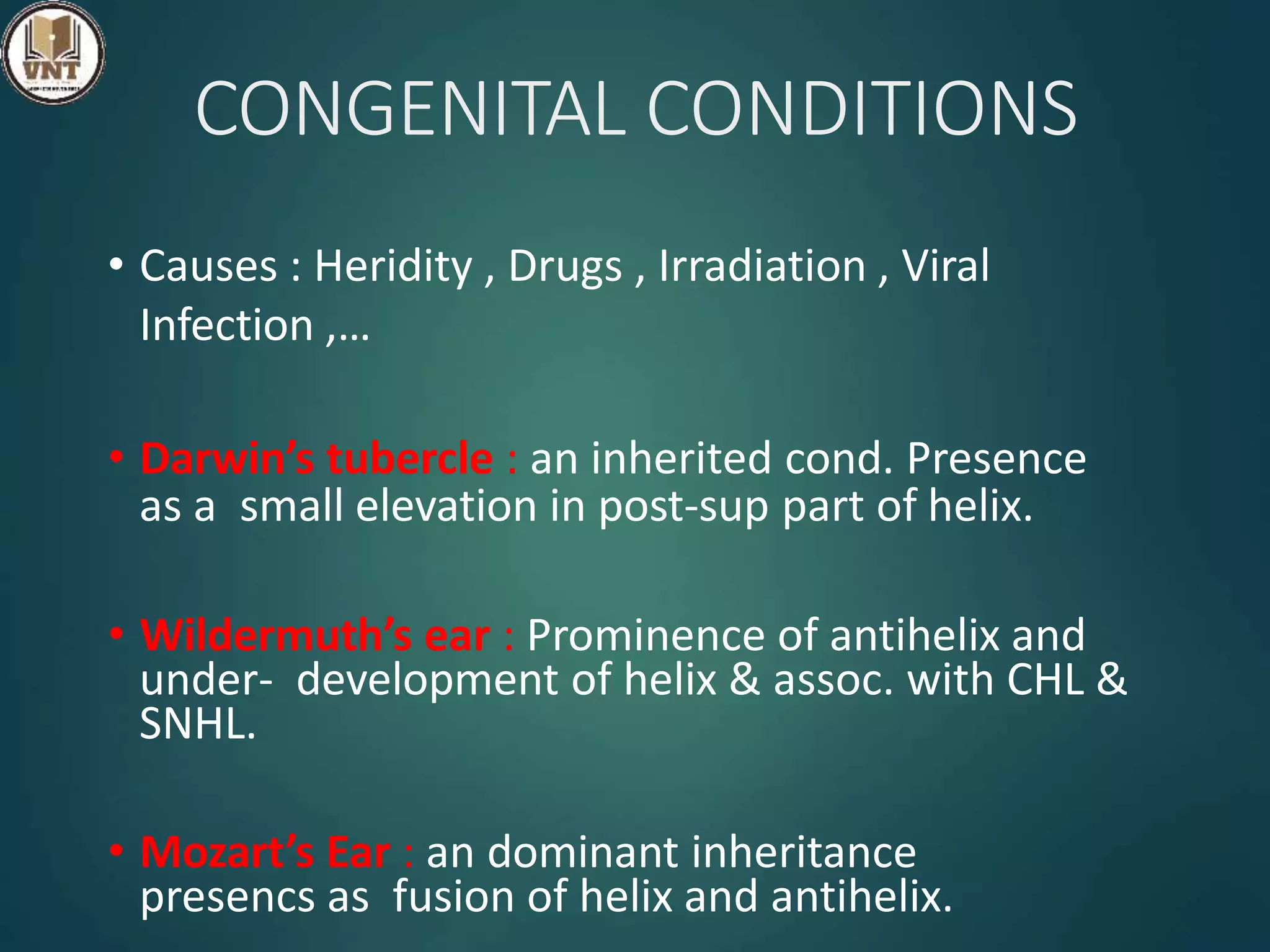
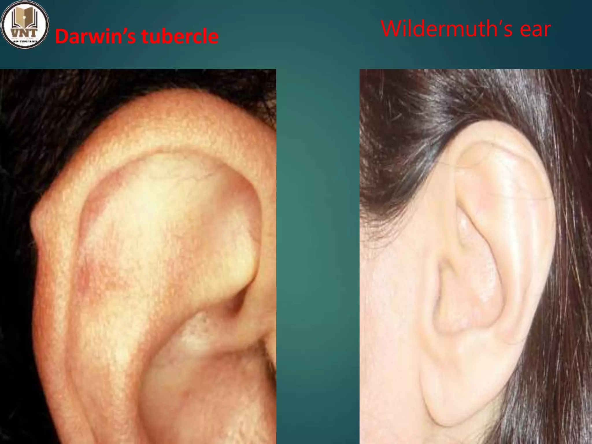
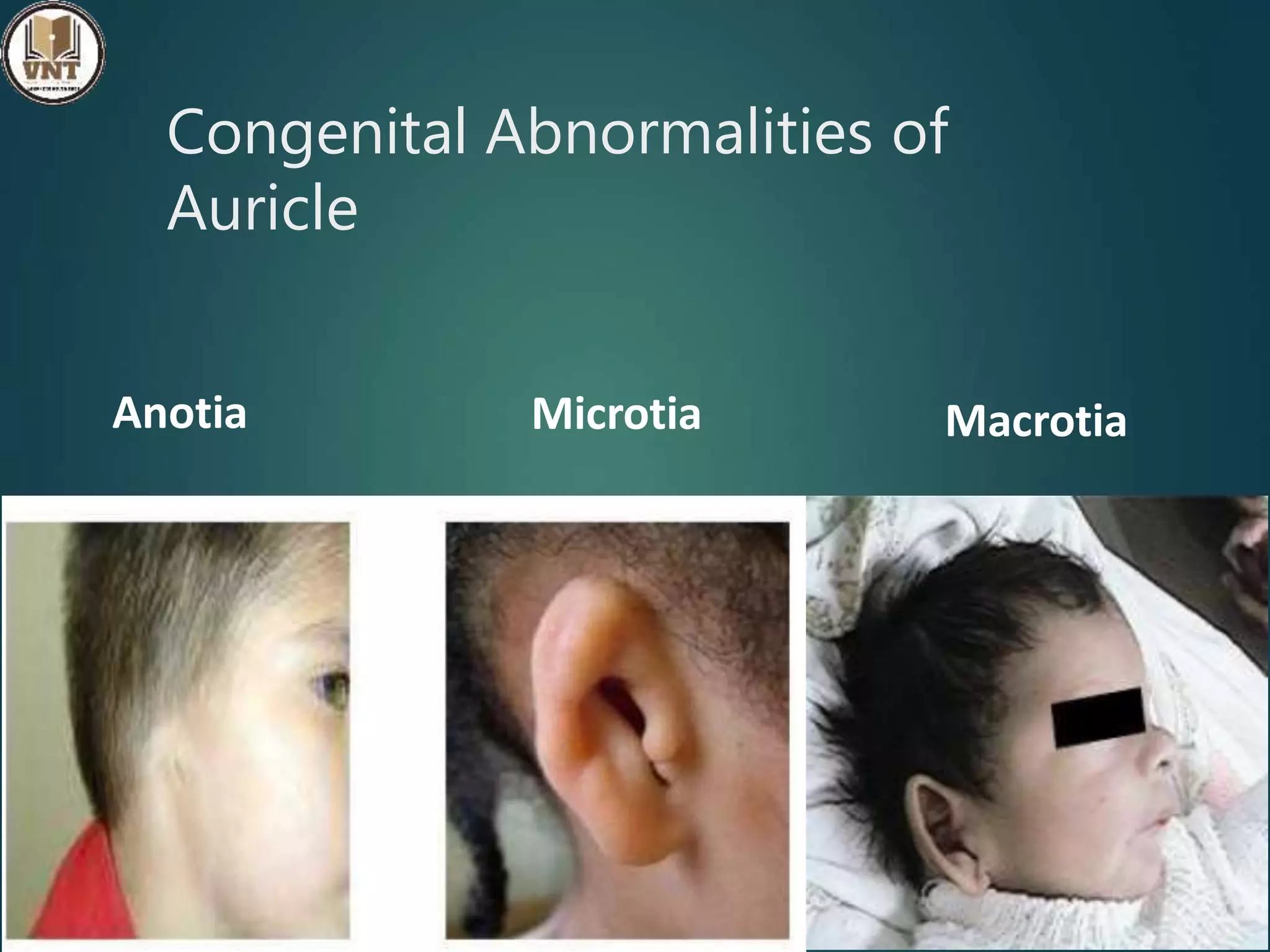
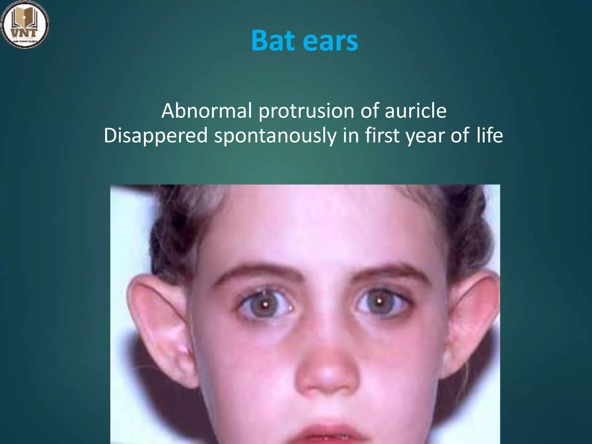
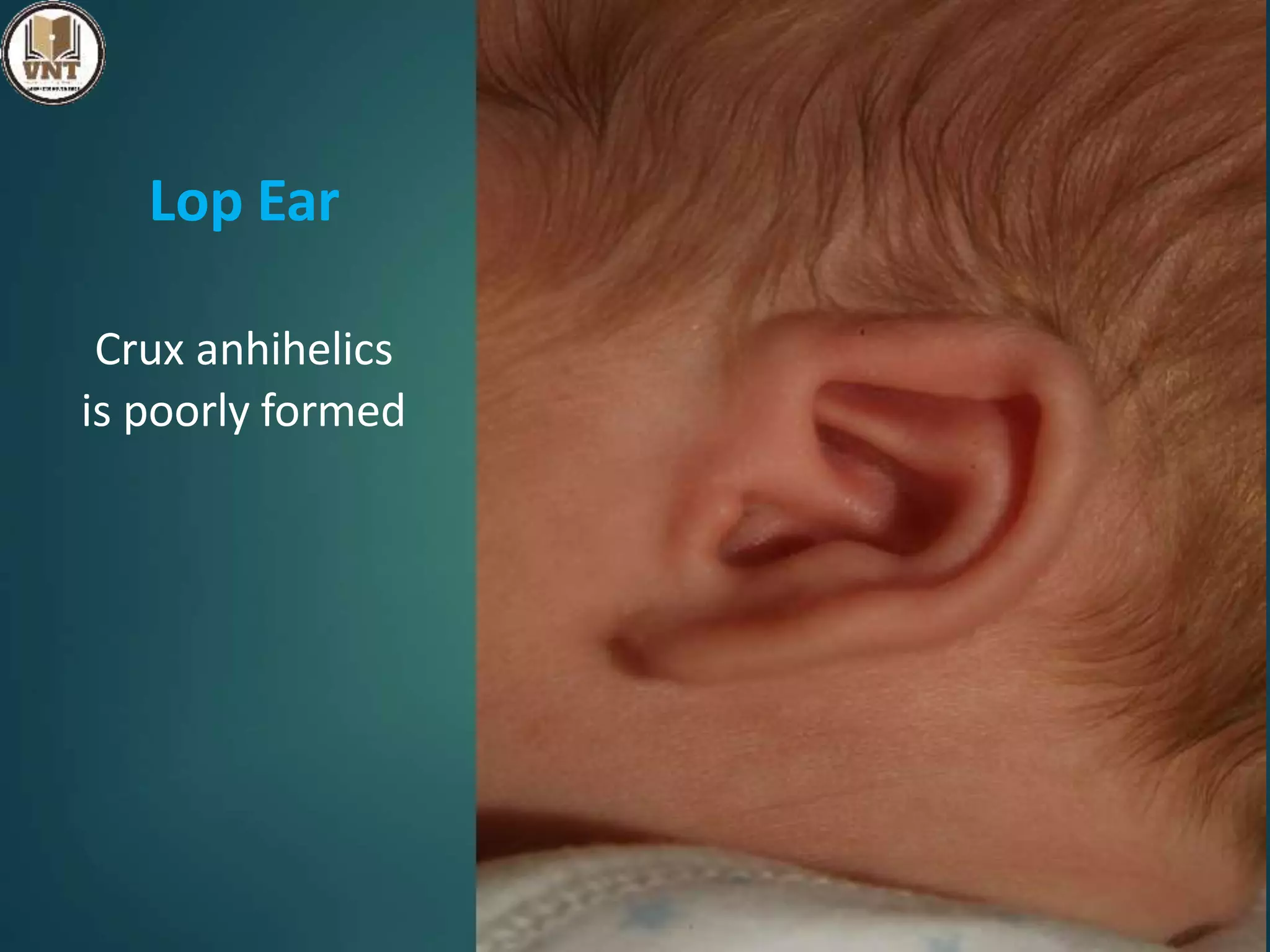
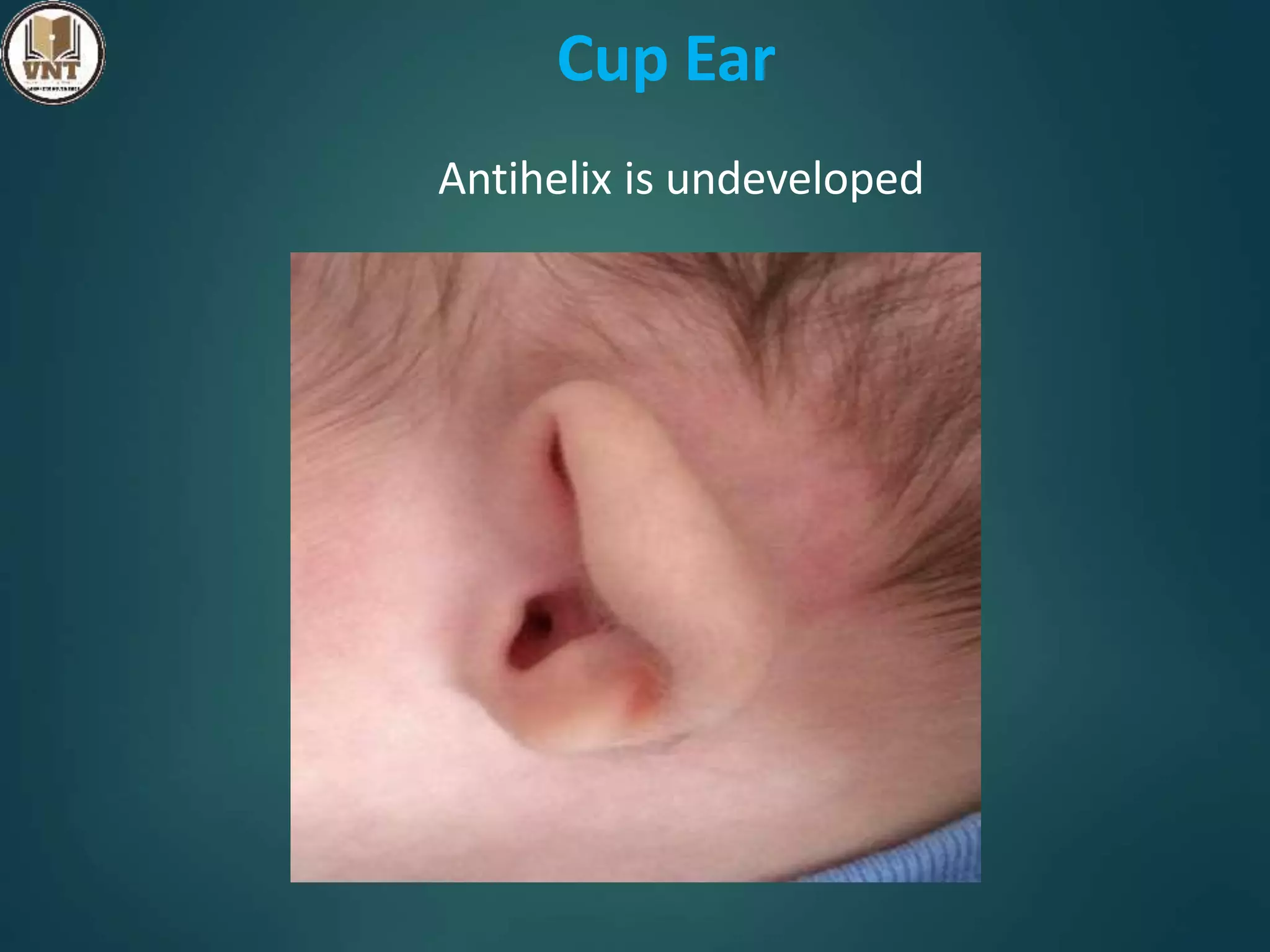
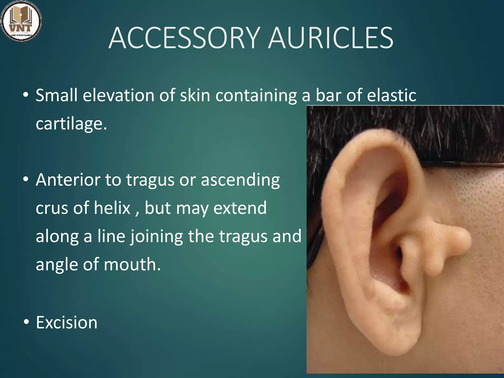
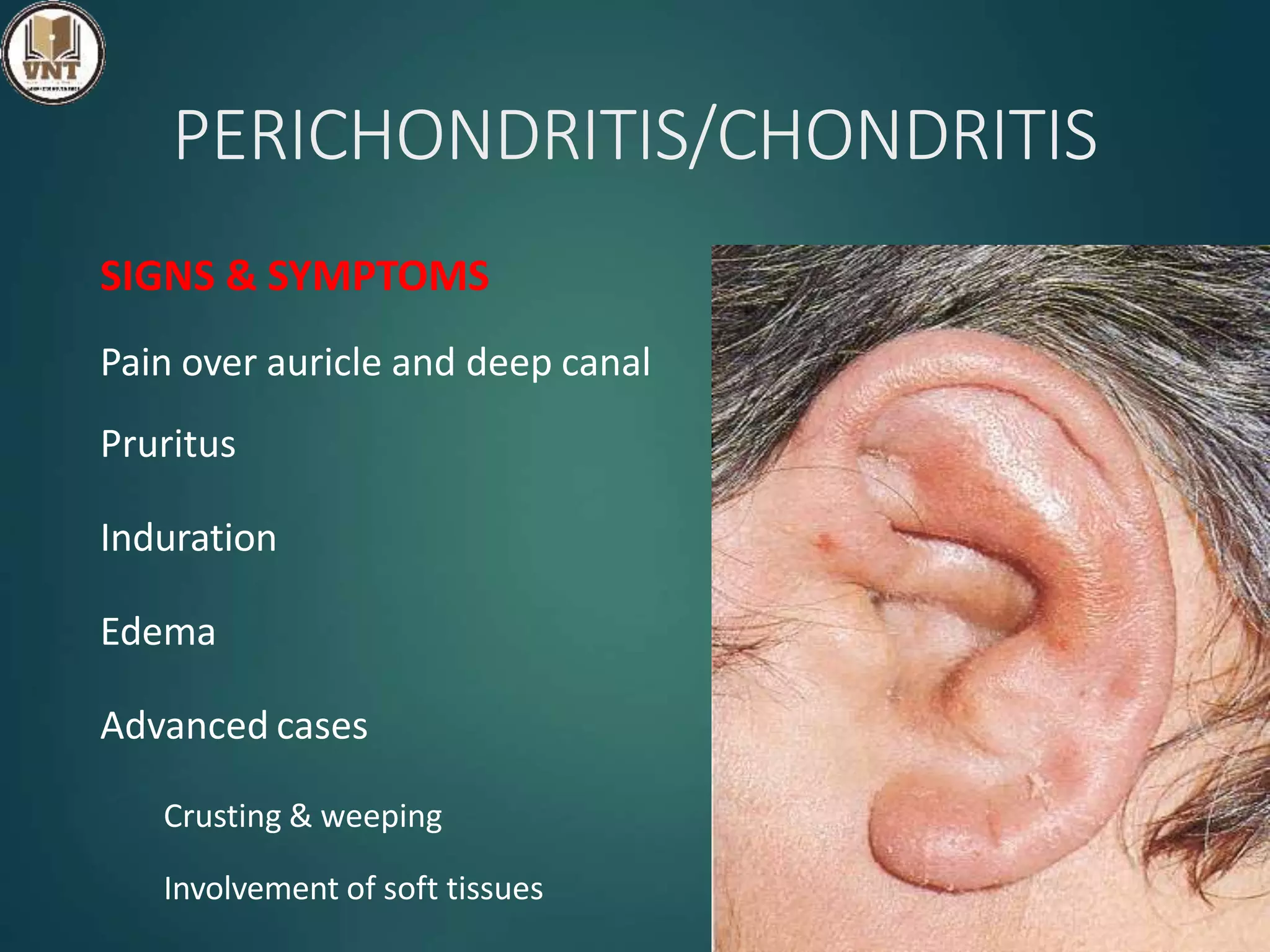
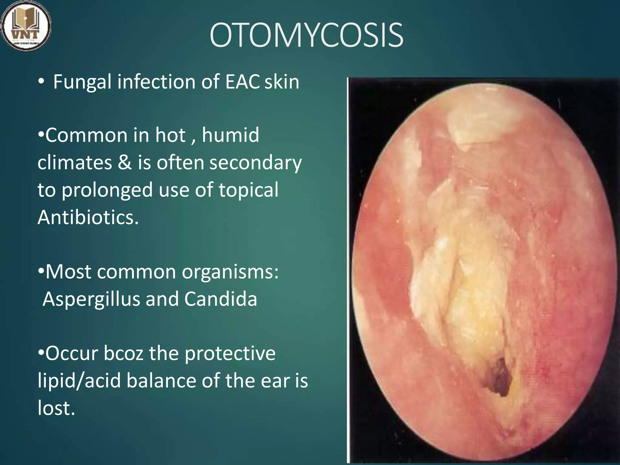
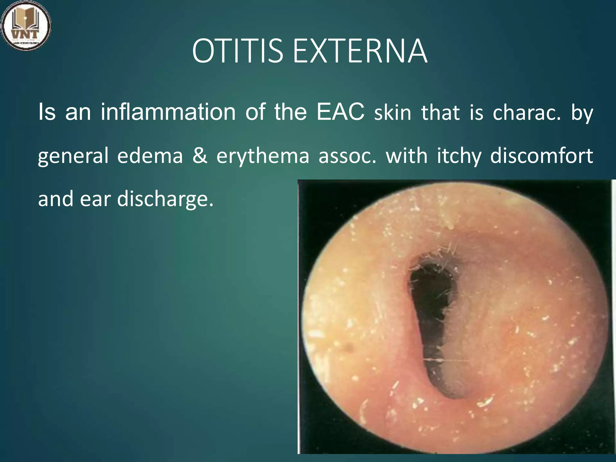
DEFORMITIES
OF
EXTERNAL EAR
BY ,
VEDANTHA VINOD
ASSISTANT LECTURER
CCON-MYSORE
CONTENTS TO BE DISCUSSED :-
Congenital abnormal conditions
External ear inflammation and its
management
CONGENITAL CONDITIONS
• Causes : Heridity , Drugs , Irradiation , Viral
Infection ,…
• Darwin’s tubercle : an inherited cond. Presence
as a small elevation in post-sup part of helix.
• Wildermuth’s ear : Prominence of antihelix and
under- development of helix & assoc. with CHL &
SNHL.
• Mozart’s Ear : an dominant inheritance
presencs as fusion of helix and antihelix.
Darwin’s tubercle Wildermuth’s ear
Congenital Abnormalities of
Auricle
Anotia Microtia Macrotia
Bat ears
Abnormal protrusion of auricle
Disappered spontanously in first year of life
Lop Ear
Crux anhihelics
is poorly formed
Cup Ear
Antihelix is undeveloped
ACCESSORY AURICLES
• Small elevation of skin containing a bar of elastic
cartilage.
• Anterior to tragus or ascending
crus of helix , but may extend
along a line joining the tragus and
angle of mouth.
• Excision
• Faulty fusion of 1st & 2nd arch
• Opening :
1)Anterior border of ascending limb of helix
2)Line extending b/w tragal notch & angle of
mouth
3)Pinna (or) Lobule
• Extend upto the level of tympanic ring.
• C/F : Asymptomatic , If infected – chr.discharge ,
recc.abscess & calculus
• Treatment : Excision ( careful for facial nerve)
PRE – AURICULAR SINUS
Tract : Line joining the angle of mandible & Sterno-
clavicular joint
Outer opening : Ant border of SCM
Inner opening : Bony Cartilagenous junction of EAC
C/F : Discharge fistula , Abscess , Ear discharge ,
Gran.tissue in EAC
Treatment : Excision of fistula
HAEMATOMA AURIS
• Caused by an extravasation of blood b/w the cartilage and the
perichondrium producing a soft doughy swelling of the pinna
• If untreated , blood clot becomes organised and the ear remains
permanently thickened – Cauliflower Ear
• Aspiration with wide bore needle
• Incision (along the margin of helix) & Evacuation of clot
HAEMATOMA AURIS
INFECTION CONDITION OF
EXTERNAL EAR
PERICHONDRITIS/CHONDRITIS
• Infection or inflammation of perichondrium / cartilage of
Auricle & EAC
• Classification
• Erysipelas of External ear ( Inf. of overlying skin)
• Cellulitis of External ear (Inf. of soft tissue )
• Perichondritis ( Inf. Involving perichondrium)
• Chondritis ( Inf. Involving cartilage )
PERICHONDRITIS/CHONDRITIS
•Result of trauma to auricle
• Laceration of auricle , Surgery to ext.ear ,
frostbite , burns , chemical injury , inf. of
hematoma of pinna , high piercing of auricle
for insertion of ear rings.
•Spontaneous (overt diabetes)
•Org : Pseudomonas Aeruginosa , Staph.
Aureus
PERICHONDRITIS/CHONDRITIS
PATHOLOGY :
 Hyperplasia of dermal layers ,
 Thickened subcutaneous tissue ,
 Intense infiltration with PML ,
 Thickening of perichondrium ,
 Destruction of cartilage by phagocytes.
PERICHONDRITIS/CHONDRITIS
SIGNS & SYMPTOMS
Pain over auricle and deep canal
Pruritus
Induration
Edema
Advanced cases
Crusting & weeping
Involvement of soft tissues
PERICHONDRITIS/CHONDRITIS
• TREATMENT :
 Topical & oral antibiotics
 Discharge (or) Abscess – Drainage
 Sub-perichondrial Abscess – I & D
 Irrigating with 1.5 % acetic acid & garamycin
PERICHONDRITIS/CHONDRITIS
PREVENTION
• By careful ear piercings away from cartilaginous
pinna.
• Avoid Surgery in and around ear – to prevent
from trauma
• Hematoma of auricle to drain properly.
• Meticulous management of burn injuries with
prophylatic antibodies against gram neg.
bacteria.
• Removal of eschars and crusts.
FURUNCULOSIS
• Acute localized infection of single hair
follicle.
• Lateral 1/3 of posterosuperior canal
• Obstructed apopilosebaceous unit
• Pathogen: S. aureus
FURUNCULOSIS
SIGNS
• Edema
• Erythema
• Tenderness
• Occasional fluctuance
DD - Ac.mastoiditis
FURUNCULOSIS
SYMPTOMS
• Localized pain
• Ear blockage
• Exudates a scanty sero-sanguinous
discharge
• Pinna & tragus – tender on palpation
• Pruritus
• Hearing loss (if lesion occludes canal)
TREATMENT
• Local heat
• Analgesics
• Oral & systemic anti-staphylococcal antibiotics
• Topical ( antibiotics , Hygroscopic Dehydrating agents)
• Incision and drainage reserved for localized abscess
• IV antibiotics for soft tissue extension
• For recurrent : Eradication theraphy with nasal mupirocin ,
oral flucloxacillin (14 days), Bacterial interferance theraphy
OTOMYCOSIS
• Fungal infection of EAC skin
•Common in hot , humid
climates & is often secondary
to prolonged use of topical
Antibiotics.
•Most common organisms:
Aspergillus and Candida
•Occur bcoz the protective
lipid/acid balance of the ear is
lost.
OTOMYCOSIS
SYMPTOMS :
• Often indistinguishable from bacterial OE
• Pruritus deep within the ear
• Dull pain
• Hearing loss (obstructive)
• Tinnitus
OTOMYCOSIS
• Canal erythema
• Mild edema
• White, grey ,green , yellow or black fungal debris
( wet newspaper)
Aspergillus Candida
OTOMYCOSIS
TREATMENT
• Thorough aural toilet & removal of debris
• Topical antifungals
• Resistant otomycosis – Exclude fungal inf. anywhere
including Athelete’s foot .
• Immunotheraphy with Trichophyton , Epidermophyton &
oidomycetes extracts and dust mite , is the treatment of
choice.
OTITIS EXTERNA
Is an inflammation of the EAC skin that is charac. by
general edema & erythema assoc. with itchy discomfort
and ear discharge.
• Predisposing factors :
• Anatomical ( narrow / obstructed ear canal) ,
• Dermatological ( Eczema , Sebhorrhoeic dermatitis )
• Allergic ( Atopy , Non–atopy , Exposure to top.med)
• Physiological ( Humid environment , Imm.compramised)
• Traumatic ( Skin maceration , ear probing , rad.theraphy )
• Microbiological ( P.aeruginosa , Active COM , Fungi )
OTITIS EXTERNA
OTITIS EXTERNA
• Any cond. that disturbs the lipid/acid balance of the
ear will predispose.
• Secondary Bacterial Infection :
• MR – Staph aureus , Pseud aeruginosa ,
Streptococci , other gram (-)ve organisms.
• Bathing :
• In fresh water lakes containing Pseud.aeruginosa
“swimmer’s ear”
Edema of stratum corneum and plugging of apo-pilo
sebaceous unit
Starts the itch / scratch cycle
Symptoms: Pruritus and Sense of fullness
Signs: Mild edema
ACUTE OTITIS EXTERNA
Progressive infection
Symptoms
• Pain
• Increased pruritus
Signs
• Erythema
• Increasing edema
• Canal debris, discharge
AOE: SEVERE
STAGE
• Severe pain, worse with
ear movement
Signs
• Lumen obliteration
• Purulent otorrhea
• Involvement of
periauricular soft tissue
AOE: TREATMENT
Frequent canal cleaning ( Aural Toilet )
Topical Medications ( IG pack )
Pain control ( NSAIDS )
Instructions for prevention
 Avoidance of water pentration into ear
 Cotton wool with petroleum jelly
 Custom made ear moulds
COE : SIGNS & SYMPTOMS
• Unrelenting pruritus
• Dryness of canal skin
• Hypertrophied skin
• Mucopurulent
otorrhea
CHRONIC OTITIS EXTERNA
COE: TREATMENT
• Topical antibiotics, frequent cleanings
• Topical Steroids
• Surgical intervention
• Failure of medical treatment
• Toenlarge and resurface the EAC
GRANULAR MYRINGITIS
Localized chronic inflammation of pars tensa with granulation
tissue with possible involvement of EAC
Causes : High temp , swimming , lack of hygeine , local
irritants , foreign body , bacterial & fungal infections
Common organisms: Pseudomonas , Proteus , Staph aureus
& Candida albicans
Sequela of Acute myringitis, Previous OE, TM Perforation
GRANULAR MYRINGITIS
Myringitis Externa Granulosa
Has granulation on lateral surface of drum & medial
part of the ear canal skin
Granular Myringitis
Involves only the ear drum
GRANULAR MYRINGITIS
PATHOLOGY
• Odematous granulation tissue with capillaries and
diffuse infiltration of chronic inflammatory cells
• Injury involving lamina propria of the tympanic
membrance supresses epithelization – development
of granulation tissue
GRANULAR MYRINGITIS
PATHOLOGY
• Odematous granulation tissue with capillaries and
diffuse infiltration of chronic inflammatory cells
• Injury involving lamina propria of the tympanic
membrance supresses epithelization – development
of granulation tissue
GRANULAR MYRINGITIS
SIGNS & SYMPTOMS
• Foul smelling discharge from one ear
• Slight irritation or fullness
• No hearing loss
• No significant pain
• TM obscured by pus
• Posterio-superior granulations
• No TM perforations
GRANULAR MYRINGITIS
• Careful and frequent debridement
• Specific anti-microbial drops or powder with or without
steroids for 2 weeks
• Removal of granulation by physical methods
• Appln of caustic agents – Chromic acid , 0.5 % formalin ,
silver nitrate
• Laser evaporation of granulation
BULLOUS MYRINGITIS
• Myringitis Bullosa Hemorrhagica – finding of vesicles in
the superficial layer of TM
• Confined b/w outer epithelium & lamina propria of
tympanic membrane
• Viral infection ( Influenza ) , Mycoplasma pnuemoniae
• Primarily involves younger children
BULLOUS MYRINGITIS
• Inflammation limited to TM & nearby canal
• Multiple reddened,
inflamed blebs
• Hemorrhagic vesicles
BULLOUS MYRINGITIS
• Sudden , unilateral throbbing pain
• Blood stained discahrge
• Hearing loss
Otoscopy
• Serous (or) sero-sanginous discharge blisters in TM &
medial part of Ear canal
BULLOUS MYRINGITIS: TREATMENT
 Self-limiting
 Analgesics
 Topical antibiotics to prevent secondary infection
 Incision of blebs is unnecessary
NECROTIZING OTITIS EXTERNA
• is the clinical cond. of idiopathic necrosis of a localised
area of the bone of the tympanic ring , with secondary
inflammation of the overlying soft tissue and skin.
• Causative organism : Staph aureus
• TM is suspectible to osteonecrosis
poor vascular supply
bcoz’ of its relatively
• Repeated local trauma – ear bud abuse , pricking of ear ,
use of hearing aids.
NECROTIZING OTITIS EXTERNA
• Poorly controlled diabetic with h/o OE
• Deep-seated aural pain
• Chronic otorrhea
• Aural fullness
• Pruritis
• Hearing loss
NECROTIZING OTITIS EXTERNA
• Small area of deficient skin and soft tissue in EAC
revealing a segment of necrotic bone
• Purulent secretions
• Occluded canal and obscured TM
• Cranial nerve involvement
NECROTIZING OTITIS EXTERNA
• Pus swab
• CT Scan – extent of bone necrosis
• Brush cytology & Biopsy – to exclude neoplasm
• Audiometry
• Syphillis & TB should be excluded.
NECROTIZING OTITIS EXTERNA
• Intravenous antibiotics for at least 4 weeks
• Local canal debridement until healed
• Pain control
• Use of topical agents - controversial
• Hyperbaric oxygen – necrosis beyond tympanic plate
• Surgical debridement
DISCUSSED CONTENTS :-
 Congenital abnormal conditions
 External ear inflammation and its management
THANK YOU

Diseases of external ear

  • 1.
    DEFORMITIES OF EXTERNAL EAR BY , VEDANTHAVINOD ASSISTANT LECTURER CCON-MYSORE
  • 2.
    CONTENTS TO BEDISCUSSED :- Congenital abnormal conditions External ear inflammation and its management
  • 3.
    CONGENITAL CONDITIONS • Causes: Heridity , Drugs , Irradiation , Viral Infection ,… • Darwin’s tubercle : an inherited cond. Presence as a small elevation in post-sup part of helix. • Wildermuth’s ear : Prominence of antihelix and under- development of helix & assoc. with CHL & SNHL. • Mozart’s Ear : an dominant inheritance presencs as fusion of helix and antihelix.
  • 4.
  • 5.
  • 6.
    Bat ears Abnormal protrusionof auricle Disappered spontanously in first year of life
  • 7.
  • 8.
  • 9.
    ACCESSORY AURICLES • Smallelevation of skin containing a bar of elastic cartilage. • Anterior to tragus or ascending crus of helix , but may extend along a line joining the tragus and angle of mouth. • Excision
  • 10.
    • Faulty fusionof 1st & 2nd arch • Opening : 1)Anterior border of ascending limb of helix 2)Line extending b/w tragal notch & angle of mouth 3)Pinna (or) Lobule • Extend upto the level of tympanic ring. • C/F : Asymptomatic , If infected – chr.discharge , recc.abscess & calculus • Treatment : Excision ( careful for facial nerve)
  • 11.
  • 12.
    Tract : Linejoining the angle of mandible & Sterno- clavicular joint Outer opening : Ant border of SCM Inner opening : Bony Cartilagenous junction of EAC C/F : Discharge fistula , Abscess , Ear discharge , Gran.tissue in EAC Treatment : Excision of fistula
  • 13.
    HAEMATOMA AURIS • Causedby an extravasation of blood b/w the cartilage and the perichondrium producing a soft doughy swelling of the pinna • If untreated , blood clot becomes organised and the ear remains permanently thickened – Cauliflower Ear • Aspiration with wide bore needle • Incision (along the margin of helix) & Evacuation of clot
  • 14.
  • 15.
  • 16.
    PERICHONDRITIS/CHONDRITIS • Infection orinflammation of perichondrium / cartilage of Auricle & EAC • Classification • Erysipelas of External ear ( Inf. of overlying skin) • Cellulitis of External ear (Inf. of soft tissue ) • Perichondritis ( Inf. Involving perichondrium) • Chondritis ( Inf. Involving cartilage )
  • 17.
    PERICHONDRITIS/CHONDRITIS •Result of traumato auricle • Laceration of auricle , Surgery to ext.ear , frostbite , burns , chemical injury , inf. of hematoma of pinna , high piercing of auricle for insertion of ear rings. •Spontaneous (overt diabetes) •Org : Pseudomonas Aeruginosa , Staph. Aureus
  • 18.
    PERICHONDRITIS/CHONDRITIS PATHOLOGY :  Hyperplasiaof dermal layers ,  Thickened subcutaneous tissue ,  Intense infiltration with PML ,  Thickening of perichondrium ,  Destruction of cartilage by phagocytes.
  • 19.
    PERICHONDRITIS/CHONDRITIS SIGNS & SYMPTOMS Painover auricle and deep canal Pruritus Induration Edema Advanced cases Crusting & weeping Involvement of soft tissues
  • 20.
    PERICHONDRITIS/CHONDRITIS • TREATMENT : Topical & oral antibiotics  Discharge (or) Abscess – Drainage  Sub-perichondrial Abscess – I & D  Irrigating with 1.5 % acetic acid & garamycin
  • 21.
    PERICHONDRITIS/CHONDRITIS PREVENTION • By carefulear piercings away from cartilaginous pinna. • Avoid Surgery in and around ear – to prevent from trauma • Hematoma of auricle to drain properly. • Meticulous management of burn injuries with prophylatic antibodies against gram neg. bacteria. • Removal of eschars and crusts.
  • 22.
    FURUNCULOSIS • Acute localizedinfection of single hair follicle. • Lateral 1/3 of posterosuperior canal • Obstructed apopilosebaceous unit • Pathogen: S. aureus
  • 23.
    FURUNCULOSIS SIGNS • Edema • Erythema •Tenderness • Occasional fluctuance DD - Ac.mastoiditis
  • 24.
    FURUNCULOSIS SYMPTOMS • Localized pain •Ear blockage • Exudates a scanty sero-sanguinous discharge • Pinna & tragus – tender on palpation • Pruritus • Hearing loss (if lesion occludes canal)
  • 25.
    TREATMENT • Local heat •Analgesics • Oral & systemic anti-staphylococcal antibiotics • Topical ( antibiotics , Hygroscopic Dehydrating agents) • Incision and drainage reserved for localized abscess • IV antibiotics for soft tissue extension • For recurrent : Eradication theraphy with nasal mupirocin , oral flucloxacillin (14 days), Bacterial interferance theraphy
  • 26.
    OTOMYCOSIS • Fungal infectionof EAC skin •Common in hot , humid climates & is often secondary to prolonged use of topical Antibiotics. •Most common organisms: Aspergillus and Candida •Occur bcoz the protective lipid/acid balance of the ear is lost.
  • 27.
    OTOMYCOSIS SYMPTOMS : • Oftenindistinguishable from bacterial OE • Pruritus deep within the ear • Dull pain • Hearing loss (obstructive) • Tinnitus
  • 28.
    OTOMYCOSIS • Canal erythema •Mild edema • White, grey ,green , yellow or black fungal debris ( wet newspaper)
  • 29.
  • 30.
    OTOMYCOSIS TREATMENT • Thorough auraltoilet & removal of debris • Topical antifungals • Resistant otomycosis – Exclude fungal inf. anywhere including Athelete’s foot . • Immunotheraphy with Trichophyton , Epidermophyton & oidomycetes extracts and dust mite , is the treatment of choice.
  • 31.
    OTITIS EXTERNA Is aninflammation of the EAC skin that is charac. by general edema & erythema assoc. with itchy discomfort and ear discharge.
  • 32.
    • Predisposing factors: • Anatomical ( narrow / obstructed ear canal) , • Dermatological ( Eczema , Sebhorrhoeic dermatitis ) • Allergic ( Atopy , Non–atopy , Exposure to top.med) • Physiological ( Humid environment , Imm.compramised) • Traumatic ( Skin maceration , ear probing , rad.theraphy ) • Microbiological ( P.aeruginosa , Active COM , Fungi ) OTITIS EXTERNA
  • 33.
    OTITIS EXTERNA • Anycond. that disturbs the lipid/acid balance of the ear will predispose. • Secondary Bacterial Infection : • MR – Staph aureus , Pseud aeruginosa , Streptococci , other gram (-)ve organisms. • Bathing : • In fresh water lakes containing Pseud.aeruginosa “swimmer’s ear”
  • 34.
    Edema of stratumcorneum and plugging of apo-pilo sebaceous unit Starts the itch / scratch cycle Symptoms: Pruritus and Sense of fullness Signs: Mild edema ACUTE OTITIS EXTERNA
  • 35.
    Progressive infection Symptoms • Pain •Increased pruritus Signs • Erythema • Increasing edema • Canal debris, discharge
  • 36.
    AOE: SEVERE STAGE • Severepain, worse with ear movement Signs • Lumen obliteration • Purulent otorrhea • Involvement of periauricular soft tissue
  • 37.
    AOE: TREATMENT Frequent canalcleaning ( Aural Toilet ) Topical Medications ( IG pack ) Pain control ( NSAIDS ) Instructions for prevention  Avoidance of water pentration into ear  Cotton wool with petroleum jelly  Custom made ear moulds
  • 38.
    COE : SIGNS& SYMPTOMS • Unrelenting pruritus • Dryness of canal skin • Hypertrophied skin • Mucopurulent otorrhea CHRONIC OTITIS EXTERNA
  • 39.
    COE: TREATMENT • Topicalantibiotics, frequent cleanings • Topical Steroids • Surgical intervention • Failure of medical treatment • Toenlarge and resurface the EAC
  • 40.
    GRANULAR MYRINGITIS Localized chronicinflammation of pars tensa with granulation tissue with possible involvement of EAC Causes : High temp , swimming , lack of hygeine , local irritants , foreign body , bacterial & fungal infections Common organisms: Pseudomonas , Proteus , Staph aureus & Candida albicans Sequela of Acute myringitis, Previous OE, TM Perforation
  • 41.
    GRANULAR MYRINGITIS Myringitis ExternaGranulosa Has granulation on lateral surface of drum & medial part of the ear canal skin Granular Myringitis Involves only the ear drum
  • 42.
    GRANULAR MYRINGITIS PATHOLOGY • Odematousgranulation tissue with capillaries and diffuse infiltration of chronic inflammatory cells • Injury involving lamina propria of the tympanic membrance supresses epithelization – development of granulation tissue
  • 43.
    GRANULAR MYRINGITIS PATHOLOGY • Odematousgranulation tissue with capillaries and diffuse infiltration of chronic inflammatory cells • Injury involving lamina propria of the tympanic membrance supresses epithelization – development of granulation tissue
  • 44.
    GRANULAR MYRINGITIS SIGNS &SYMPTOMS • Foul smelling discharge from one ear • Slight irritation or fullness • No hearing loss • No significant pain • TM obscured by pus • Posterio-superior granulations • No TM perforations
  • 45.
    GRANULAR MYRINGITIS • Carefuland frequent debridement • Specific anti-microbial drops or powder with or without steroids for 2 weeks • Removal of granulation by physical methods • Appln of caustic agents – Chromic acid , 0.5 % formalin , silver nitrate • Laser evaporation of granulation
  • 46.
    BULLOUS MYRINGITIS • MyringitisBullosa Hemorrhagica – finding of vesicles in the superficial layer of TM • Confined b/w outer epithelium & lamina propria of tympanic membrane • Viral infection ( Influenza ) , Mycoplasma pnuemoniae • Primarily involves younger children
  • 47.
    BULLOUS MYRINGITIS • Inflammationlimited to TM & nearby canal • Multiple reddened, inflamed blebs • Hemorrhagic vesicles
  • 48.
    BULLOUS MYRINGITIS • Sudden, unilateral throbbing pain • Blood stained discahrge • Hearing loss Otoscopy • Serous (or) sero-sanginous discharge blisters in TM & medial part of Ear canal
  • 49.
    BULLOUS MYRINGITIS: TREATMENT Self-limiting  Analgesics  Topical antibiotics to prevent secondary infection  Incision of blebs is unnecessary
  • 50.
    NECROTIZING OTITIS EXTERNA •is the clinical cond. of idiopathic necrosis of a localised area of the bone of the tympanic ring , with secondary inflammation of the overlying soft tissue and skin. • Causative organism : Staph aureus • TM is suspectible to osteonecrosis poor vascular supply bcoz’ of its relatively • Repeated local trauma – ear bud abuse , pricking of ear , use of hearing aids.
  • 51.
    NECROTIZING OTITIS EXTERNA •Poorly controlled diabetic with h/o OE • Deep-seated aural pain • Chronic otorrhea • Aural fullness • Pruritis • Hearing loss
  • 52.
    NECROTIZING OTITIS EXTERNA •Small area of deficient skin and soft tissue in EAC revealing a segment of necrotic bone • Purulent secretions • Occluded canal and obscured TM • Cranial nerve involvement
  • 53.
    NECROTIZING OTITIS EXTERNA •Pus swab • CT Scan – extent of bone necrosis • Brush cytology & Biopsy – to exclude neoplasm • Audiometry • Syphillis & TB should be excluded.
  • 54.
    NECROTIZING OTITIS EXTERNA •Intravenous antibiotics for at least 4 weeks • Local canal debridement until healed • Pain control • Use of topical agents - controversial • Hyperbaric oxygen – necrosis beyond tympanic plate • Surgical debridement
  • 55.
    DISCUSSED CONTENTS :- Congenital abnormal conditions  External ear inflammation and its management
  • 56.