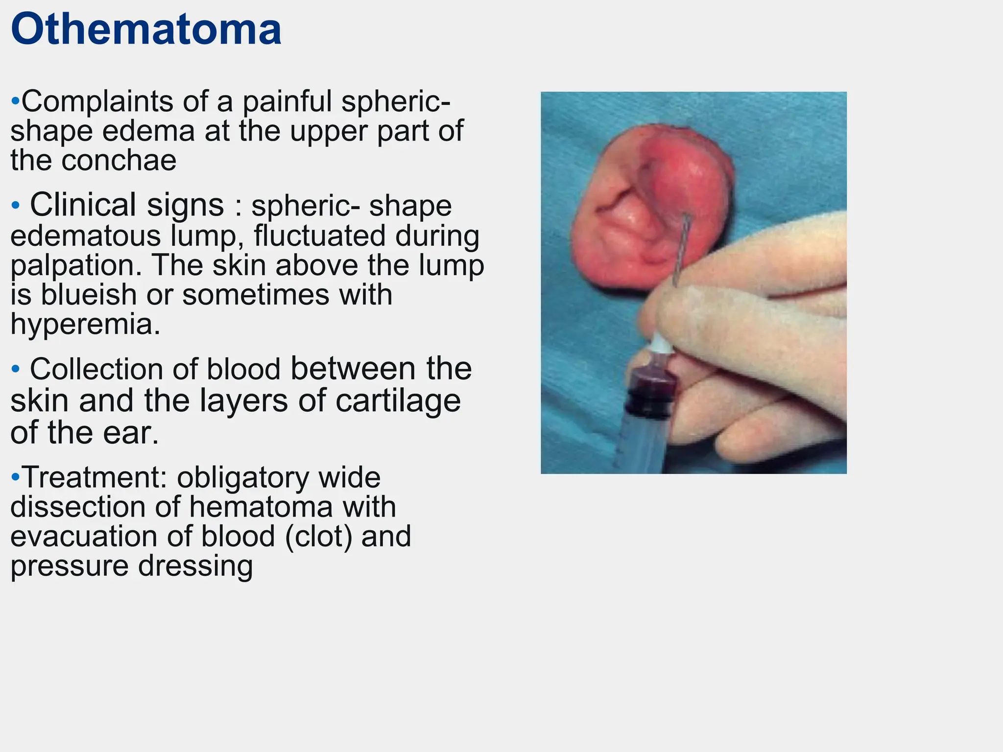
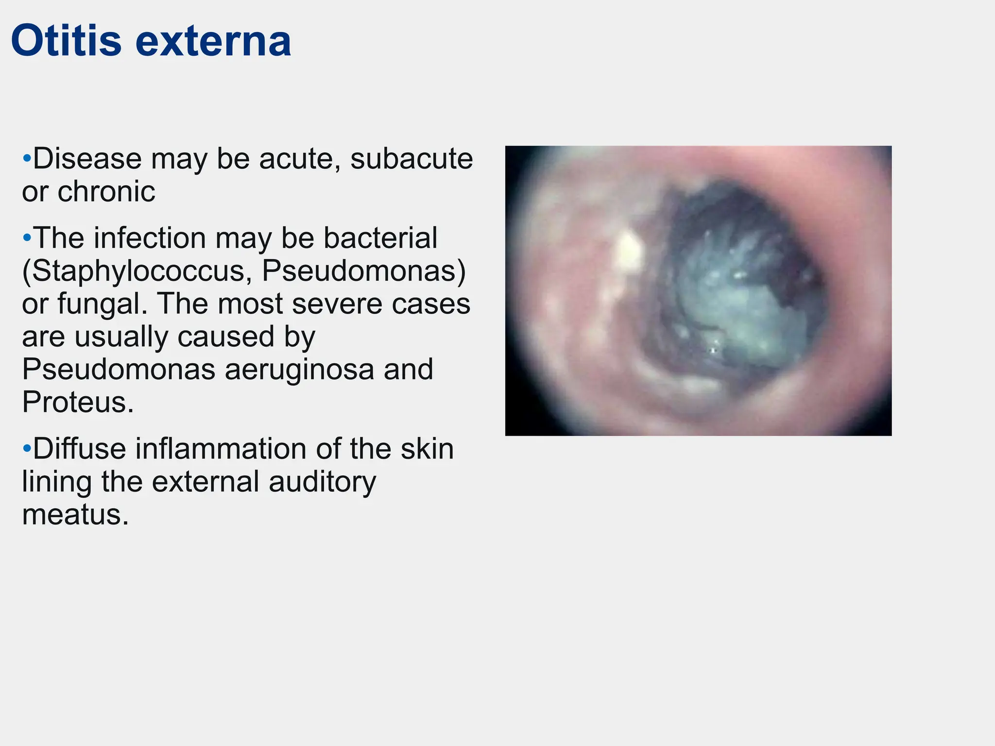
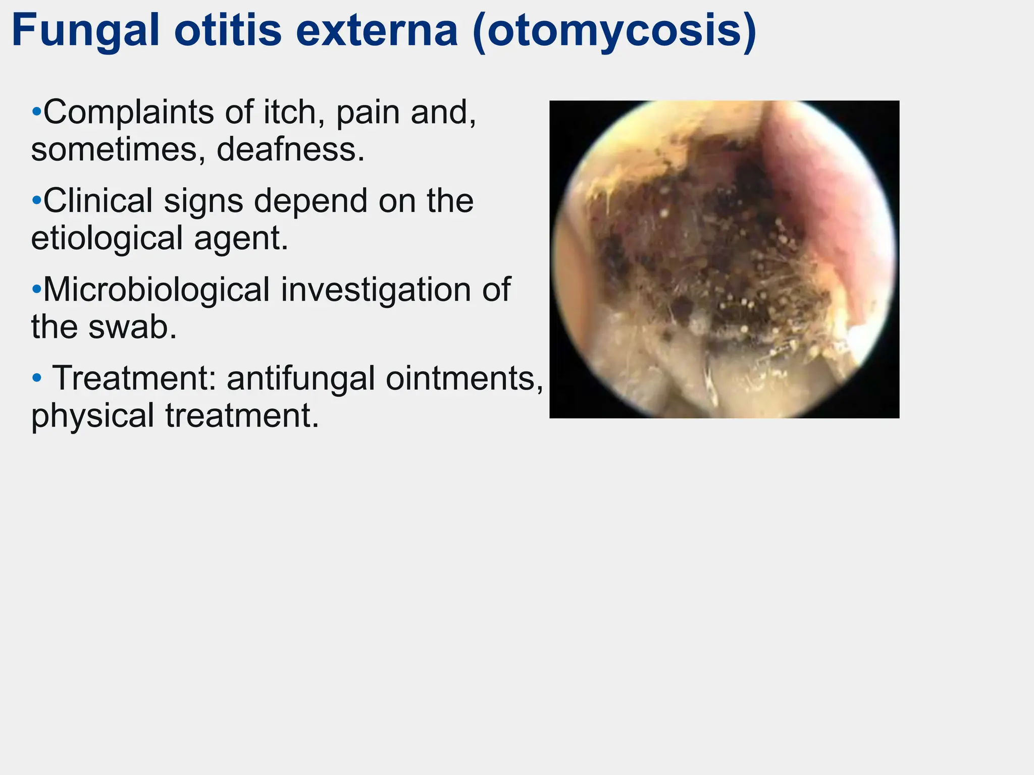
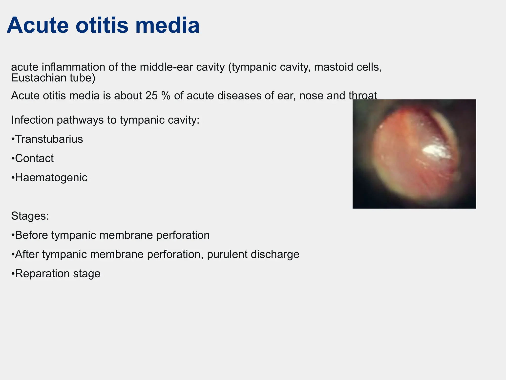
External ear diseases. Acute and chronic
otitis media. Otogenic intracranial
complications.
Othematoma
•Complaints of a painful spheric-
shape edema at the upper part of
the conchae
• Clinical signs : spheric- shape
edematous lump, fluctuated during
palpation. The skin above the lump
is blueish or sometimes with
hyperemia.
• Collection of blood between the
skin and the layers of cartilage
of the ear.
•Treatment: obligatory wide
dissection of hematoma with
evacuation of blood (clot) and
pressure dressing
Perichondritis
•Complaints of pain, edema, hyperemia.
Lobe is intact.
•In some cases there is a fluctuation.
•Inflammation of the perichondrium.
•There are two forms: catarrhal and
purulent.
•Treatment of catarrhal form is
conservative (physical therapy and
antibiotics).
•Treatment of purulent form is surgical
(dissection and damaged cartilage
resection).
•In severe cases perichondritis may lead
to pinna deformity.
Erysipelas
•Cause: Streptococcal infection
may enter the skin through minor
trauma or chap.
•Complaints of pain, edema, well-
demarcated rash of the pinna.
•Treatment involves either oral or
intravenous antibiotics,
using penicillins, clindamycin,
or erythromycin. Physical treatment
can be used.
Eczema
•The most common predictor is
purulent discharge in patients with
chronic otitis media. Risk factors
include diabetes mellitus and allergy.
•There are two different types of
eczema:«dry» and «moist» eczema.
•The skin of the pinna, external
auditory canal and postauricular region
may be involved in a process.
•Symptoms: persistent itching (patients
damage the skin due to scratching and
provoke the disease elongation)
•Treatment: ointments with
corticosteroids.
Furunculosis
•Causes: local (different traumas of
the external auditory canal) and
common (diabetes mellitus).
•There are two stages: infiltration and
suppuration.
•Symptoms: severe pain, that is made
much worse by movement of the
pinna or pressure on the tragus or
mouth opening and chewing.
•There is often no visible lesion during
infiltration stage but the introduction of
an aural speculum causes intense
pain.
•If the furuncle is larger, it will be seen
as a red swelling in the outer meatus.
At a more advanced stage, the
furuncle will be seen to be pointing or
may present as a fluctuant abscess.
•Treatment may be conservative
(infiltration stage) or surgical
(suppuration stage).
Otitis externa
•Disease may be acute, subacute
or chronic
•The infection may be bacterial
(Staphylococcus, Pseudomonas)
or fungal. The most severe cases
are usually caused by
Pseudomonas aeruginosa and
Proteus.
•Diffuse inflammation of the skin
lining the external auditory
meatus.
Fungal otitis externa (otomycosis)
•Complaints of itch, pain and,
sometimes, deafness.
•Clinical signs depend on the
etiological agent.
•Microbiological investigation of
the swab.
• Treatment: antifungal ointments,
physical treatment.
Malignant otitis externa
(necrotizing otitis externa)
•Soft tissue, auricular cartilage and bone
are involved in a pathology.
•The process may spread into the cranial
base and even to the other side.
•More common in elderly patients with
diabetes mellitus or immunodeficiency.
•Treatment: antibiotics, in case of resistant
to antibiotics or significant tissue damage
(surgical excision of the damaged
tissue).
Acute otitis media
acute inflammation of the middle-ear cavity (tympanic cavity, mastoid cells,
Eustachian tube)
Acute otitis media is about 25 % of acute diseases of ear, nose and throat
Infection pathways to tympanic cavity:
•Transtubarius
•Contact
•Haematogenic
Stages:
•Before tympanic membrane perforation
•After tympanic membrane perforation, purulent discharge
•Reparation stage
Pre-perforation stage
•Symptoms: pain, deafness,
tinnitus.
•Otoscope signs: redness of
the tympanic membrane (at
the beginning localized only
around the malleus handle,
after that radial and then total);
bulging of tympanic membrane
with loss of landmarks.
Perforative stage
After perforation or paracentesis purulent
discharge is decreasing or discontinued,
pain resolves, but the deafness and
tinnitus still present.
•Otoscopy: redness and fullness of the
drum, perforation with otorrhoea, which
will often be blood-stained. Profuse and
mucoid at first, later becoming thick and
yellow.
Treatment of acute otitis media
• The restoration of the Eustachian tube function (nasal
vasoconstrictors).
• Antibiotic therapy.
• Topical treatment (ear drops).
• At the stage of purulent discharge topical antibiotics
and corticosteroids.
• Paracentesis (miringotomy)– the incision at the
posterior-inferior part of the tympanic membrane.
Paracentesis
Indications: bulging of the tympanic
membrane at the stage of pre-perforative
acute purulent otitis media
Mastoiditis– acute purulent inflamation ot
the mastoid
• primary (posttraumatic, specific infections,
infectious disease)
• secondary (complication of acute otitis media
or the exaceboration of the chronic otitis)
Signs and symptoms of
mastoiditis
• Pain worsening
• Conductive hearing loss
• Local signs:
-redness of tympanic membrane,
- swelling and hanging of the posterior-
superior external auditory canal wall,
- purulent discharge ( «reservoir» symptom)–
not obliratory,
- swelling in the postauricular region, with
obliteration of the sulcus
- pinna is pushed down and forward
• Radiological features:
- opacification of the mastoid air cells
- erosion of mastoid air cell bony septum may
be present in coalescent mastoiditis
Occasional features of acute mastoiditis
• Subperiosteal abscess over the mastoid
process.
• Bezold’s abscess —pus breaks through
the mastoid tip and forms an abscess in
the neck.
• Zygomatic mastoiditis —results in
swelling over the zygoma.
Types of surgical treatment
• Partial antromastoidectomy (mastoid trepanation for
pathological tissues removing and tympanic cavity
drainage)
• Expanded antromastoidectomy (trepanation of all
mastoid cells with the dura mater and/or sigmoid sinus
wall revision)
• Antrotomy (in case pf so called antritis in children
under 1 year old)
Antromastoidectomy
Indications to expanded
antromastoidectomy
• Intracranial complications (otogenic meningitis,
sigmoid sinus trombosis (otogenic sepsis), brain
abscess ( temporal lobe abscess or cerebellum
abscess, occurred as an outcome of acute purulent
otitis media)

Acute and chronic middle otitis prophylaxis

  • 1.
    External ear diseases.Acute and chronic otitis media. Otogenic intracranial complications.
  • 2.
    Othematoma •Complaints of apainful spheric- shape edema at the upper part of the conchae • Clinical signs : spheric- shape edematous lump, fluctuated during palpation. The skin above the lump is blueish or sometimes with hyperemia. • Collection of blood between the skin and the layers of cartilage of the ear. •Treatment: obligatory wide dissection of hematoma with evacuation of blood (clot) and pressure dressing
  • 3.
    Perichondritis •Complaints of pain,edema, hyperemia. Lobe is intact. •In some cases there is a fluctuation. •Inflammation of the perichondrium. •There are two forms: catarrhal and purulent. •Treatment of catarrhal form is conservative (physical therapy and antibiotics). •Treatment of purulent form is surgical (dissection and damaged cartilage resection). •In severe cases perichondritis may lead to pinna deformity.
  • 4.
    Erysipelas •Cause: Streptococcal infection mayenter the skin through minor trauma or chap. •Complaints of pain, edema, well- demarcated rash of the pinna. •Treatment involves either oral or intravenous antibiotics, using penicillins, clindamycin, or erythromycin. Physical treatment can be used.
  • 5.
    Eczema •The most commonpredictor is purulent discharge in patients with chronic otitis media. Risk factors include diabetes mellitus and allergy. •There are two different types of eczema:«dry» and «moist» eczema. •The skin of the pinna, external auditory canal and postauricular region may be involved in a process. •Symptoms: persistent itching (patients damage the skin due to scratching and provoke the disease elongation) •Treatment: ointments with corticosteroids.
  • 6.
    Furunculosis •Causes: local (differenttraumas of the external auditory canal) and common (diabetes mellitus). •There are two stages: infiltration and suppuration. •Symptoms: severe pain, that is made much worse by movement of the pinna or pressure on the tragus or mouth opening and chewing. •There is often no visible lesion during infiltration stage but the introduction of an aural speculum causes intense pain. •If the furuncle is larger, it will be seen as a red swelling in the outer meatus. At a more advanced stage, the furuncle will be seen to be pointing or may present as a fluctuant abscess. •Treatment may be conservative (infiltration stage) or surgical (suppuration stage).
  • 7.
    Otitis externa •Disease maybe acute, subacute or chronic •The infection may be bacterial (Staphylococcus, Pseudomonas) or fungal. The most severe cases are usually caused by Pseudomonas aeruginosa and Proteus. •Diffuse inflammation of the skin lining the external auditory meatus.
  • 8.
    Fungal otitis externa(otomycosis) •Complaints of itch, pain and, sometimes, deafness. •Clinical signs depend on the etiological agent. •Microbiological investigation of the swab. • Treatment: antifungal ointments, physical treatment.
  • 9.
    Malignant otitis externa (necrotizingotitis externa) •Soft tissue, auricular cartilage and bone are involved in a pathology. •The process may spread into the cranial base and even to the other side. •More common in elderly patients with diabetes mellitus or immunodeficiency. •Treatment: antibiotics, in case of resistant to antibiotics or significant tissue damage (surgical excision of the damaged tissue).
  • 10.
    Acute otitis media acuteinflammation of the middle-ear cavity (tympanic cavity, mastoid cells, Eustachian tube) Acute otitis media is about 25 % of acute diseases of ear, nose and throat Infection pathways to tympanic cavity: •Transtubarius •Contact •Haematogenic Stages: •Before tympanic membrane perforation •After tympanic membrane perforation, purulent discharge •Reparation stage
  • 11.
    Pre-perforation stage •Symptoms: pain,deafness, tinnitus. •Otoscope signs: redness of the tympanic membrane (at the beginning localized only around the malleus handle, after that radial and then total); bulging of tympanic membrane with loss of landmarks.
  • 12.
    Perforative stage After perforationor paracentesis purulent discharge is decreasing or discontinued, pain resolves, but the deafness and tinnitus still present. •Otoscopy: redness and fullness of the drum, perforation with otorrhoea, which will often be blood-stained. Profuse and mucoid at first, later becoming thick and yellow.
  • 13.
    Treatment of acuteotitis media • The restoration of the Eustachian tube function (nasal vasoconstrictors). • Antibiotic therapy. • Topical treatment (ear drops). • At the stage of purulent discharge topical antibiotics and corticosteroids. • Paracentesis (miringotomy)– the incision at the posterior-inferior part of the tympanic membrane.
  • 14.
    Paracentesis Indications: bulging ofthe tympanic membrane at the stage of pre-perforative acute purulent otitis media
  • 15.
    Mastoiditis– acute purulentinflamation ot the mastoid • primary (posttraumatic, specific infections, infectious disease) • secondary (complication of acute otitis media or the exaceboration of the chronic otitis)
  • 16.
    Signs and symptomsof mastoiditis • Pain worsening • Conductive hearing loss • Local signs: -redness of tympanic membrane, - swelling and hanging of the posterior- superior external auditory canal wall, - purulent discharge ( «reservoir» symptom)– not obliratory, - swelling in the postauricular region, with obliteration of the sulcus - pinna is pushed down and forward • Radiological features: - opacification of the mastoid air cells - erosion of mastoid air cell bony septum may be present in coalescent mastoiditis
  • 17.
    Occasional features ofacute mastoiditis • Subperiosteal abscess over the mastoid process. • Bezold’s abscess —pus breaks through the mastoid tip and forms an abscess in the neck. • Zygomatic mastoiditis —results in swelling over the zygoma.
  • 18.
    Types of surgicaltreatment • Partial antromastoidectomy (mastoid trepanation for pathological tissues removing and tympanic cavity drainage) • Expanded antromastoidectomy (trepanation of all mastoid cells with the dura mater and/or sigmoid sinus wall revision) • Antrotomy (in case pf so called antritis in children under 1 year old)
  • 19.
  • 20.
    Indications to expanded antromastoidectomy •Intracranial complications (otogenic meningitis, sigmoid sinus trombosis (otogenic sepsis), brain abscess ( temporal lobe abscess or cerebellum abscess, occurred as an outcome of acute purulent otitis media)