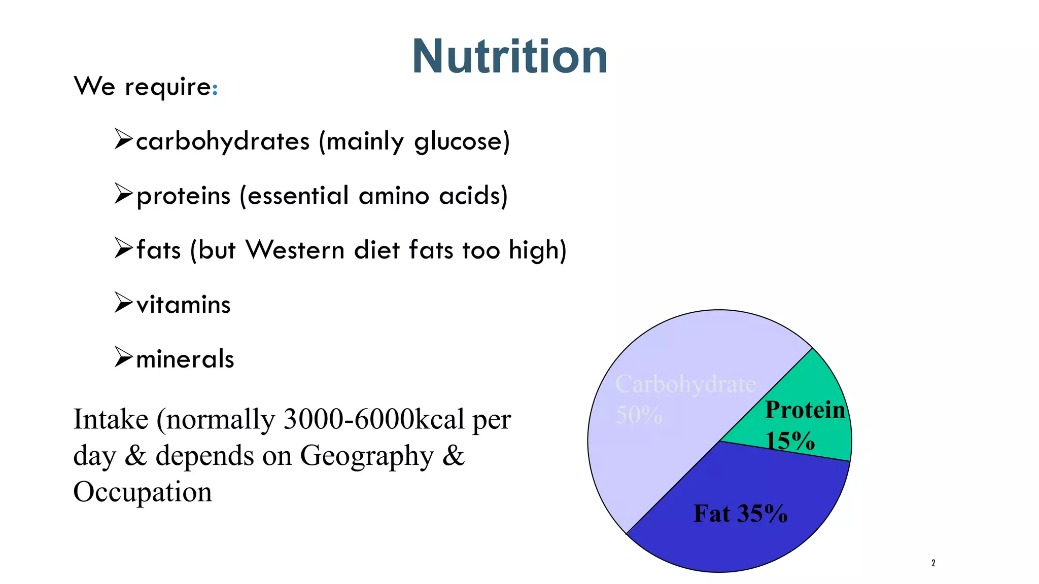
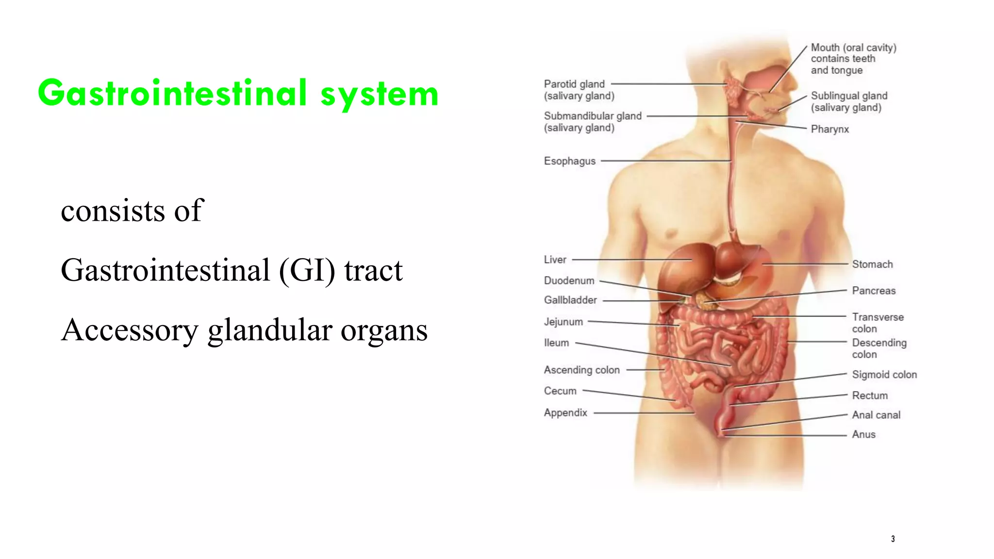
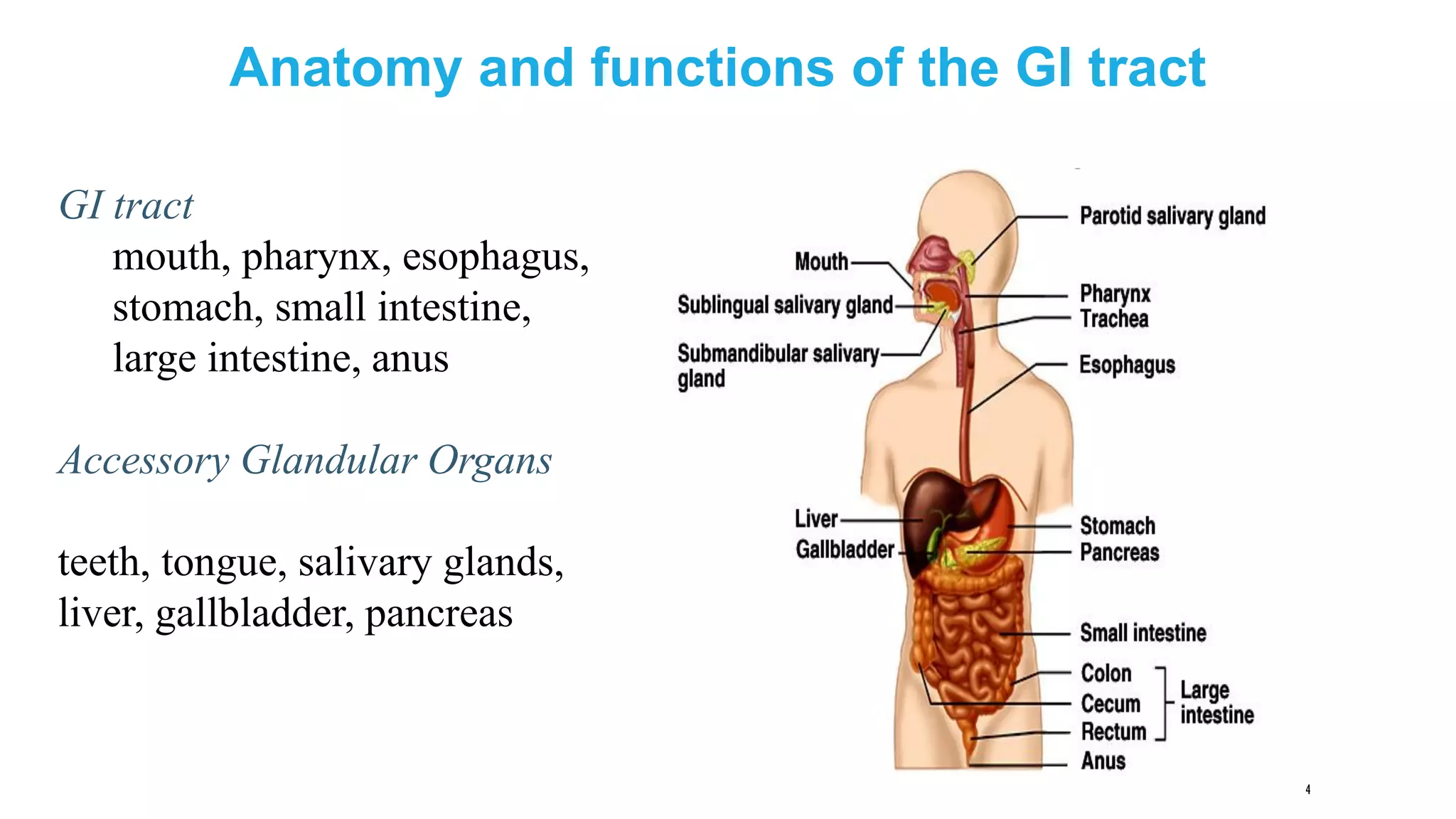
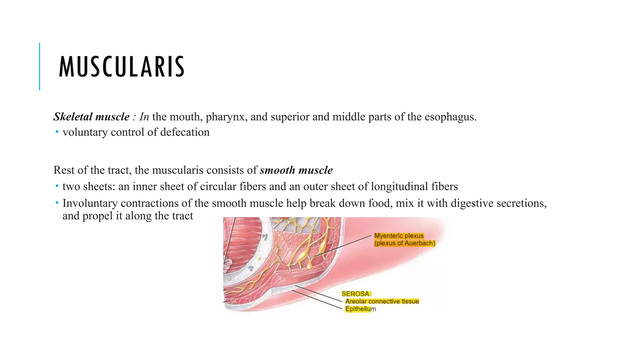
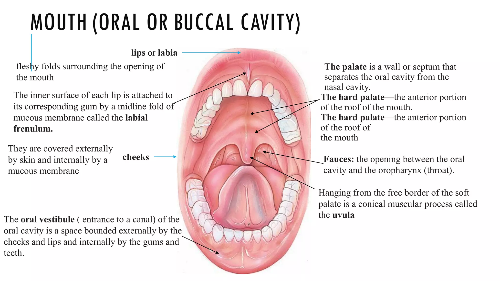
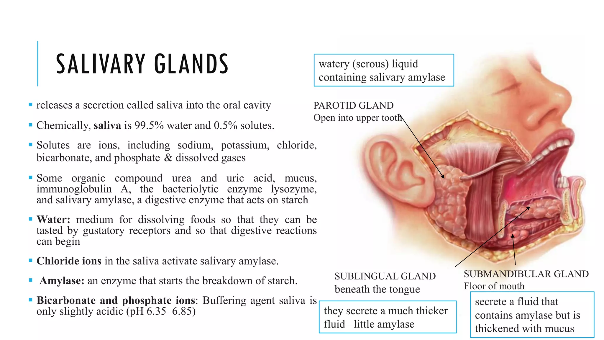
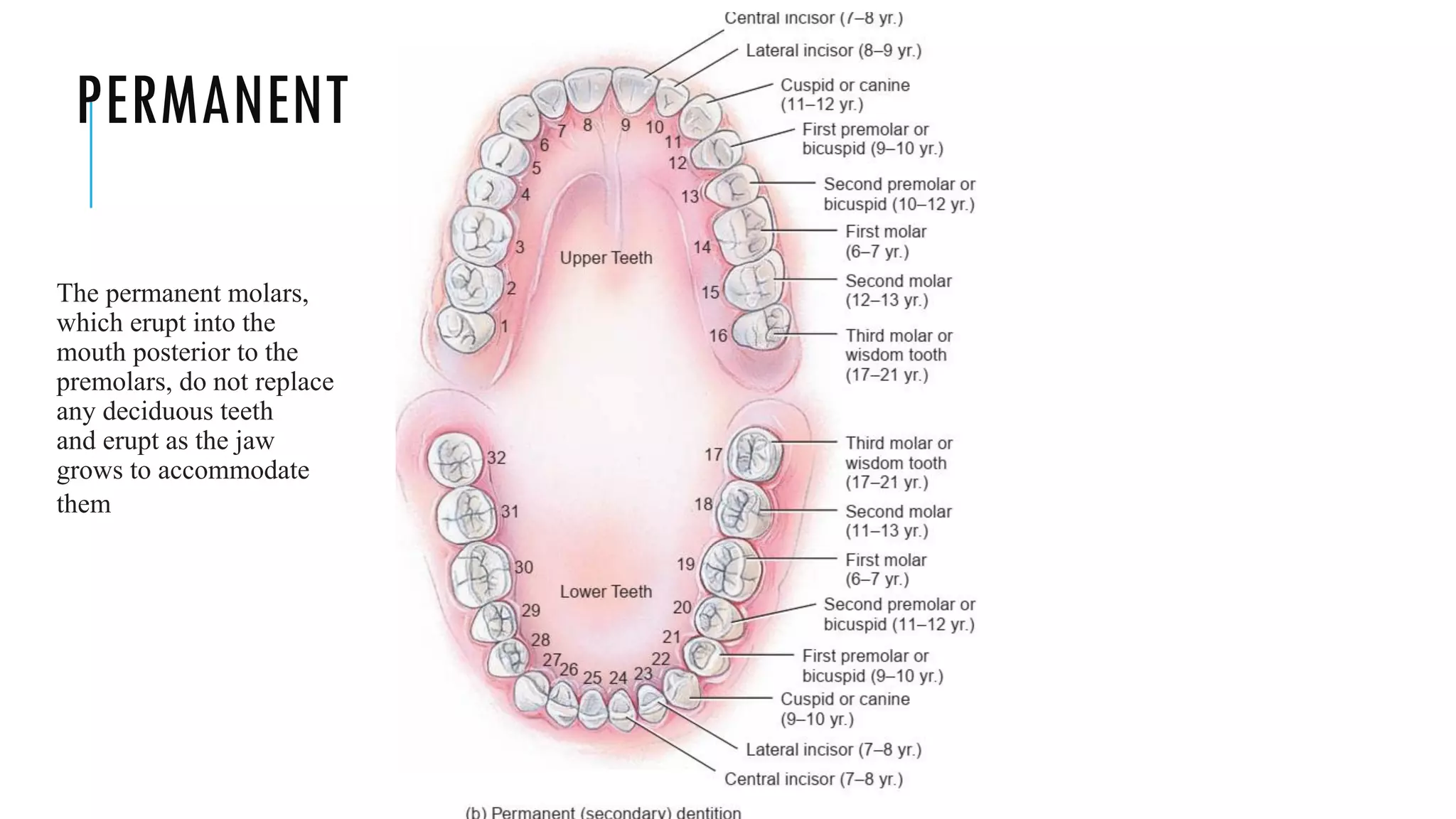
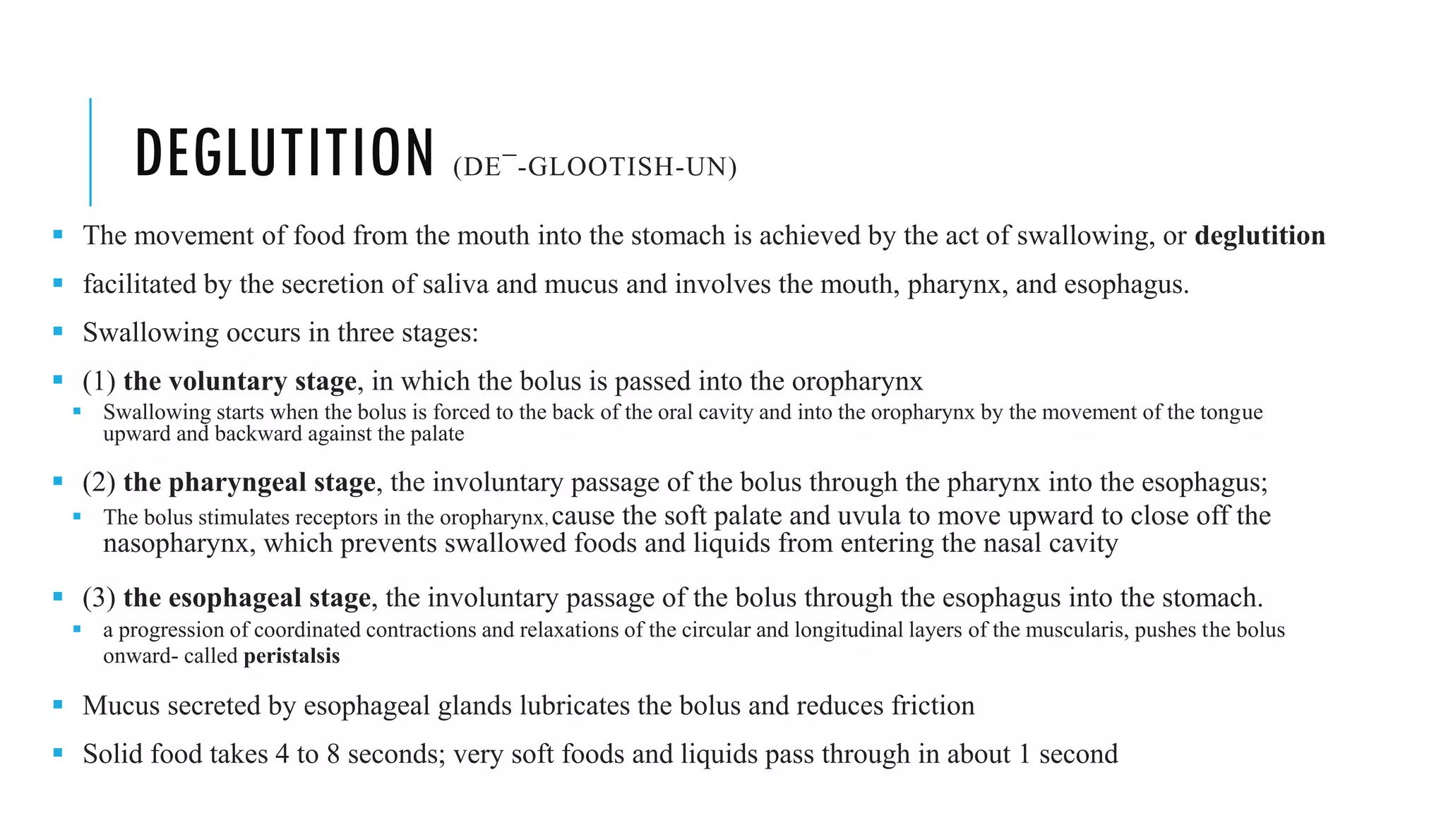
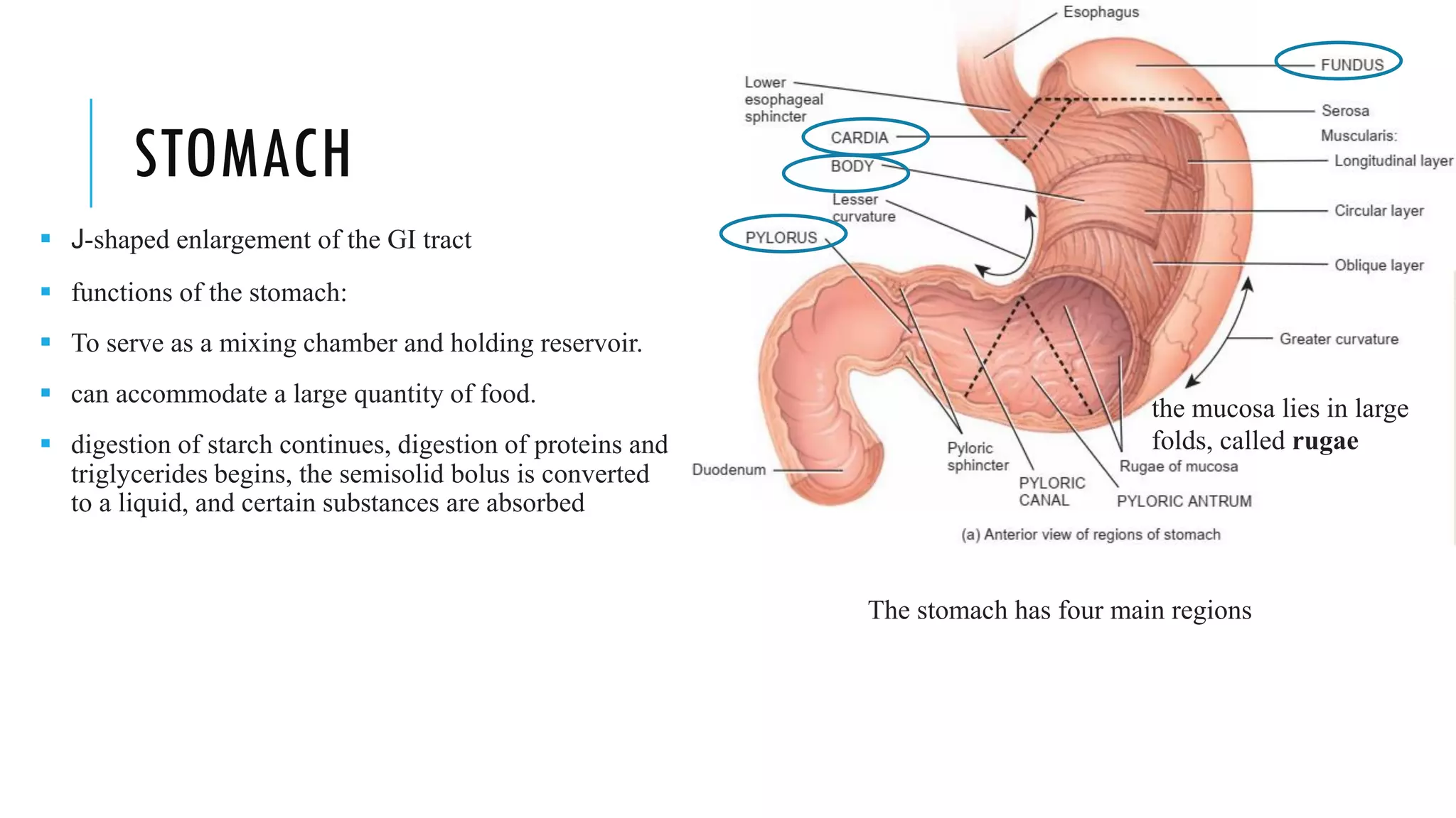
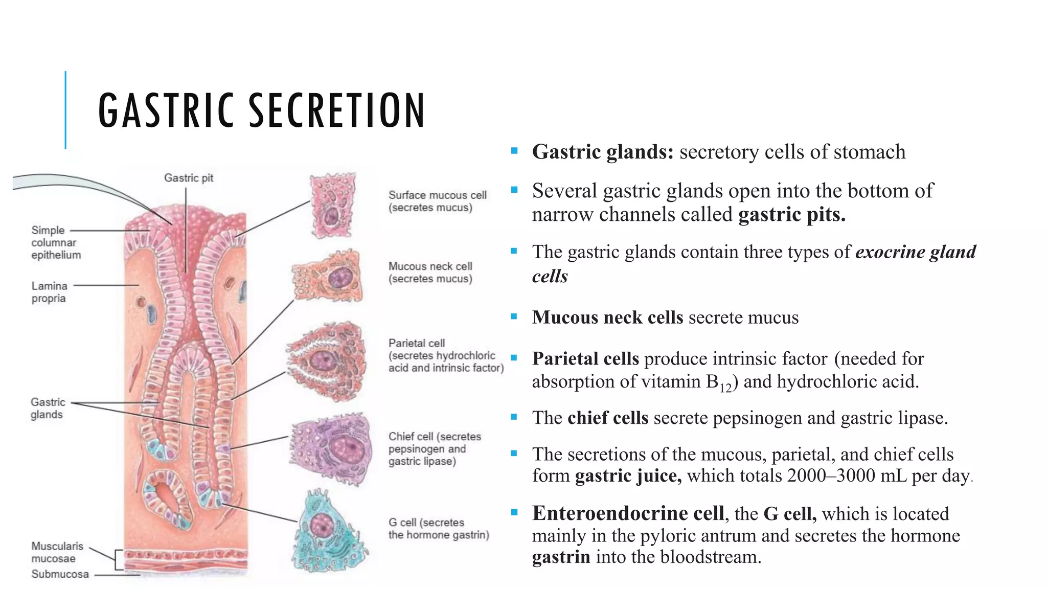
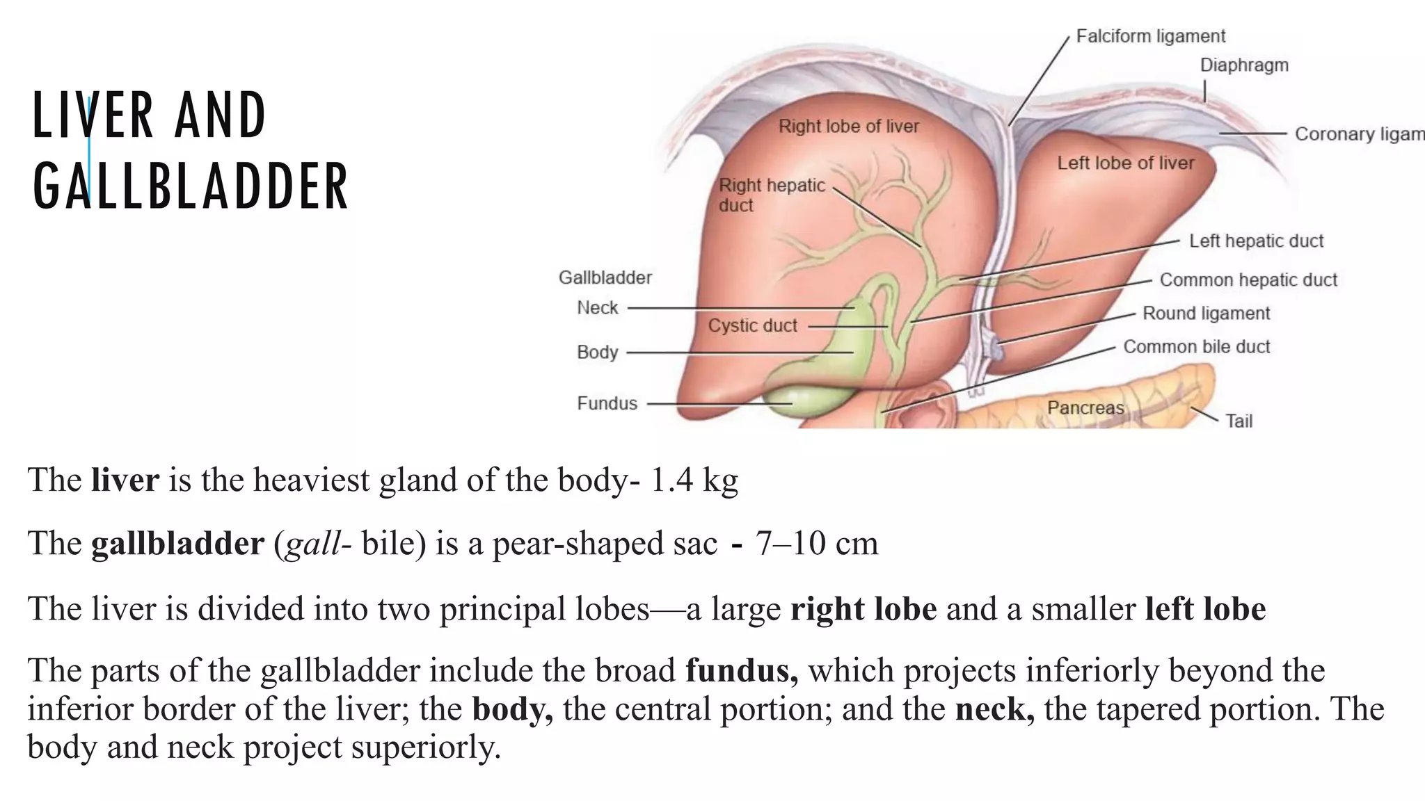
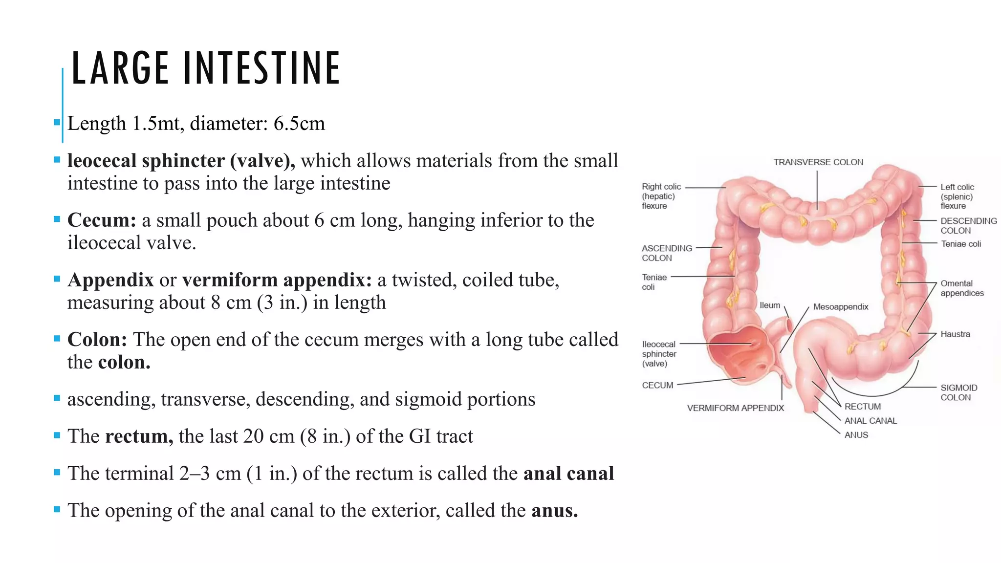
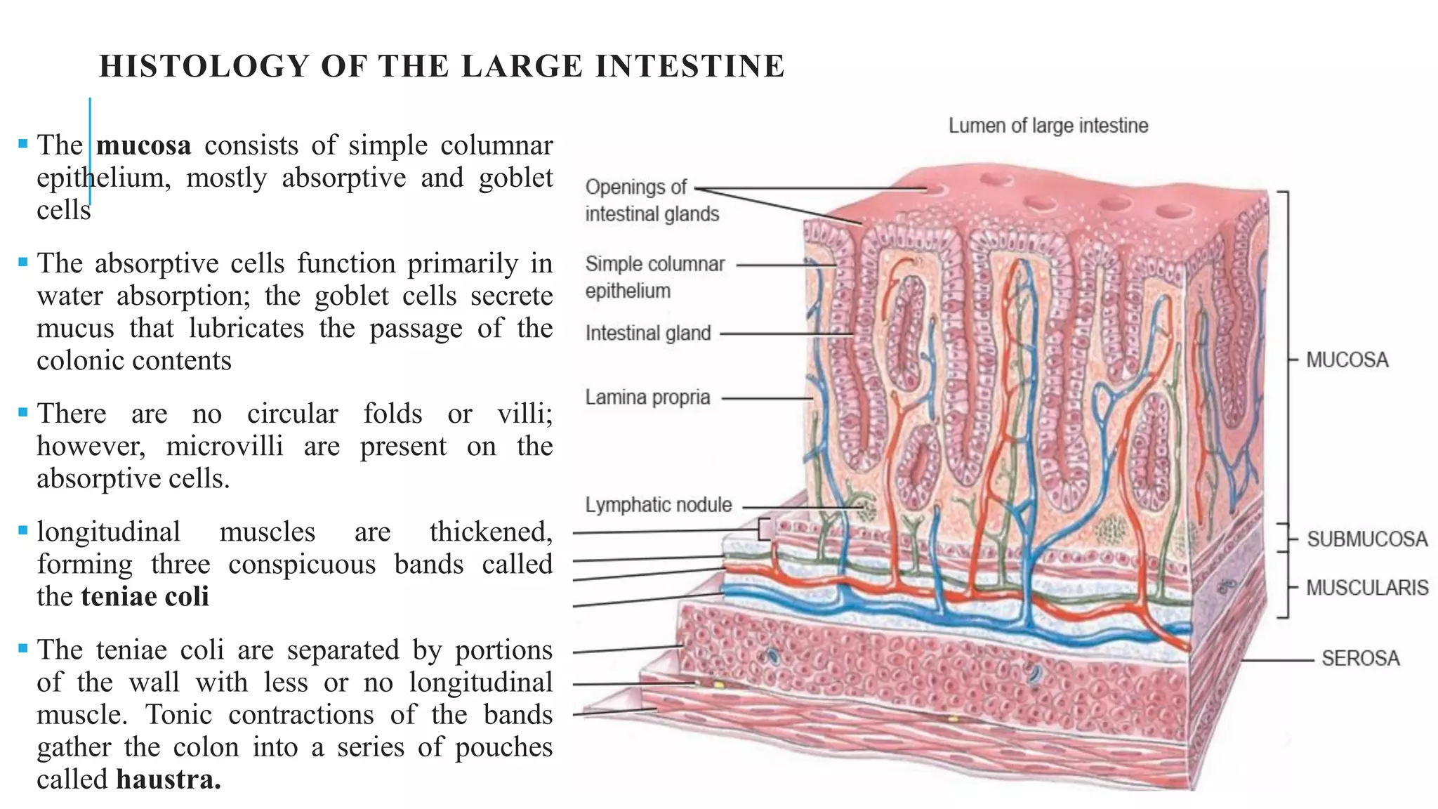
This document provides information on digestion and nutrition. It discusses the nutrients required by the body and the roles and structures of the gastrointestinal tract and accessory organs involved in digestion. The gastrointestinal tract consists of the mouth, esophagus, stomach, small intestine, large intestine and anus. Accessory organs include teeth, salivary glands, liver, gallbladder and pancreas. Food is broken down mechanically and chemically by these organs to absorb nutrients into the bloodstream.
![TEETH [DENTES]
▪ are accessory digestive organs
▪ gingivae or gums
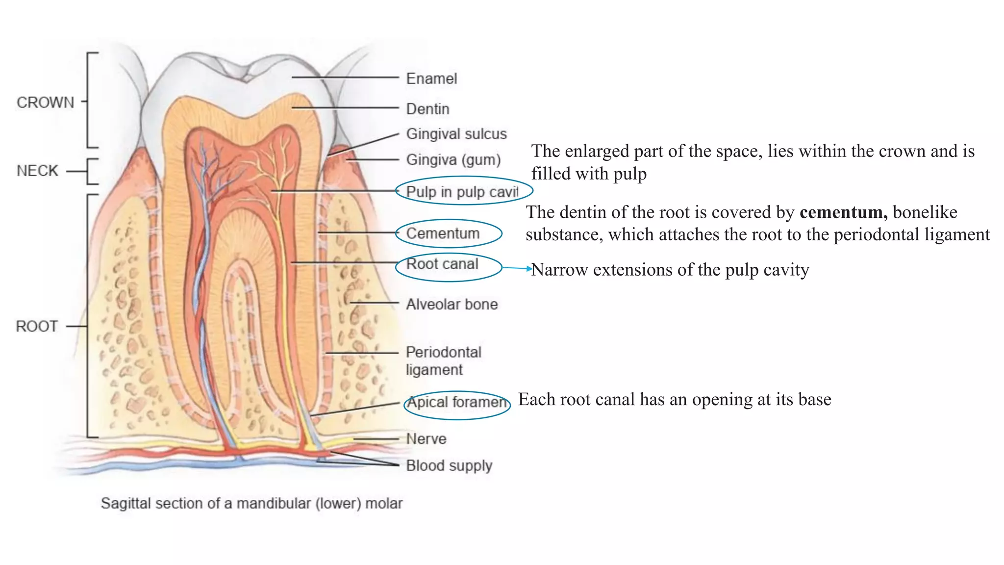
▪ A typical tooth has three major external regions: the
crown, root, and neck.
▪ Dentin: forms the majority of the tooth. Dentin
consists of a calcified connective tissue that gives the
tooth its basic shape and rigidity. It is harder than
bone because of its higher content of calcium salts
(70% of dry weight).
▪ Enamel: The dentin of the crown is covered by
enamel. Made up of calcium phosphate and calcium
carbonate.
▪ harder than bone
▪ It serves to protect the tooth from the wear and tear
of chewing
▪ Protects against acid](https://image.slidesharecdn.com/unit2-digestion-230401061500-f7823f81/75/Unit-2-digestion-pdf-15-2048.jpg)


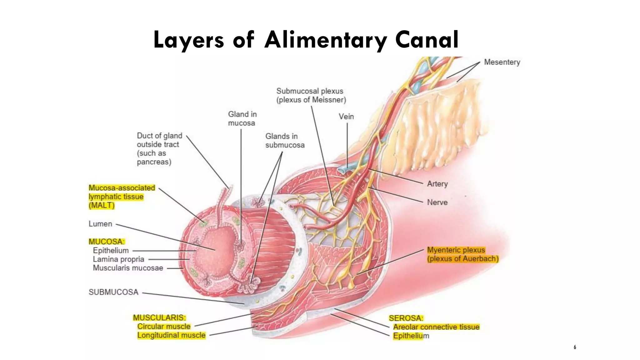
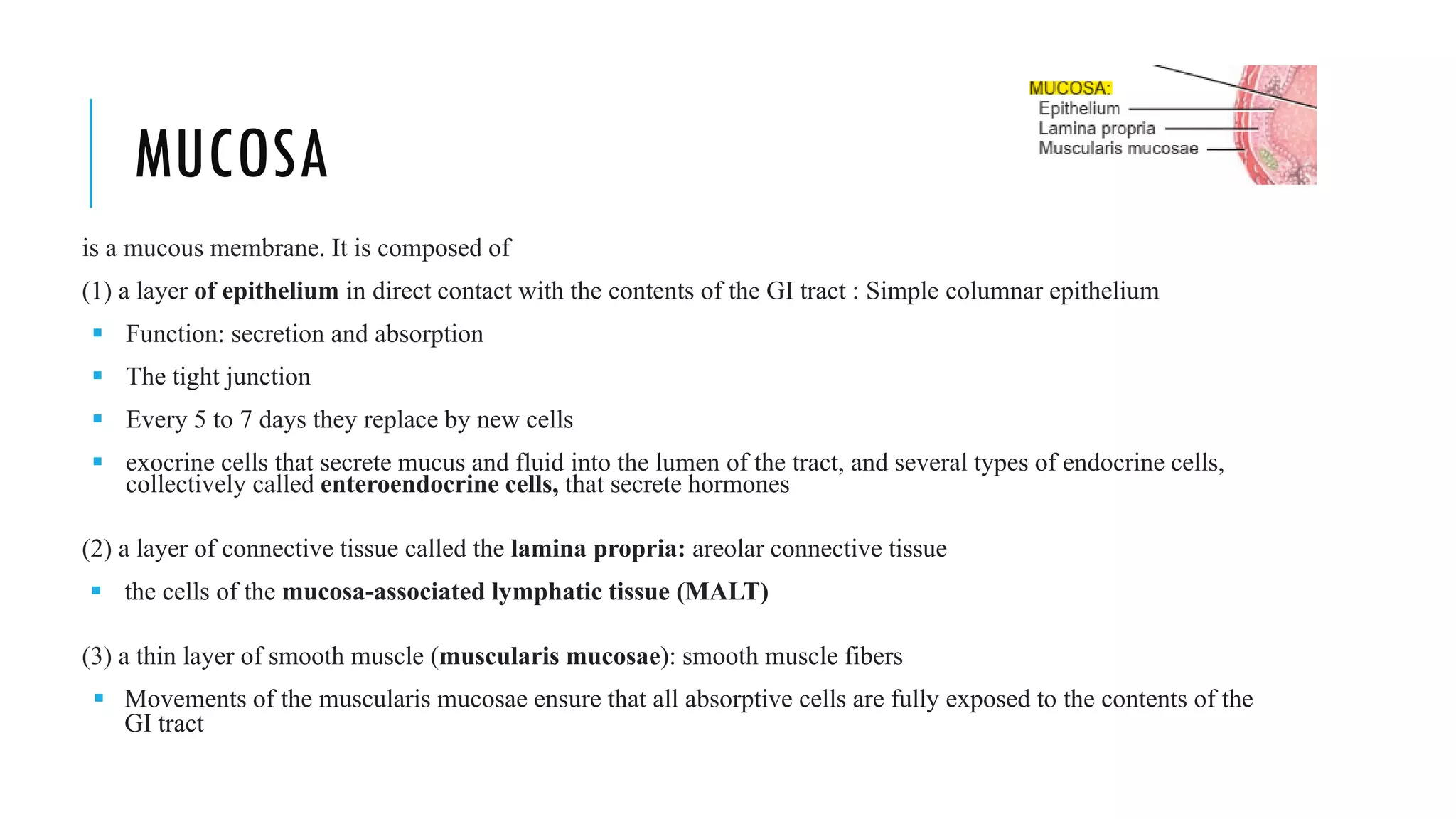
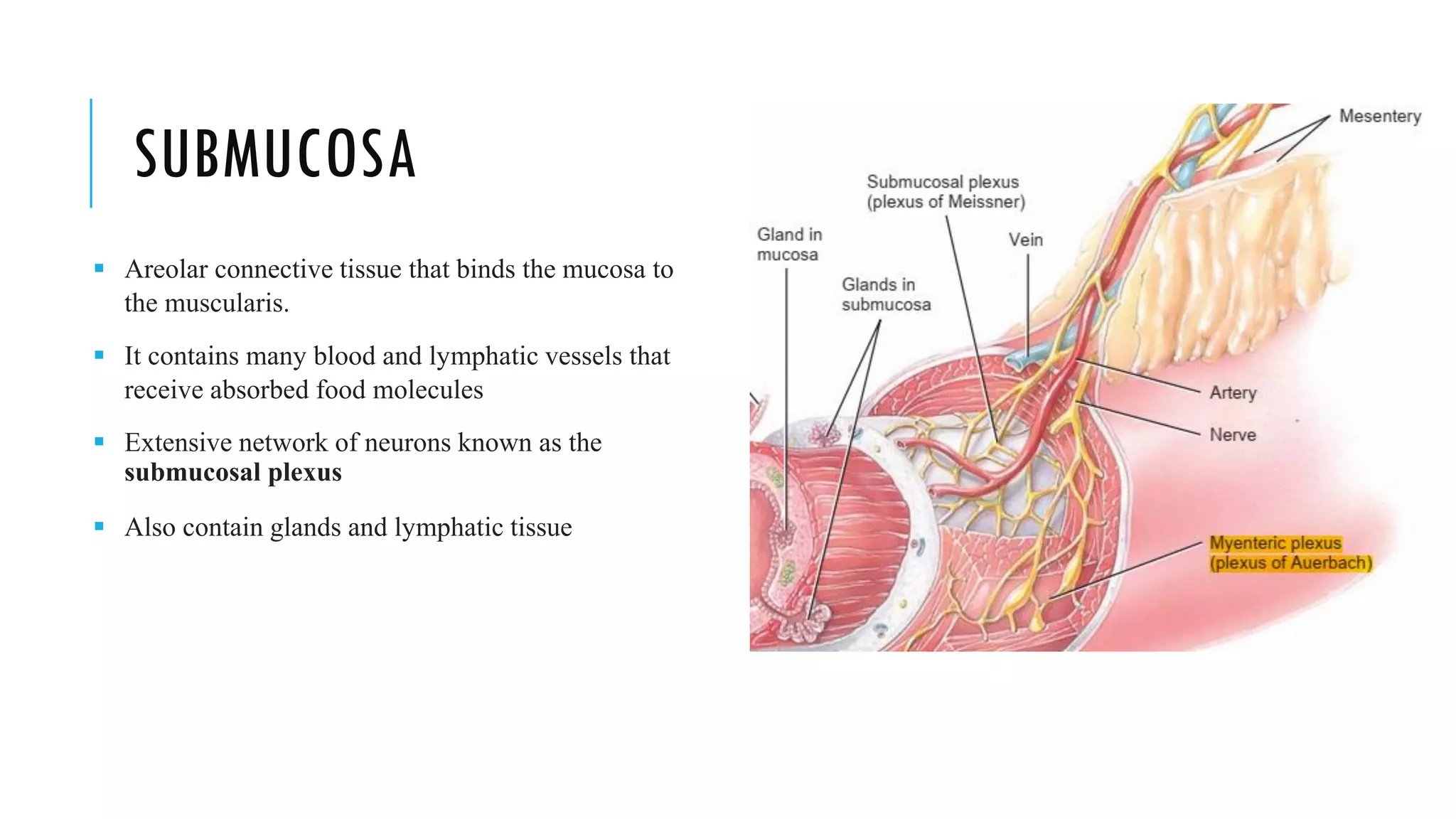
![HISTOLOGY OF THE SMALL INTESTINE
The mucosa is composed of a layer of epithelium (columnar), lamina propria, and
muscularis mucosae
▪ Absorptive cells of the epithelium digest and absorb nutrients in small intestinal
chyme
▪ goblet cells, which secrete mucus
▪ crypts of Lieberkühn (intestinal glands) secrete intestinal juice
▪ paneth cells: secrete lysozyme: regulate the microbial population in the small
intestine
▪ enteroendocrine cells: S cells, CCK cells, and K cells, which secrete the hormones
secretin, cholecystokinin, Glucose dependent insulinotropic peptide [GIP]
The lamina propria
mucosa-associated lymphoid tissue (MALT) & Peyer’s patches: Groups of
lymphatic nodules
The submucosa
Brunner’s glands: secrete an alkaline mucus that helps neutralize gastric acid
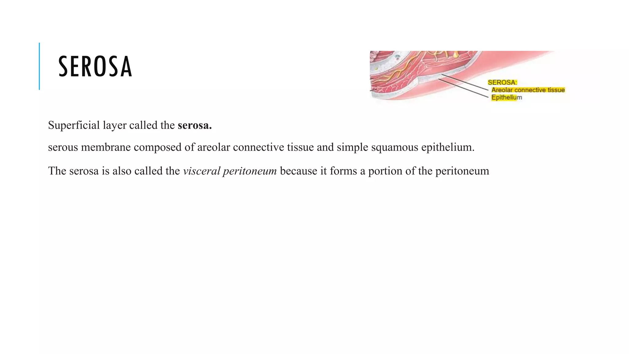
The serosa (or visceral peritoneum) completely surrounds the small intestine.](https://image.slidesharecdn.com/unit2-digestion-230401061500-f7823f81/75/Unit-2-digestion-pdf-35-2048.jpg)
![CHEMICAL DIGESTION & ABSORPTION IN THE SMALL
INTESTINE
▪ chyme entering the small intestine contains partially digested carbohydrates, proteins, and lipids
▪ The completion of the digestion of carbohydrates, proteins, and lipids is a collective effort of pancreatic juice, bile,
and intestinal juice in the small intestine
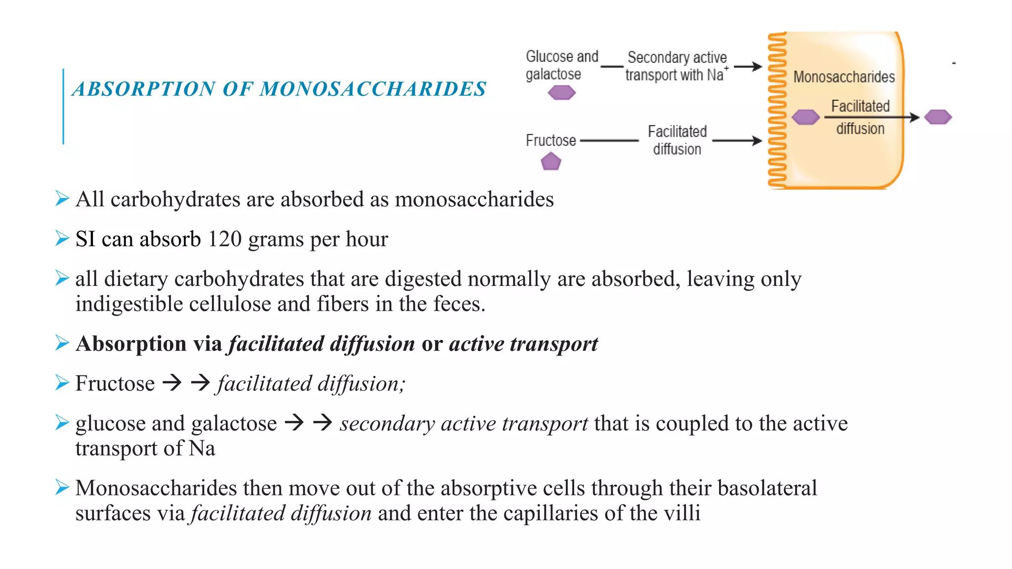
▪ Digestion of Carbohydrates
▪ Unbroken starch are cleaved by pancreatic amylase
▪ Smaller fragments of starch [α–dextrins] will be cleaved by a brush-border enzyme called α-dextrinase,
clipping off one glucose unit at a time
▪ Three brush-border enzymes digest the disaccharides into monosaccharides.
▪ Sucrase breaks sucrose into a molecule of glucose and a molecule of fructose;
▪ lactase digests lactose into a molecule of glucose and a molecule of galactose;
▪ Maltase splits maltose and maltotriose into two or three molecules of glucose,
▪ Pancreatic amylase can act on starch and glycogen
▪ They can not act on cellulose [from plant fiber],
▪ roughage – undigested cellulose](https://image.slidesharecdn.com/unit2-digestion-230401061500-f7823f81/75/Unit-2-digestion-pdf-39-2048.jpg)
![ABSORPTION OF LIPIDS IN SI
▪ All dietary lipids are absorbed via simple diffusion
Short-chain fatty acids are hydrophobic, they are very small in size →pass through the absorptive cells
via simple diffusion → villi
▪ Long-chain fatty acids and monoglycerides are large and hydrophobic and have difficulty being
suspended in the watery environment of the intestinal chyme.
▪ The bile salts in intestinal chyme surround the long-chain fatty acids and monoglycerides, forming tiny
spheres called micelles [2-10nm with 20-50 bile salts]
▪ The micelles move from the interior of the small intestinal lumen to the brush border of the absorptive
cells. At that point, the long-chain fatty acids and monoglycerides diffuse out of the micelles into the
absorptive cells, leaving the micelles behind in the chyme
• Inside the absorptive cells, long-chain fatty
acids and monoglycerides are recombined to
form triglycerides, which aggregate into
globules along with phospholipids and
cholesterol and become coated with proteins,
called chylomicrons
• Chylomicrons leave the absorptive cell via
exocytosis.](https://image.slidesharecdn.com/unit2-digestion-230401061500-f7823f81/75/Unit-2-digestion-pdf-43-2048.jpg)
![ENTEROHEPATIC CIRCULATION
▪ Chylomicron→ lymphatic vessels→ blood capillary→
hepatic portal vein→ liver or adipose tissue.
▪ Lipoprotein lipase [enzyme attached to the apical
surface of capillary endothelial cells] , that breaks down
triglycerides in chylomicrons and other lipoproteins into
fatty acids and glycerol
▪ The fatty acids diffuse into hepatocytes and adipose
cells and combine with glycerol during resynthesis of
triglycerides & chylomicrons remain in the blood
▪ 90–95% of the bile salts are reabsorbed by active
transport in the final segment of the small intestine
(ileum) and returned by the blood to the liver through
the hepatic portal system for recycling.
▪ This cycle of bile salt secretion by hepatocytes into bile,
reabsorption by the ileum, and re-secretion into bile is
called the enterohepatic circulation.](https://image.slidesharecdn.com/unit2-digestion-230401061500-f7823f81/75/Unit-2-digestion-pdf-44-2048.jpg)
![CHEMICAL DIGESTION IN THE LARGE INTESTINE
▪ The final stage of digestion occurs in the colon through the activity of bacteria that inhabit
the lumen.
▪ Mucus is secreted by the glands of the large intestine, but no enzymes are secreted
▪ Bacteria ferment any remaining carbohydrates and release hydrogen, carbon dioxide, and
methane gases. [flatulence when it is excessive gas]
Bacteria also convert any remaining proteins to amino acids and break down the amino acids
into simpler substances: indole, skatole, hydrogen sulfide, and fatty acids
▪ Some secreted in feces contributes to their odor, the rest is absorbed and transported to the
liver, where these compounds are converted to less toxic compounds and excreted in the urine
▪ Bacteria also decompose bilirubin to simpler pigments, including stercobilin, which gives
feces their brown color. Bacterial products that are absorbed in the colon include several
vitamins needed for normal metabolism, among them some B vitamins and vitamin K.](https://image.slidesharecdn.com/unit2-digestion-230401061500-f7823f81/75/Unit-2-digestion-pdf-51-2048.jpg)


![Human_Digestive_System[1].pptx by medical with us.pptx](https://cdn.slidesharecdn.com/ss_thumbnails/humandigestivesystem1-250517060919-d012c22d-thumbnail.jpg?width=640&height=640&fit=bounds)


