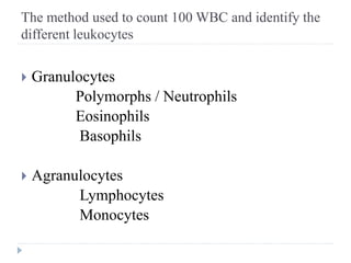
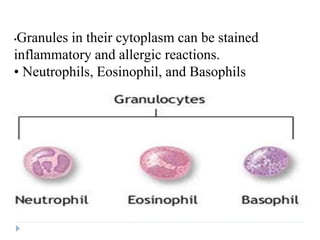
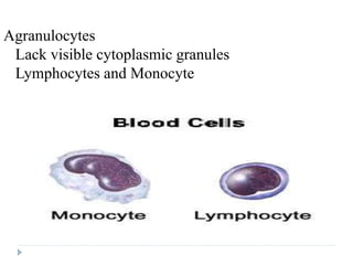
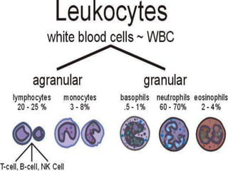
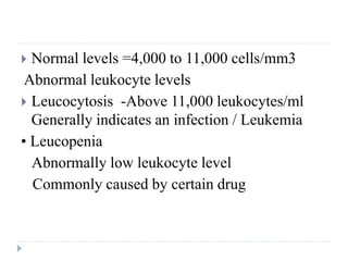
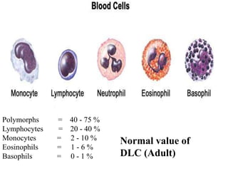
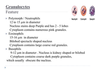
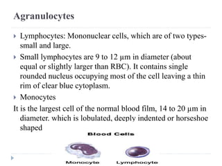
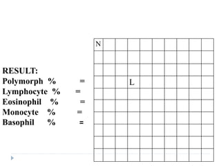
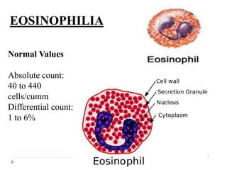
Differential Leucocyte count (TLC)
& Eosinophilia
Dr Sandeep singh
OBJECTIVES
 Types of WBC and their counts
 Formation of WBC
 Morphology, life span, function.
 Applied aspects.
 To draw a peripheral blood smear, stain it
with Leishman's stain and do the
differential count( DLC) of the given
blood sample.
INTRODUCTION
 Crucial in the body’s defense against pathogens
Capable of phagocytosis.
 These are complete cells, with a nucleus and
organelles
 They lack Hb so they are colorless (i.e. white)
 Able to move into and out of blood vessels
• Can respond to chemicals released by damaged
tissues.
The method used to count 100 WBC and identify the
different leukocytes
 Granulocytes
Polymorphs / Neutrophils
Eosinophils
Basophils
 Agranulocytes
Lymphocytes
Monocytes
•Granules in their cytoplasm can be stained
inflammatory and allergic reactions.
• Neutrophils, Eosinophil, and Basophils
Agranulocytes
Lack visible cytoplasmic granules
Lymphocytes and Monocyte
 Normal levels =4,000 to 11,000 cells/mm3
Abnormal leukocyte levels
 Leucocytosis -Above 11,000 leukocytes/ml
Generally indicates an infection / Leukemia
• Leucopenia
Abnormally low leukocyte level
Commonly caused by certain drug
Polymorphs = 40 - 75 %
Lymphocytes = 20 - 40 %
Monocytes = 2 - 10 %
Eosinophils = 1 - 6 %
Basophils = 0 - 1 %
Normal value of
DLC (Adult)
Granulocytes
Feature
 Polymorph / Neutrophils
12 to 15 µm in diameter
Nucleus stains deep Purple and has 2 - 5 lobes
Cytoplasm contains numerous pink granules.
 Eosinophils
15-16 µm in diameter
Bilobed spectacle shaped nucleus
Cytoplasm contains large coarse red granules.
 Basophils
9-12 µm in diameter . Nucleus is kidney shaped or bilobed
Cytoplasm contains coarse dark purple granules,
which usually obscure the nucleus.
Agranulocytes
 Lymphocytes: Mononuclear cells, which are of two types-
small and large.
 Small lymphocytes are 9 to 12 µm in diameter (about
equal or slightly larger than RBC). It contains single
rounded nucleus occupying most of the cell leaving a thin
rim of clear blue cytoplasm.
 Monocytes
It is the largest cell of the normal blood film, 14 to 20 µm in
diameter. which is lobulated, deeply indented or horseshoe
shaped
Preparation of Blood Film

Differential leukocyte count is performed on stained
blood film.
 Material required
 Glass slides
 Blood sample
 Leishman's stain
 Buffer water (pH = 6.8)
 Cedar wood oil
 Microscope with oil immersion objective
Glass slides should be clean and dry.
 Place a small drop of blood on one end of a clean slide, in the
middle. Hold a second slide (i.e., spreader) above the first, right
angle of 30 to 450.
 Move the spreader forward rapidly, thus spreading the blood
over the slide in a thin film.
 Leave the slide to air dry. A properly made film should dry
quickly; if the film is not dried rapidly there will be rouleaux
formation of RBC and the cells will appear shrunken.
 A good blood film should not cover the entire surface of the
slide. It should be even, smooth and should be having three
parts i.e., head, body and tail.
 Dried film should be stained with Leishman's stain.
Leishman's staining and DLC
 Pour the Leishman's stain over the film to cover it completely. Keep it for
two minutes.
 After one minute, buffer water is added gently to the stain already on the
blood film to cover the slide completely (it is approximately double the
volume of stain).
 Allow the diluted stain to act for about 8 to 10 minutes.
 Wash the film in buffer water or distilled water. The film should be rose
pink in colour.
 Allow the film dry and focus under low power of microscope. Then put a
drop of cedar wood oil and see under oil immersion objective.
 Count the WBCs in a zig-zag fashion across the breadth of film, in the body
and tail junction area of film, until 100 leukocytes are counted.
 To record them draw a large square and divide it into 100 small squares.
Identify one cell and write in each square. Count different WBCs coming
across. The result thus obtained is directly in percentage.
N
L
RESULT:
Polymorph % =
Lymphocyte % =
Eosinophil % =
Monocyte % =
Basophil % =
Normal Values
Absolute count:
40 to 440
cells/cumm
Differential count:
1 to 6%
EOSINOPHILIA
Physiological:
Diurnal variations are seen.
Parasitic infestations: due to allergic reaction to parasites
Allergic disorders: Bronchial asthma, Urticaria, Hay fever, Drug
hypersensitivity.
Drugs: Chlopromazine, Penicillin, and Streptomycin.
Skin diseases: Pemphigus, Dermatitis herpetiformis, Erythema
multiforme, Psoriasis, Eczema, Exfoliative Dermatitis, and Scabies.
Pulmonary diseases: Löeffler’s syndrome, and Tropical
eosinophilia.
Haematopoietic diseases: Chronic myeloid leukaemia,
Polycythaemia vera, Hodgkin’s disease.
Miscellaneous conditions: Rheumatoid arthritis, Polyarteritis
nodosa, Systemic Lupus Erythematosus (SLE), Ulcerative colitis,
and Irradiation.
Causes of Eosinophilia
Eosinopenia
 Decrease in the count of eosinophils is known as
eosinopenia.
 Causes:
 Uraemia
 After ACTH therapy
 After steroid therapy
 After stress caused by burn or postoperative conditions
 Few hours after ECT (electroconvulsive therapy)
 In some stages of acute infections.
Differential leucocyte count & eosinophilia

Differential leucocyte count & eosinophilia

  • 1.
    Differential Leucocyte count(TLC) & Eosinophilia Dr Sandeep singh
  • 2.
    OBJECTIVES  Types ofWBC and their counts  Formation of WBC  Morphology, life span, function.  Applied aspects.  To draw a peripheral blood smear, stain it with Leishman's stain and do the differential count( DLC) of the given blood sample.
  • 3.
    INTRODUCTION  Crucial inthe body’s defense against pathogens Capable of phagocytosis.  These are complete cells, with a nucleus and organelles  They lack Hb so they are colorless (i.e. white)  Able to move into and out of blood vessels • Can respond to chemicals released by damaged tissues.
  • 5.
    The method usedto count 100 WBC and identify the different leukocytes  Granulocytes Polymorphs / Neutrophils Eosinophils Basophils  Agranulocytes Lymphocytes Monocytes
  • 6.
    •Granules in theircytoplasm can be stained inflammatory and allergic reactions. • Neutrophils, Eosinophil, and Basophils
  • 7.
    Agranulocytes Lack visible cytoplasmicgranules Lymphocytes and Monocyte
  • 9.
     Normal levels=4,000 to 11,000 cells/mm3 Abnormal leukocyte levels  Leucocytosis -Above 11,000 leukocytes/ml Generally indicates an infection / Leukemia • Leucopenia Abnormally low leukocyte level Commonly caused by certain drug
  • 10.
    Polymorphs = 40- 75 % Lymphocytes = 20 - 40 % Monocytes = 2 - 10 % Eosinophils = 1 - 6 % Basophils = 0 - 1 % Normal value of DLC (Adult)
  • 11.
    Granulocytes Feature  Polymorph /Neutrophils 12 to 15 µm in diameter Nucleus stains deep Purple and has 2 - 5 lobes Cytoplasm contains numerous pink granules.  Eosinophils 15-16 µm in diameter Bilobed spectacle shaped nucleus Cytoplasm contains large coarse red granules.  Basophils 9-12 µm in diameter . Nucleus is kidney shaped or bilobed Cytoplasm contains coarse dark purple granules, which usually obscure the nucleus.
  • 12.
    Agranulocytes  Lymphocytes: Mononuclearcells, which are of two types- small and large.  Small lymphocytes are 9 to 12 µm in diameter (about equal or slightly larger than RBC). It contains single rounded nucleus occupying most of the cell leaving a thin rim of clear blue cytoplasm.  Monocytes It is the largest cell of the normal blood film, 14 to 20 µm in diameter. which is lobulated, deeply indented or horseshoe shaped
  • 13.
    Preparation of BloodFilm  Differential leukocyte count is performed on stained blood film.  Material required  Glass slides  Blood sample  Leishman's stain  Buffer water (pH = 6.8)  Cedar wood oil  Microscope with oil immersion objective
  • 14.
    Glass slides shouldbe clean and dry.  Place a small drop of blood on one end of a clean slide, in the middle. Hold a second slide (i.e., spreader) above the first, right angle of 30 to 450.  Move the spreader forward rapidly, thus spreading the blood over the slide in a thin film.  Leave the slide to air dry. A properly made film should dry quickly; if the film is not dried rapidly there will be rouleaux formation of RBC and the cells will appear shrunken.  A good blood film should not cover the entire surface of the slide. It should be even, smooth and should be having three parts i.e., head, body and tail.  Dried film should be stained with Leishman's stain.
  • 15.
    Leishman's staining andDLC  Pour the Leishman's stain over the film to cover it completely. Keep it for two minutes.  After one minute, buffer water is added gently to the stain already on the blood film to cover the slide completely (it is approximately double the volume of stain).  Allow the diluted stain to act for about 8 to 10 minutes.  Wash the film in buffer water or distilled water. The film should be rose pink in colour.  Allow the film dry and focus under low power of microscope. Then put a drop of cedar wood oil and see under oil immersion objective.  Count the WBCs in a zig-zag fashion across the breadth of film, in the body and tail junction area of film, until 100 leukocytes are counted.  To record them draw a large square and divide it into 100 small squares. Identify one cell and write in each square. Count different WBCs coming across. The result thus obtained is directly in percentage.
  • 16.
    N L RESULT: Polymorph % = Lymphocyte% = Eosinophil % = Monocyte % = Basophil % =
  • 17.
    Normal Values Absolute count: 40to 440 cells/cumm Differential count: 1 to 6% EOSINOPHILIA
  • 18.
    Physiological: Diurnal variations areseen. Parasitic infestations: due to allergic reaction to parasites Allergic disorders: Bronchial asthma, Urticaria, Hay fever, Drug hypersensitivity. Drugs: Chlopromazine, Penicillin, and Streptomycin. Skin diseases: Pemphigus, Dermatitis herpetiformis, Erythema multiforme, Psoriasis, Eczema, Exfoliative Dermatitis, and Scabies. Pulmonary diseases: Löeffler’s syndrome, and Tropical eosinophilia. Haematopoietic diseases: Chronic myeloid leukaemia, Polycythaemia vera, Hodgkin’s disease. Miscellaneous conditions: Rheumatoid arthritis, Polyarteritis nodosa, Systemic Lupus Erythematosus (SLE), Ulcerative colitis, and Irradiation. Causes of Eosinophilia
  • 19.
    Eosinopenia  Decrease inthe count of eosinophils is known as eosinopenia.  Causes:  Uraemia  After ACTH therapy  After steroid therapy  After stress caused by burn or postoperative conditions  Few hours after ECT (electroconvulsive therapy)  In some stages of acute infections.