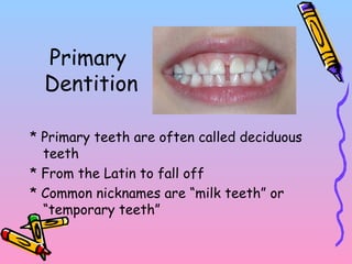
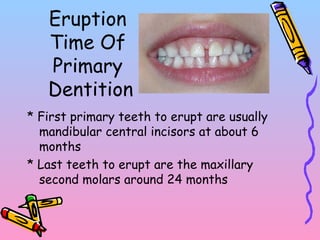
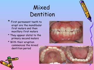
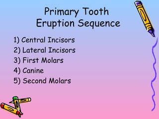
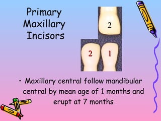
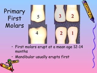
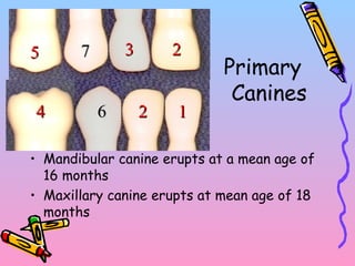
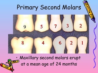
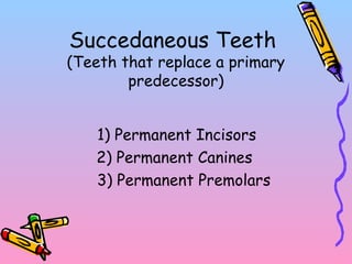
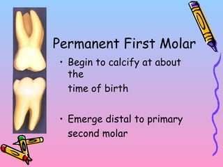
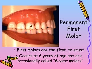
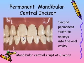
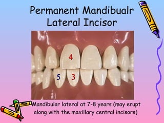
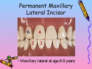
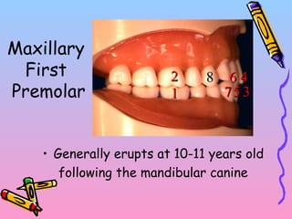
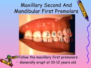
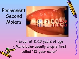
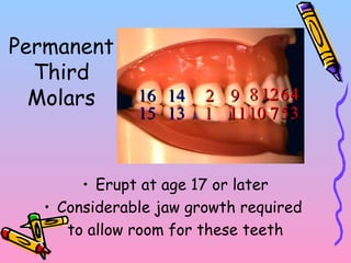
Differences between primary and permanent dentitionAkshMinhas
A longitudinal radiological study of children (N = 549) who participated in a comprehensive preventive maintenance program showed that caries related events in the approximal surfaces of permanent teeth differed from those in deciduous teeth. Changes in the approximal surfaces of the younger permanent teeth were more pronounced than of the older primary teeth and differed significantly from 1 year to 2.5 years. These findings can be explained by posteruptive maturation of tooth enamel.
Difference between primary and permanent teethprincesoni3954
The presentation features the basic difference between primary and permanent dentition. The differences are tabulated under the headings of crown, roor and pulp.
A detailed look at the differences between the human primary and permanent dentition. Hope you find this informative. for further queries, please contact at dr.mathewthomasm@gmail.com.
Dentin dysplasia (DD) is a rare hereditary disturbance is inherited as an autosomal dominant trait.
unknown etiology that affects approximately 1 :100,000.
In 1972, Witkop classified it into type I and type II which affect both dentitions.DD Type I
Radicular dentin dysplasia
Characterized by:-
1.Both dentitions are affected.
2.Normal appearing crowns
3.No or only rudimentary root development (rootless teeth)
4.Incomplete or total obliteration of the pulp chamber.
5.Teeth may exhibit extreme mobility and exfoliate prematurely.DD type II
coronal dentin dysplasia
Characterized by:-
1.partial pulpal obliteration.
2.Thistle-tube-or flame-shaped coronal pulp chambers
3. Thread-like root canals
4. Usually the absence of periapical radiolucencies.
5. In this type of anomaly, teeth roots are of normal shape and contour.The enamel and the immediately subjacent dentin appear normal.
Deeper layers of dentin show an atypical tubular pattern with an amorphous, atubular area, and irregular organization.
Normal dentinal tubule formation appears to have been blocked so that new dentine forms around obstacles and takes on the characteristic appearances described as “lava flowing around boulders”The radiograph revealed features of dentine dysplasia type I with normal appearance of crown but no root development Autosomal Dominant Disorder:
Manifested in heterozygous states
At least one parent of index case is usually affected
Both males and females are affected.
Clinical feature can be modified by variation in penetrance and expressivity. Some individual inherit the mutant gene but are phenotpically normal. This is reffered to as “incomplete penetrance”.
In many condition the age of onset is delayed.
Inheritance Pattern:
Typical pattern is a heterozygous affected parent with a homozygous unaffected parent.
Every child has one chance in two of having the disease
Both sexes are affected equally..Autosomal Recessive Disorder
Largest category of Mendelian disorder
Usually does not affect the parent of the affected individual, but sibling may show the disease.
Complete penetrance is common.
Onset is frequently early in life.
Usually affect enzymatic proteins.
Pattern Of Inheritance:
Typical pattern is two heterozygous unaffected (carrier) parent.
The triat does not usually affect the parent, but siblings may show the disease
Siblings have one chance in four of being affected
Both sexes affected equally.
Fundamentals in tooth preparation, Now many indications for treatment for teeth are not due to caries and, therefore, the preparation of the tooth is no longer referred to as cavity preparation but as tooth preparation, and the term cavity is used only as a historical reference.
NOMENCLATURE
Differences between primary and permanent teeth and importanceKarishma Sirimulla
This is a small brief presentation and contains basic differences between primary and permanent dentition an also an added note on importance of young permanent molar and its management clinically
Dental anatomy is a field of anatomy dedicated to the study of human tooth structures. The development, appearance, and classification of teeth fall within its purview.
ARTIFICIAL INTELLIGENCE IN HEALTHCARE.pdfAnujkumaranit
Artificial intelligence (AI) refers to the simulation of human intelligence processes by machines, especially computer systems. It encompasses tasks such as learning, reasoning, problem-solving, perception, and language understanding. AI technologies are revolutionizing various fields, from healthcare to finance, by enabling machines to perform tasks that typically require human intelligence.
Lung Cancer: Artificial Intelligence, Synergetics, Complex System Analysis, S...Oleg Kshivets
RESULTS: Overall life span (LS) was 2252.1±1742.5 days and cumulative 5-year survival (5YS) reached 73.2%, 10 years – 64.8%, 20 years – 42.5%. 513 LCP lived more than 5 years (LS=3124.6±1525.6 days), 148 LCP – more than 10 years (LS=5054.4±1504.1 days).199 LCP died because of LC (LS=562.7±374.5 days). 5YS of LCP after bi/lobectomies was significantly superior in comparison with LCP after pneumonectomies (78.1% vs.63.7%, P=0.00001 by log-rank test). AT significantly improved 5YS (66.3% vs. 34.8%) (P=0.00000 by log-rank test) only for LCP with N1-2. Cox modeling displayed that 5YS of LCP significantly depended on: phase transition (PT) early-invasive LC in terms of synergetics, PT N0—N12, cell ratio factors (ratio between cancer cells- CC and blood cells subpopulations), G1-3, histology, glucose, AT, blood cell circuit, prothrombin index, heparin tolerance, recalcification time (P=0.000-0.038). Neural networks, genetic algorithm selection and bootstrap simulation revealed relationships between 5YS and PT early-invasive LC (rank=1), PT N0—N12 (rank=2), thrombocytes/CC (3), erythrocytes/CC (4), eosinophils/CC (5), healthy cells/CC (6), lymphocytes/CC (7), segmented neutrophils/CC (8), stick neutrophils/CC (9), monocytes/CC (10); leucocytes/CC (11). Correct prediction of 5YS was 100% by neural networks computing (area under ROC curve=1.0; error=0.0).
CONCLUSIONS: 5YS of LCP after radical procedures significantly depended on: 1) PT early-invasive cancer; 2) PT N0--N12; 3) cell ratio factors; 4) blood cell circuit; 5) biochemical factors; 6) hemostasis system; 7) AT; 8) LC characteristics; 9) LC cell dynamics; 10) surgery type: lobectomy/pneumonectomy; 11) anthropometric data. Optimal diagnosis and treatment strategies for LC are: 1) screening and early detection of LC; 2) availability of experienced thoracic surgeons because of complexity of radical procedures; 3) aggressive en block surgery and adequate lymph node dissection for completeness; 4) precise prediction; 5) adjuvant chemoimmunoradiotherapy for LCP with unfavorable prognosis.
Ozempic: Preoperative Management of Patients on GLP-1 Receptor Agonists Saeid Safari
Preoperative Management of Patients on GLP-1 Receptor Agonists like Ozempic and Semiglutide
ASA GUIDELINE
NYSORA Guideline
2 Case Reports of Gastric Ultrasound
Factory Supply Best Quality Pmk Oil CAS 28578–16–7 PMK Powder in Stockrebeccabio
Factory Supply Best Quality Pmk Oil CAS 28578–16–7 PMK Powder in Stock
Telegram: bmksupplier
signal: +85264872720
threema: TUD4A6YC
You can contact me on Telegram or Threema
Communicate promptly and reply
Free of customs clearance, Double Clearance 100% pass delivery to USA, Canada, Spain, Germany, Netherland, Poland, Italy, Sweden, UK, Czech Republic, Australia, Mexico, Russia, Ukraine, Kazakhstan.Door to door service
Hot Selling Organic intermediates
Title: Sense of Smell
Presenter: Dr. Faiza, Assistant Professor of Physiology
Qualifications:
MBBS (Best Graduate, AIMC Lahore)
FCPS Physiology
ICMT, CHPE, DHPE (STMU)
MPH (GC University, Faisalabad)
MBA (Virtual University of Pakistan)
Learning Objectives:
Describe the primary categories of smells and the concept of odor blindness.
Explain the structure and location of the olfactory membrane and mucosa, including the types and roles of cells involved in olfaction.
Describe the pathway and mechanisms of olfactory signal transmission from the olfactory receptors to the brain.
Illustrate the biochemical cascade triggered by odorant binding to olfactory receptors, including the role of G-proteins and second messengers in generating an action potential.
Identify different types of olfactory disorders such as anosmia, hyposmia, hyperosmia, and dysosmia, including their potential causes.
Key Topics:
Olfactory Genes:
3% of the human genome accounts for olfactory genes.
400 genes for odorant receptors.
Olfactory Membrane:
Located in the superior part of the nasal cavity.
Medially: Folds downward along the superior septum.
Laterally: Folds over the superior turbinate and upper surface of the middle turbinate.
Total surface area: 5-10 square centimeters.
Olfactory Mucosa:
Olfactory Cells: Bipolar nerve cells derived from the CNS (100 million), with 4-25 olfactory cilia per cell.
Sustentacular Cells: Produce mucus and maintain ionic and molecular environment.
Basal Cells: Replace worn-out olfactory cells with an average lifespan of 1-2 months.
Bowman’s Gland: Secretes mucus.
Stimulation of Olfactory Cells:
Odorant dissolves in mucus and attaches to receptors on olfactory cilia.
Involves a cascade effect through G-proteins and second messengers, leading to depolarization and action potential generation in the olfactory nerve.
Quality of a Good Odorant:
Small (3-20 Carbon atoms), volatile, water-soluble, and lipid-soluble.
Facilitated by odorant-binding proteins in mucus.
Membrane Potential and Action Potential:
Resting membrane potential: -55mV.
Action potential frequency in the olfactory nerve increases with odorant strength.
Adaptation Towards the Sense of Smell:
Rapid adaptation within the first second, with further slow adaptation.
Psychological adaptation greater than receptor adaptation, involving feedback inhibition from the central nervous system.
Primary Sensations of Smell:
Camphoraceous, Musky, Floral, Pepperminty, Ethereal, Pungent, Putrid.
Odor Detection Threshold:
Examples: Hydrogen sulfide (0.0005 ppm), Methyl-mercaptan (0.002 ppm).
Some toxic substances are odorless at lethal concentrations.
Characteristics of Smell:
Odor blindness for single substances due to lack of appropriate receptor protein.
Behavioral and emotional influences of smell.
Transmission of Olfactory Signals:
From olfactory cells to glomeruli in the olfactory bulb, involving lateral inhibition.
Primitive, less old, and new olfactory systems with different path
Pulmonary Thromboembolism - etilogy, types, medical- Surgical and nursing man...VarunMahajani
Disruption of blood supply to lung alveoli due to blockage of one or more pulmonary blood vessels is called as Pulmonary thromboembolism. In this presentation we will discuss its causes, types and its management in depth.
Couples presenting to the infertility clinic- Do they really have infertility...Sujoy Dasgupta
Dr Sujoy Dasgupta presented the study on "Couples presenting to the infertility clinic- Do they really have infertility? – The unexplored stories of non-consummation" in the 13th Congress of the Asia Pacific Initiative on Reproduction (ASPIRE 2024) at Manila on 24 May, 2024.
New Directions in Targeted Therapeutic Approaches for Older Adults With Mantl...i3 Health
i3 Health is pleased to make the speaker slides from this activity available for use as a non-accredited self-study or teaching resource.
This slide deck presented by Dr. Kami Maddocks, Professor-Clinical in the Division of Hematology and
Associate Division Director for Ambulatory Operations
The Ohio State University Comprehensive Cancer Center, will provide insight into new directions in targeted therapeutic approaches for older adults with mantle cell lymphoma.
STATEMENT OF NEED
Mantle cell lymphoma (MCL) is a rare, aggressive B-cell non-Hodgkin lymphoma (NHL) accounting for 5% to 7% of all lymphomas. Its prognosis ranges from indolent disease that does not require treatment for years to very aggressive disease, which is associated with poor survival (Silkenstedt et al, 2021). Typically, MCL is diagnosed at advanced stage and in older patients who cannot tolerate intensive therapy (NCCN, 2022). Although recent advances have slightly increased remission rates, recurrence and relapse remain very common, leading to a median overall survival between 3 and 6 years (LLS, 2021). Though there are several effective options, progress is still needed towards establishing an accepted frontline approach for MCL (Castellino et al, 2022). Treatment selection and management of MCL are complicated by the heterogeneity of prognosis, advanced age and comorbidities of patients, and lack of an established standard approach for treatment, making it vital that clinicians be familiar with the latest research and advances in this area. In this activity chaired by Michael Wang, MD, Professor in the Department of Lymphoma & Myeloma at MD Anderson Cancer Center, expert faculty will discuss prognostic factors informing treatment, the promising results of recent trials in new therapeutic approaches, and the implications of treatment resistance in therapeutic selection for MCL.
Target Audience
Hematology/oncology fellows, attending faculty, and other health care professionals involved in the treatment of patients with mantle cell lymphoma (MCL).
Learning Objectives
1.) Identify clinical and biological prognostic factors that can guide treatment decision making for older adults with MCL
2.) Evaluate emerging data on targeted therapeutic approaches for treatment-naive and relapsed/refractory MCL and their applicability to older adults
3.) Assess mechanisms of resistance to targeted therapies for MCL and their implications for treatment selection
micro teaching on communication m.sc nursing.pdfAnurag Sharma
Microteaching is a unique model of practice teaching. It is a viable instrument for the. desired change in the teaching behavior or the behavior potential which, in specified types of real. classroom situations, tends to facilitate the achievement of specified types of objectives.
Title: Sense of Taste
Presenter: Dr. Faiza, Assistant Professor of Physiology
Qualifications:
MBBS (Best Graduate, AIMC Lahore)
FCPS Physiology
ICMT, CHPE, DHPE (STMU)
MPH (GC University, Faisalabad)
MBA (Virtual University of Pakistan)
Learning Objectives:
Describe the structure and function of taste buds.
Describe the relationship between the taste threshold and taste index of common substances.
Explain the chemical basis and signal transduction of taste perception for each type of primary taste sensation.
Recognize different abnormalities of taste perception and their causes.
Key Topics:
Significance of Taste Sensation:
Differentiation between pleasant and harmful food
Influence on behavior
Selection of food based on metabolic needs
Receptors of Taste:
Taste buds on the tongue
Influence of sense of smell, texture of food, and pain stimulation (e.g., by pepper)
Primary and Secondary Taste Sensations:
Primary taste sensations: Sweet, Sour, Salty, Bitter, Umami
Chemical basis and signal transduction mechanisms for each taste
Taste Threshold and Index:
Taste threshold values for Sweet (sucrose), Salty (NaCl), Sour (HCl), and Bitter (Quinine)
Taste index relationship: Inversely proportional to taste threshold
Taste Blindness:
Inability to taste certain substances, particularly thiourea compounds
Example: Phenylthiocarbamide
Structure and Function of Taste Buds:
Composition: Epithelial cells, Sustentacular/Supporting cells, Taste cells, Basal cells
Features: Taste pores, Taste hairs/microvilli, and Taste nerve fibers
Location of Taste Buds:
Found in papillae of the tongue (Fungiform, Circumvallate, Foliate)
Also present on the palate, tonsillar pillars, epiglottis, and proximal esophagus
Mechanism of Taste Stimulation:
Interaction of taste substances with receptors on microvilli
Signal transduction pathways for Umami, Sweet, Bitter, Sour, and Salty tastes
Taste Sensitivity and Adaptation:
Decrease in sensitivity with age
Rapid adaptation of taste sensation
Role of Saliva in Taste:
Dissolution of tastants to reach receptors
Washing away the stimulus
Taste Preferences and Aversions:
Mechanisms behind taste preference and aversion
Influence of receptors and neural pathways
Impact of Sensory Nerve Damage:
Degeneration of taste buds if the sensory nerve fiber is cut
Abnormalities of Taste Detection:
Conditions: Ageusia, Hypogeusia, Dysgeusia (parageusia)
Causes: Nerve damage, neurological disorders, infections, poor oral hygiene, adverse drug effects, deficiencies, aging, tobacco use, altered neurotransmitter levels
Neurotransmitters and Taste Threshold:
Effects of serotonin (5-HT) and norepinephrine (NE) on taste sensitivity
Supertasters:
25% of the population with heightened sensitivity to taste, especially bitterness
Increased number of fungiform papillae
4. Primary
Dentition
* Primary teeth emerge in children between
the ages of 6 months and 2 years
* Beginning at age 6 these teeth are
gradually replaced by the permanent teeth
5. Primary
Dentition
* Primary teeth are often called deciduous
teeth
* From the Latin to fall off
* Common nicknames are “milk teeth” or
“temporary teeth”
6. Primary Dentition
* The teeth function an average of 8 years
for maxillary teeth
* The teeth function an average of 7.6 years
for mandibular teeth
* Hold an important role in reserving space
for permanent teeth
7. Important Functions Of
Sound Primary Teeth
1) Efficient mastication of food
2) Maintenance of a normal facial appearance
3) Formulation of clear speech
4) Maintenance of proper diet
5) Avoidance of infection and pain
6) Maintenance of space and arch continuity
8. Eruption
Time Of
Primary
Dentition
* First primary teeth to erupt are usually
mandibular central incisors at about 6
months
* Last teeth to erupt are the maxillary
second molars around 24 months
9. Mixed
Dentition
* First permanent teeth to
erupt are the mandibular
first molars and then
maxillary first molars
* They appear distal to the
primary second molars
* With their eruption
commences the mixed
dentition period
10. Mixed Dentition
* The roots of primary teeth are completely
formed 1 year after emergence of the
crown into the mouth
* The roots are short lived beginning to
resorb in just three years
* After exfoliation, the permanent teeth
emerge within a few months
11. General
Characteristics Of
Primary Teeth
1) Primary teeth are smaller in size than
permanent teeth
2) They are whiter in color than the
analogous permanent teeth
3) They are less mineralized than permanent
teeth
12. General
Characteristics Of
Primary Teeth
4) They have shorter crowns with respect to
their roots
5) Crowns have a marked constriction at the
cervix
6) The enamel seems to bulge close to the
cervical line, rather than gradually tapering
13. General
Characteristics Of
Primary Teeth
7) The layers of enamel and dentin are
thinner than on permanent teeth
8) The pulp cavities are proportionally larger
9) Primary teeth exhibit fewer anomalies
10) The crowns appear bulbous, often with
labial or buccal cingula
15. General
Characteristics
Of The
Anterior Teeth
1) Usually no depressions on the labial
surface of the crowns of incisors
2) There are no mamelons on the incisal
edges
3) Facial cervical ridges are prominent
16. General
Characteristics
Of The
Anterior Teeth
4) Cingula are prominent and occupy 1/3
cervicoincisal length
5) The roots are long in proportion to the
crown length, and narrow mesiodistally
6) The roots bend labially in their apical 1/3
to 1/2 as much as 10 degrees
17. Proximal Aspect Of
Primary Incisors
Root Shape
* The roots of maxillary incisors are S-shaped,
bending toward the lingual in the cervical third
to half and then bend labially in the apical half
* The roots of mandibular incisors are straight in
the cervical half and bend labially in their
apical half
* This bend help provide space for the developing
teeth
18. Primary
Canines
Uniqueness of Contact Areas:
* Mesial and distal contacts are in the middle
third of the crowns, with the mesial being
located more cervical than the distal (a
condition unique to the tooth and one other,
which tooth?)
D
19. Primary Canines
Primary canine root:
* Are bulky in the middle and cervical
thirds, tapering in the apical third where
they bend labially
21. 1) The molar crowns are wider mesiodistally, yet shorter
cervico-occlusally
2) The second molars are much larger than the first molars
3) The molar crowns have a narrow chewing surface
4) The molar occlusal anatomy is shallow (cusps are short)
5) The buccal cusp tips are not pointed and their slopes meet at
very obtuse angles
General
Characteristics
Of The
Posterior Teeth
22. 6) There are few grooves or depressions in the crowns
7) The root furcations are near the crown, with little or no root
trunk
8) The roots are widely spread beyond the outlines of the
crown
9) The roots are thin and slender
10) The second molar roots are spread more widely than the
first molar roots
General
Characteristics
Of The
Posterior Teeth
23. Primary Molars
* Primary first molars are smaller than primary
second molars
* Primary first molars are quite unique in their shape
* Primary second molars have considerable likeness
to permanent first molars
* Maxillary molars have three roots and mandibular
possess two
25. Primary Mandibular
First Molar
Uniqueness for primary mandibular first molar:
1. Resembles no other tooth in either primary or
permanent dentition*
2. Two roots: Mesial and distal. With the Mesial
root wider and longer than the distal (opposite
of permanent mandibular molars (in length))
27. Eruption and Resportion
• Eruption: Historically denotes the eruption of tooth
through the gingiva
• Resorption: The permanent tooth in its follicle
attempts to force its way into the position held by its
predecessor
• The pressure brought to bear against
primary root causes resorption of the root
until exfoliated
28. Dental Age
• Has been assessed on the basis of the
number of teeth at each chronological age
• Calcification of the organic matrix, root
formation, and tooth eruption are
important indicators of dental age
• Dentition is the single best physiological
indicator of chronological age in juveniles
30. Primary
Mandibular
Incisors
• Centrals erupt at a mean age of 6 months
• Laterals erupt at a mean age of 7 months
(occasionally, all lower incisors will erupt
before the maxillary incisors)
1122
46. Maxillary Second And
Mandibular First Premolars
• Follow the maxillary first premolars
• Generally erupt at 10-12 years old
7711
22
33
44
55
668899
1010
47. Mandibular Second Premolar
And Maxillary Canine
• Follow the maxillary second premolars and
mandibular first premolars
• Generally erupt at age 11-12 years
11
22
33
44
55
668899
10101111
1212
77
48. Permanent
Second
Molars
• Erupt at 11-13 years of age
• Mandibular usually erupts first
called "12-year molar"
11
22
33
44
55
668899
10101111
1212
771313
1414
49. Permanent
Third
Molars
• Erupt at age 17 or later
• Considerable jaw growth required
to allow room for these teeth
11
22
33
44
55
668899
10101111
1212
771313
1414
1515
1616