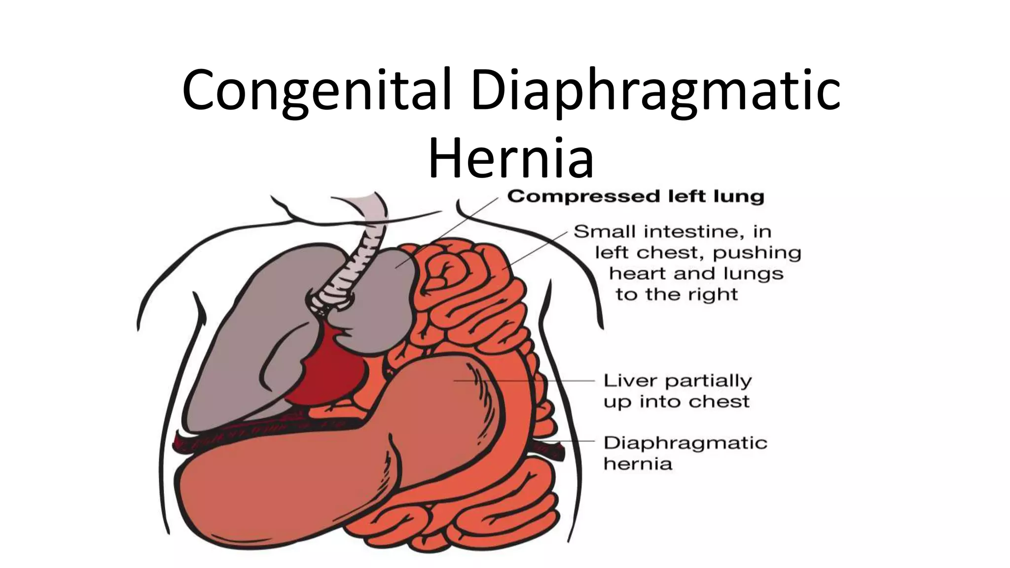
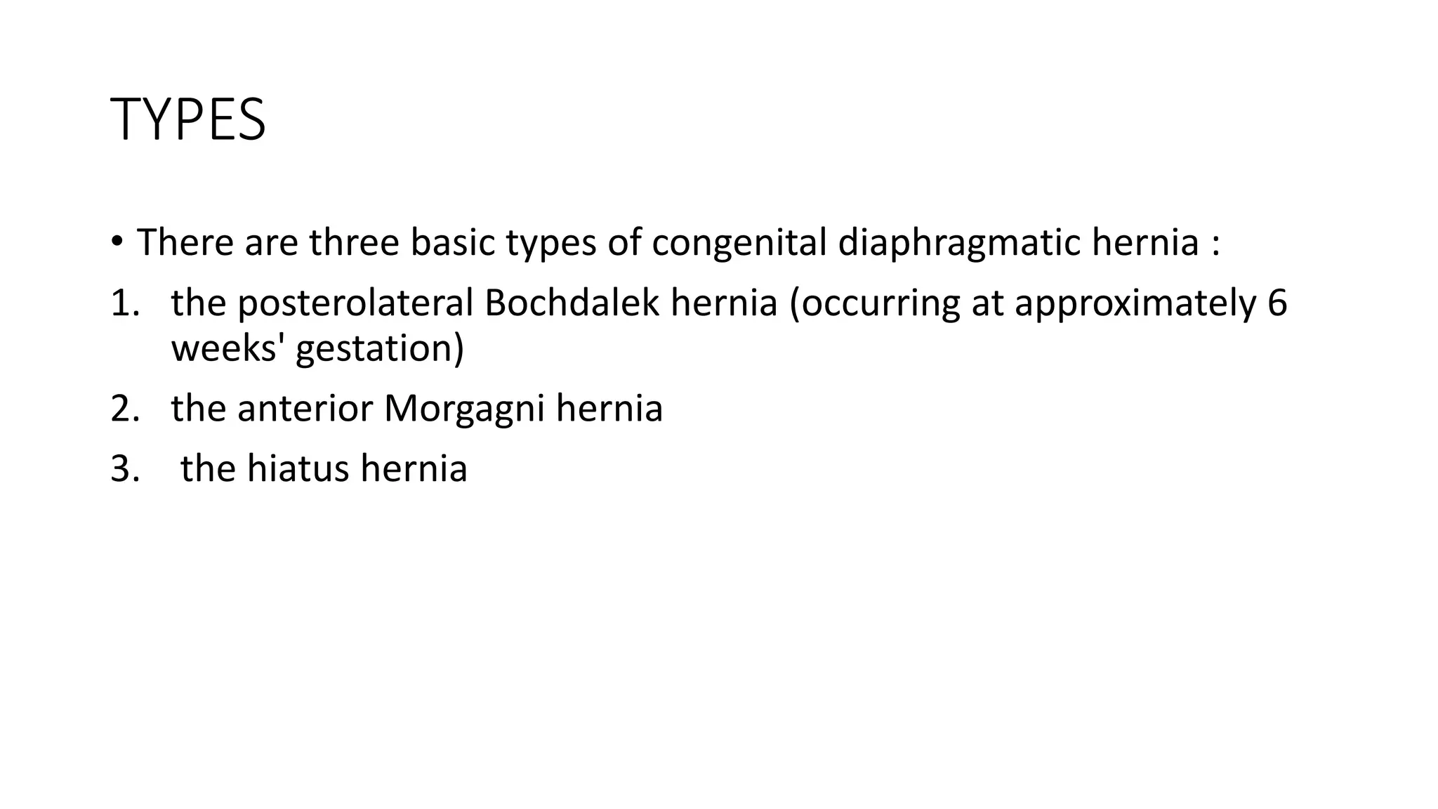
Congenital Diaphragmatic
Hernia
Development of Diaphragm
Develops from 4 sources
• Septum transversum
• Pleuroperitoneal membrane
• Medial dorsal portion of primary oesophageal mesentry
• Marginal ingrowths of the body wall
• In 1761, Morgagni described the classical anterior diaphragmatic
hernia, which today bears his name—Morgagni hernia.
• In 1848, Bochdalek described both right and left posterolateral CDH.
To this day, these CDHs are commonly referred to as Bochdalek
hernias.
TYPES
• There are three basic types of congenital diaphragmatic hernia :
1. the posterolateral Bochdalek hernia (occurring at approximately 6
weeks' gestation)
2. the anterior Morgagni hernia
3. the hiatus hernia
ETIOLOGY
• CDH occurs when the muscular entities of the diaphragm fail to develop
normally, resulting in displacement of abdominal components into the
thorax.
• Bochdalek hernia
• Bochdalek hernias[6] make up the majority of cases of CDH. The major
problem in these hernias is posterolateral defects of the diaphragm, which
results in either failure in the development of the pleuroperitoneal folds or
improper or absent migration of the diaphragmatic musculature.
• As many as 90% of patients with CDH present in the neonatal period or
within the first year of life. These cases have a mortality of 45-50%. Most of
the morbidity and mortality of CDH relates to hypoplasia of the lung and
pulmonary hypertension on the affected side.
• Morgagni hernia
• Morgagni hernia [7, 8, 9] is a less common CDH, accounting for only 5-
10% of CDH cases. The foramen of a Morgagni hernia occurs in the
anterior midline through the sternocostal hiatus of the diaphragm,
with 90% of cases occurring on the right side.
Pathophysiiology
• In CDH,Airway generations and terminal bronchioles are markedly
decreased, alveolar septa are thickened, and there is decreased
complexity of the respiratory acinus and alveolar volume
• Arterial medial wall thickness is increased and peripheral
muscularisation of smaller pre-acinar arteries occurs
• Overall, the lungs are hypoplastic, with lower than normal DNA and
protein content, more so in the ipsilateral than contralateral lung
Antenatal presentation and imaging characteristics
of CDH
• Antenatal ultrasound screening identifies >70% of cases of CDH.
• Left-sided CDH typically presents with a mediastinal shift to the right,
caused by herniation of the stomach and intestines.
• In right-sided CDH, part of the liver is visible in the chest. Because the
liver is echogenic, it may be difficult to differentiate from the lung.
Doppler studies of the umbilical vein and hepatic vessels or location
of the gall bladder may be used as additional landmarks.
• MRI is useful to confirm the diagnosis of CDH in cases of equivocal
sonographic findings, to characterise the content of the herniated
tissues.
• Diaphragmatic eventration, characterised by defective diaphragmatic
muscularisation, can be challenging to differentiate from CDH
antenatally.
a) Coronal and b) axial fetal magnetic resonance images, and c) axial antenatal ultrasound of a
left-sided congenital diaphragmatic hernia at gestational age 26 weeks.
S. Kotecha et al. Eur Respir J 2012;39:820-829
©2012 by European Respiratory Society
Antenatal management and prognosis of CDH
• Ultrasonographic lung size assessment is best undertaken by the use
of the observed/expected lung-to-head ratio
• The LHR predicts survival, with a trend for better prediction at 32–33
weeks rather than 22–23 weeks, and short-term morbidity
• Right-sided lesions have worse outcome
• Three-dimensional ultrasound and MRI both permit absolute
volumetry, but MRI is superior mostly because of better visualisation
of the ipsilateral lung . MRI can quantify the extent of liver occupying
the thorax.
• In all cases, delivery should be planned at a tertiary perinatal centre;
in those with a poor prognosis (e.g. having other congenital
abnormalities), termination of pregnancy may be considered.
• In utero anatomical repair improves lung development, but requires
open fetal surgery and is not currently offered if there is liver
herniation
• Alternatively, tracheal occlusion (TO) has been used to promote lung
growth
• Antenatal TO prevents egress of lung fluid, which increases airway
pressure, causing cell proliferation, increased alveolar airspace and
maturation of pulmonary vasculature.
• TO is possible by percutaneous fetoscopic endoluminal TO
(FETO) via a 3.3-mm cannula without general anaesthesia
• When compared with the predicted survival rate after expectant
management, temporary FETO in fetuses with left CDH increased
survival from 24% to 49% (left-sided) and from 0% to 35% (right-
sided)
CLINICAL PRESENTATION OF CDH
• Clinical manifestations of congenital diaphragmatic hernia (CDH) include the
following:
• Early diagnosis - Right-side heart; decreased breath sounds on the affected side;
scaphoid abdomen; bowel sounds in the thorax, respiratory distress, and/or
cyanosis on auscultation; CDH can often be diagnosed in utero with
ultrasonography (US), magnetic resonance imaging (MRI), or both
• Late diagnosis - Chest mass on chest radiography, gastric volvulus, splenic
volvulus, or large-bowel obstruction
• Congenital hernias (neonatal onset) - Respiratory distress and/or cyanosis occurs
within the first 24 hours of life; CDH may not be diagnosed for several years if the
defect is small enough that it does not cause significant pulmonary dysfunction
• Congenital hernias (childhood or adult onset) - Obstructive symptoms from
protrusion of the colon, chest pain, tightness or fullness the in chest, sepsis
following strangulation or perforation, and many respiratory symptoms occur
INVESTIGATIONS FOR CDH
• This will demonstrate an opacified
• hemithorax with a contralateral
• shift of the mediastinum. Bowel
• gas may be seen in the chest. The
• oesophageal portion of the
• nasogastric tube is deviated to
• the right in infants with left-sided
• CDH and to the left if the lesion is on the right.
• Echocardiography should be undertaken to exclude congenital cardiac
lesions in infants with CDH and in any infant in whom pulmonary
hypertension is suspected. It is important to determine right
ventricular function, as the ability of the right ventricle to function
under increased afterload is an important determinant of illness
severity
POST-NATAL MANAGEMENT OF CDH
Venue and timing of delivery
• Analysis of data from 628 term infants from the CDH Study Group
registry demonstrated early-term delivery by elective Caesarean
section (37–38 versus 39–41 weeks) was associated with less use of
ECMO and a trend towards greater survival later.
Labour ward management
• Infants with CDH should be immediately intubated and ventilated,
and peak inspiratory pressures <25 cmH2O employed. Face-mask and
T-piece or bag and mask resuscitation should not be used, as this can
cause distension of the herniated bowel, increasing respiratory
embarrassment.
MANAGEMENT IN THE NEONATAL UNIT
• The best outcomes for CDH infants are achieved by early medical
stabilisation and delay of surgical repair until optimisation has
occurred
• Gentle ventilation, i.e. avoidance of high pressures (peak pressure <25
cmH2O and peak end-expiratory pressure <5 cmH2O and accepting
higher levels of arterial carbon dioxide tension (Pa,CO2) (permissive
hypercapnia up to a Pa,CO2 of 60–65 mmHg, is preferred.
MANAGEMENT IN THE NEONATAL UNIT
• Treatment in the delivery room
• No resuscitation delivered by bag and mask
Immediate intubation
PI,max <25 cmH2O (peak pressure <25 cmH2O and peak end-expiratory pressure <5 cmH2O
Nasogastric tube
• Treatment in the NICU
• Adapt ventilation to obtain preductal saturation between 85 and 95%
pH >7.20, lactate 3–5 mmol·L−1
CMV or HFOV, maximum PI,max 25–28 cmH2O in CMV and P̄aw 17 cmH2O in HFOV
Targeting blood pressure: normal value for gestational age
Consider inotropic support
• Pulmonary hypertension
• Perform echocardiograhy
iNO but stop if no response
In the chronic phase: phosphodiesterase inhibitors, endothelin antagonists, tyrosine kinase inhibitors
• ECMO indications
• Inability to maintain preductal saturation >85%
Respiratory acidosis
Inadequate oxygen delivery (lactate >5 mmol·L−1)
• Timing of surgical repair
• FI,O2 <0.5
Mean blood pressure normal for gestational age
Urine output >2 mL·kg−1·h−1
No signs of PH
SURGICAL MANAGEMENT OF CDH
• The standard surgical approach to repair the diaphragmatic defect consists
of a subcostal incision with removal of the herniated abdominal viscera
from the thorax and complete exposure of the defect.
• A true hernia sac is sometimes observed (<20% of cases) and, if present,
should be excised.
• Closure of the defect can be accomplished primarily using nonabsorbable
sutures or by means of a prosthetic patch.
• Because synthetic material lacks the capacity for growth, hernia recurrence
may reach up to 50% in some series.
• Alternatively, muscle flaps have been used or a tissue-engineered
diaphragm substitute has been proposed to close very large defects or
even complete agenesis of the diaphragm
Congenital diaphragmatic hernia: A rare cause
of obstructive jaundice
• Case History
A 19-year-old woman, with no previous past medical history, presented
to hospital with a 24-hour history of epigastric pain and shortness of
breath. A plain chest x-ray taken in the accident and emergency
department at the time demonstrated a right basal consolidation. The
patient was admitted under the medical team and treated with
intravenous antibiotics for a lower respiratory tract infection. The
patient discharged herself from hospital two days later and failed to
attend a subsequent follow-up outpatient appointment to repeat the
chest x-ray and monitor her recovery.
• Two years later the patient attended hospital with a one-week history
of jaundice (bilirubin 142µmol/l), icterus, right upper quadrant pain,
gastric acid reflux, nausea and vomiting. There was no history of
trauma. A plain chest x-ray taken on this admission once again
demonstrated a right lower lobe consolidation and an elevated right
hemidiaphragm.
• Ultrasonography of the abdomen performed on the same day
demonstrated both intra- and extrahepatic bile duct dilatation and
‘sludge’ in the gallbladder but no definitive common bile duct
calculus.
• Due to the dual findings of the chest x-ray and ultra-sonography,
double contrast computed tomography (CT) of the abdomen and
thorax was performed. This demonstrated that the entire small
bowel, caecum, mesentery and large bowel to transverse colon was
within the right chest cavity. It also demonstrated intrahepatic duct
dilatation as a result of the anatomical distortion. There was no
obvious pathology seen in the spleen, kidneys, pancreas and
adrenals, left hemidiaphragm or lung parenchyma.
• The patient was admitted for an elective repair of a diaphragmatic hernia
12 weeks following the initial presentation with obstructive jaundice. The
patient had a rooftop laparotomy incision. A large defect was identified in
the posterolateral aspect of the right hemidiaphragm and the hernia sac
contained the right colon, hepatic flexure and the whole of the small
bowel, including the second, third and fourth parts of the duodenum.
• The liver, gallbladder and right kidney were all in the correct anatomical
position. After reduction of the hernial contents, the defect was closed and
reinforced with an on-laying mesh and the right hemicolon was sutured to
the abdominal wall to further prevent recurrence. Primary closure of the
abdominal cavity was achieved and there was no evidence of abdominal
compartment syndrome postoperatively.
• The patient made an excellent postoperative recovery, spending one
day in the high dependence unit and finally being discharged from
hospital at day 5. Regular follow-up appointments in the outpatients
department showed continued improvement in diet and a reduction
in gastric reflux symptoms. The patient was discharged from the care
of the surgeons ten months after the operation, having made a full
and successful recovery.
• CDH is exceedingly rare in adulthood and has been reported to
become symptomatic in only a handful of cases. However, since its
presence can lead to serious adverse events such as acute intestinal
obstruction, or in this case obstruction of the biliary system, it should
be investigated fully and repaired rapidly. The diagnosis should be
considered in any patient presenting with abdominal pain and an
unexplained consolidation on a chest x-ray.

Congenital diaphragmatic hernia

  • 1.
  • 2.
    Development of Diaphragm Developsfrom 4 sources • Septum transversum • Pleuroperitoneal membrane • Medial dorsal portion of primary oesophageal mesentry • Marginal ingrowths of the body wall
  • 3.
    • In 1761,Morgagni described the classical anterior diaphragmatic hernia, which today bears his name—Morgagni hernia. • In 1848, Bochdalek described both right and left posterolateral CDH. To this day, these CDHs are commonly referred to as Bochdalek hernias.
  • 4.
    TYPES • There arethree basic types of congenital diaphragmatic hernia : 1. the posterolateral Bochdalek hernia (occurring at approximately 6 weeks' gestation) 2. the anterior Morgagni hernia 3. the hiatus hernia
  • 6.
    ETIOLOGY • CDH occurswhen the muscular entities of the diaphragm fail to develop normally, resulting in displacement of abdominal components into the thorax. • Bochdalek hernia • Bochdalek hernias[6] make up the majority of cases of CDH. The major problem in these hernias is posterolateral defects of the diaphragm, which results in either failure in the development of the pleuroperitoneal folds or improper or absent migration of the diaphragmatic musculature. • As many as 90% of patients with CDH present in the neonatal period or within the first year of life. These cases have a mortality of 45-50%. Most of the morbidity and mortality of CDH relates to hypoplasia of the lung and pulmonary hypertension on the affected side.
  • 7.
    • Morgagni hernia •Morgagni hernia [7, 8, 9] is a less common CDH, accounting for only 5- 10% of CDH cases. The foramen of a Morgagni hernia occurs in the anterior midline through the sternocostal hiatus of the diaphragm, with 90% of cases occurring on the right side.
  • 8.
    Pathophysiiology • In CDH,Airwaygenerations and terminal bronchioles are markedly decreased, alveolar septa are thickened, and there is decreased complexity of the respiratory acinus and alveolar volume • Arterial medial wall thickness is increased and peripheral muscularisation of smaller pre-acinar arteries occurs • Overall, the lungs are hypoplastic, with lower than normal DNA and protein content, more so in the ipsilateral than contralateral lung
  • 9.
    Antenatal presentation andimaging characteristics of CDH • Antenatal ultrasound screening identifies >70% of cases of CDH. • Left-sided CDH typically presents with a mediastinal shift to the right, caused by herniation of the stomach and intestines. • In right-sided CDH, part of the liver is visible in the chest. Because the liver is echogenic, it may be difficult to differentiate from the lung. Doppler studies of the umbilical vein and hepatic vessels or location of the gall bladder may be used as additional landmarks.
  • 10.
    • MRI isuseful to confirm the diagnosis of CDH in cases of equivocal sonographic findings, to characterise the content of the herniated tissues. • Diaphragmatic eventration, characterised by defective diaphragmatic muscularisation, can be challenging to differentiate from CDH antenatally.
  • 11.
    a) Coronal andb) axial fetal magnetic resonance images, and c) axial antenatal ultrasound of a left-sided congenital diaphragmatic hernia at gestational age 26 weeks. S. Kotecha et al. Eur Respir J 2012;39:820-829 ©2012 by European Respiratory Society
  • 12.
    Antenatal management andprognosis of CDH • Ultrasonographic lung size assessment is best undertaken by the use of the observed/expected lung-to-head ratio • The LHR predicts survival, with a trend for better prediction at 32–33 weeks rather than 22–23 weeks, and short-term morbidity • Right-sided lesions have worse outcome • Three-dimensional ultrasound and MRI both permit absolute volumetry, but MRI is superior mostly because of better visualisation of the ipsilateral lung . MRI can quantify the extent of liver occupying the thorax.
  • 13.
    • In allcases, delivery should be planned at a tertiary perinatal centre; in those with a poor prognosis (e.g. having other congenital abnormalities), termination of pregnancy may be considered. • In utero anatomical repair improves lung development, but requires open fetal surgery and is not currently offered if there is liver herniation
  • 14.
    • Alternatively, trachealocclusion (TO) has been used to promote lung growth • Antenatal TO prevents egress of lung fluid, which increases airway pressure, causing cell proliferation, increased alveolar airspace and maturation of pulmonary vasculature. • TO is possible by percutaneous fetoscopic endoluminal TO (FETO) via a 3.3-mm cannula without general anaesthesia
  • 15.
    • When comparedwith the predicted survival rate after expectant management, temporary FETO in fetuses with left CDH increased survival from 24% to 49% (left-sided) and from 0% to 35% (right- sided)
  • 16.
    CLINICAL PRESENTATION OFCDH • Clinical manifestations of congenital diaphragmatic hernia (CDH) include the following: • Early diagnosis - Right-side heart; decreased breath sounds on the affected side; scaphoid abdomen; bowel sounds in the thorax, respiratory distress, and/or cyanosis on auscultation; CDH can often be diagnosed in utero with ultrasonography (US), magnetic resonance imaging (MRI), or both • Late diagnosis - Chest mass on chest radiography, gastric volvulus, splenic volvulus, or large-bowel obstruction • Congenital hernias (neonatal onset) - Respiratory distress and/or cyanosis occurs within the first 24 hours of life; CDH may not be diagnosed for several years if the defect is small enough that it does not cause significant pulmonary dysfunction • Congenital hernias (childhood or adult onset) - Obstructive symptoms from protrusion of the colon, chest pain, tightness or fullness the in chest, sepsis following strangulation or perforation, and many respiratory symptoms occur
  • 17.
    INVESTIGATIONS FOR CDH •This will demonstrate an opacified • hemithorax with a contralateral • shift of the mediastinum. Bowel • gas may be seen in the chest. The • oesophageal portion of the • nasogastric tube is deviated to • the right in infants with left-sided • CDH and to the left if the lesion is on the right.
  • 18.
    • Echocardiography shouldbe undertaken to exclude congenital cardiac lesions in infants with CDH and in any infant in whom pulmonary hypertension is suspected. It is important to determine right ventricular function, as the ability of the right ventricle to function under increased afterload is an important determinant of illness severity
  • 19.
    POST-NATAL MANAGEMENT OFCDH Venue and timing of delivery • Analysis of data from 628 term infants from the CDH Study Group registry demonstrated early-term delivery by elective Caesarean section (37–38 versus 39–41 weeks) was associated with less use of ECMO and a trend towards greater survival later.
  • 20.
    Labour ward management •Infants with CDH should be immediately intubated and ventilated, and peak inspiratory pressures <25 cmH2O employed. Face-mask and T-piece or bag and mask resuscitation should not be used, as this can cause distension of the herniated bowel, increasing respiratory embarrassment.
  • 21.
    MANAGEMENT IN THENEONATAL UNIT • The best outcomes for CDH infants are achieved by early medical stabilisation and delay of surgical repair until optimisation has occurred • Gentle ventilation, i.e. avoidance of high pressures (peak pressure <25 cmH2O and peak end-expiratory pressure <5 cmH2O and accepting higher levels of arterial carbon dioxide tension (Pa,CO2) (permissive hypercapnia up to a Pa,CO2 of 60–65 mmHg, is preferred.
  • 22.
    MANAGEMENT IN THENEONATAL UNIT • Treatment in the delivery room • No resuscitation delivered by bag and mask Immediate intubation PI,max <25 cmH2O (peak pressure <25 cmH2O and peak end-expiratory pressure <5 cmH2O Nasogastric tube • Treatment in the NICU • Adapt ventilation to obtain preductal saturation between 85 and 95% pH >7.20, lactate 3–5 mmol·L−1 CMV or HFOV, maximum PI,max 25–28 cmH2O in CMV and P̄aw 17 cmH2O in HFOV Targeting blood pressure: normal value for gestational age Consider inotropic support • Pulmonary hypertension • Perform echocardiograhy iNO but stop if no response In the chronic phase: phosphodiesterase inhibitors, endothelin antagonists, tyrosine kinase inhibitors • ECMO indications • Inability to maintain preductal saturation >85% Respiratory acidosis Inadequate oxygen delivery (lactate >5 mmol·L−1) • Timing of surgical repair • FI,O2 <0.5 Mean blood pressure normal for gestational age Urine output >2 mL·kg−1·h−1 No signs of PH
  • 23.
    SURGICAL MANAGEMENT OFCDH • The standard surgical approach to repair the diaphragmatic defect consists of a subcostal incision with removal of the herniated abdominal viscera from the thorax and complete exposure of the defect. • A true hernia sac is sometimes observed (<20% of cases) and, if present, should be excised. • Closure of the defect can be accomplished primarily using nonabsorbable sutures or by means of a prosthetic patch. • Because synthetic material lacks the capacity for growth, hernia recurrence may reach up to 50% in some series. • Alternatively, muscle flaps have been used or a tissue-engineered diaphragm substitute has been proposed to close very large defects or even complete agenesis of the diaphragm
  • 24.
    Congenital diaphragmatic hernia:A rare cause of obstructive jaundice • Case History A 19-year-old woman, with no previous past medical history, presented to hospital with a 24-hour history of epigastric pain and shortness of breath. A plain chest x-ray taken in the accident and emergency department at the time demonstrated a right basal consolidation. The patient was admitted under the medical team and treated with intravenous antibiotics for a lower respiratory tract infection. The patient discharged herself from hospital two days later and failed to attend a subsequent follow-up outpatient appointment to repeat the chest x-ray and monitor her recovery.
  • 25.
    • Two yearslater the patient attended hospital with a one-week history of jaundice (bilirubin 142µmol/l), icterus, right upper quadrant pain, gastric acid reflux, nausea and vomiting. There was no history of trauma. A plain chest x-ray taken on this admission once again demonstrated a right lower lobe consolidation and an elevated right hemidiaphragm. • Ultrasonography of the abdomen performed on the same day demonstrated both intra- and extrahepatic bile duct dilatation and ‘sludge’ in the gallbladder but no definitive common bile duct calculus.
  • 26.
    • Due tothe dual findings of the chest x-ray and ultra-sonography, double contrast computed tomography (CT) of the abdomen and thorax was performed. This demonstrated that the entire small bowel, caecum, mesentery and large bowel to transverse colon was within the right chest cavity. It also demonstrated intrahepatic duct dilatation as a result of the anatomical distortion. There was no obvious pathology seen in the spleen, kidneys, pancreas and adrenals, left hemidiaphragm or lung parenchyma.
  • 28.
    • The patientwas admitted for an elective repair of a diaphragmatic hernia 12 weeks following the initial presentation with obstructive jaundice. The patient had a rooftop laparotomy incision. A large defect was identified in the posterolateral aspect of the right hemidiaphragm and the hernia sac contained the right colon, hepatic flexure and the whole of the small bowel, including the second, third and fourth parts of the duodenum. • The liver, gallbladder and right kidney were all in the correct anatomical position. After reduction of the hernial contents, the defect was closed and reinforced with an on-laying mesh and the right hemicolon was sutured to the abdominal wall to further prevent recurrence. Primary closure of the abdominal cavity was achieved and there was no evidence of abdominal compartment syndrome postoperatively.
  • 29.
    • The patientmade an excellent postoperative recovery, spending one day in the high dependence unit and finally being discharged from hospital at day 5. Regular follow-up appointments in the outpatients department showed continued improvement in diet and a reduction in gastric reflux symptoms. The patient was discharged from the care of the surgeons ten months after the operation, having made a full and successful recovery.
  • 30.
    • CDH isexceedingly rare in adulthood and has been reported to become symptomatic in only a handful of cases. However, since its presence can lead to serious adverse events such as acute intestinal obstruction, or in this case obstruction of the biliary system, it should be investigated fully and repaired rapidly. The diagnosis should be considered in any patient presenting with abdominal pain and an unexplained consolidation on a chest x-ray.