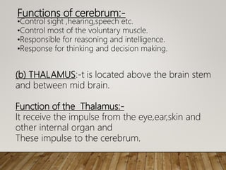
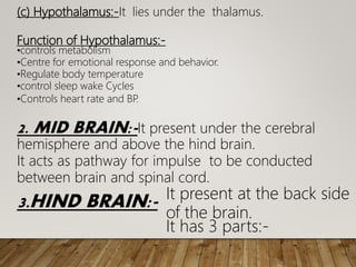
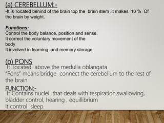
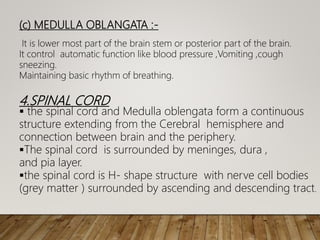
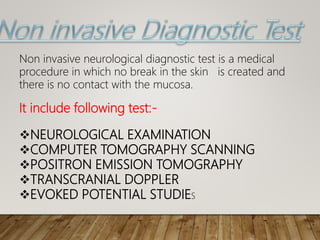
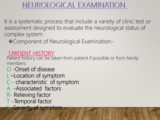
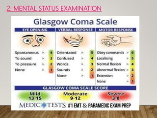
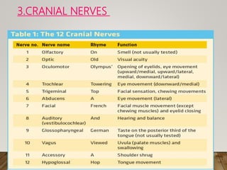
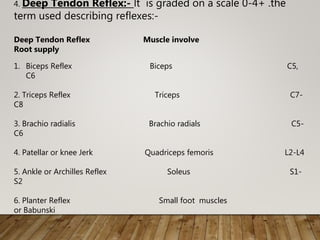
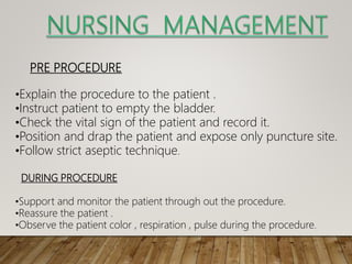
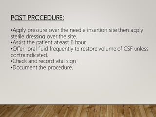
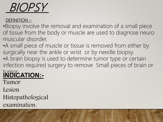
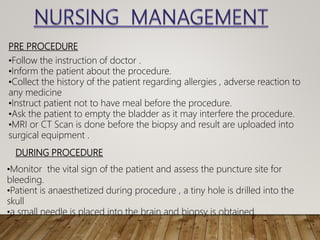
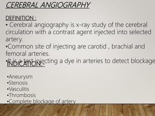
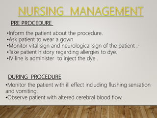
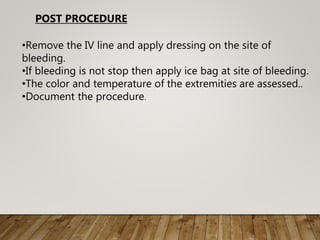
DIAGNOSTIC TEST IN
NEUROLOGICAL DISORDER AND ITS
NURSING MANAGEMENT
Bsc (h) Nursing
RAKCON
A Diagnostic test is a kind of medical test
perform to aid in diagnosis or detection of
disease in Neurological System. It may be used
to diagnose disease or measure
To progress or recovery from disease or confirm
that person is free from disease.
The term diagnostic comprises all activities in
me
•Human brain is the organ of the body in the
head that control functions like
movement,sensation and thoughts etc.
•Human brain is divided into three main parts
on the basis of function and placement.
1. FOREBRAIN:-It is anterior part of the brain.It has 3 parts:-
(a)CEREBRUM:-It is greatly folded and largest section of the
brain. Cerebrum having two halves called cerebral
hemisphere (right hemisphere and left hemisphere.
HUMAN BRAIN
Functions of cerebrum:-
•Control sight ,hearing,speech etc.
•Control most of the voluntary muscle.
•Responsible for reasoning and intelligence.
•Response for thinking and decision making.
(b) THALAMUS:-t is located above the brain stem
and between mid brain.
Function of the Thalamus:-
It receive the impulse from the eye,ear,skin and
other internal organ and
These impulse to the cerebrum.
(c) Hypothalamus:-It lies under the thalamus.
Function of Hypothalamus:-
•controls metabolism
•Centre for emotional response and behavior.
•Regulate body temperature
•control sleep wake Cycles
•Controls heart rate and ВР
.
2. MID BRAIN:-It present under the cerebral
hemisphere and above the hind brain.
It acts as pathway for impulse to be conducted
between brain and spinal cord.
3.HIND BRAIN:- It present at the back side
of the brain.
It has 3 parts:-
(a) CEREBELLUM:-
-It is located behind of the brain top the brain stem .it makes 10 % Of
the brain by weight.
Functions:
Control the body balance, position and sense.
It correct the voluntary movement of the
body
It involved in learning and memory storage.
(b) PONS
It located above the medulla oblangata
“Pons” means bridge connect the cerebellum to the rest of
the brain
FUNCTION:-
It Contains nuclei that deals with respiration,swallowing,
bladder control, hearing , equillibrium
It control sleep.
(c) MEDULLA OBLANGATA :-
It is lower most part of the brain stem or posterior part of the brain.
It control automatic function like blood pressure ,Vomiting ,cough
sneezing.
Maintaining basic rhythm of breathing.
4.SPINAL CORD
 the spinal cord and Medulla oblengata form a continuous
structure extending from the Cerebral hemisphere and
connection between brain and the periphery.
The spinal cord is surrounded by meninges, dura ,
and pia layer.
the spinal cord is H- shape structure with nerve cell bodies
(grey matter ) surrounded by ascending and descending tract.
Definition-A diagnostic test is a test is used to identify the
cause or condition of a disease in nervous system .
Diagnostic tests are vital tools that help the physicians to
confirm or role out neurological disorder or other medical
condition.
Purposes
•To diagnose a disease.
• To Screen for risk factors
•To monitor the disease , is disease controlled.
•Prognosis allows clinicians to assess the patient has for
developing disease in the future.
Non Invasive
 Invasive
TYPE OF NEUROLOGICAL DIAGNOSTIC TEST
It include following test:-
NEUROLOGICAL EXAMINATION
COMPUTER TOMOGRAPHY SCANNING
POSITRON EMISSION TOMOGRAPHY
TRANSCRANIAL DOPPLER
EVOKED POTENTIAL STUDIES
Non invasive neurological diagnostic test is a medical
procedure in which no break in the skin is created and
there is no contact with the mucosa.
It is a systematic process that include a variety of clinic test or
assessment designed to evaluate the neurological status of
complex system.
Component of Neurological Examination:-
1.PATIENT HISTORY
Patient history can be taken from patient if possible or from family
members.
O -Onset of disease
L –Location of symptom
С - characteristic of symptom
А -Associated factors
R- Relieving factor
T –Temporal factor
S- Severity of symptom
2. MENTAL STATUS EXAMINATION
3.CRANIAL NERVES
4. Deep Tendon Reflex:- lt is graded on a scale 0-4+ .the
term used describing reflexes:-
Deep Tendon Reflex Muscle involve
Root supply
1. Biceps Reflex Biceps C5,
C6
2. Triceps Reflex Triceps C7-
C8
3. Brachio radialis Brachio radials C5-
C6
4. Patellar or knee Jerk Quadriceps femoris L2-L4
5. Ankle or Archilles Reflex Soleus S1-
S2
6. Planter Reflex Small foot muscles
or Babunski
X-RAY
•An X-ray is a quick and painless procedure commonly used to
produce images of the inside of the body.
•X-rays are a type of radiation that can
pass through the body.
•As they pass through the body, the energy from X-rays is absorbed at
different rates by different parts of the body. A detector on the other
side of the body picks up the X-rays after they've passed through and
turns them into an image.
Contrast material
Before some types of X-rays, you're given a liquid called
contrast medium. Contrast mediums, such as barium and
iodine, help outline a specific area of your body on the
X-ray image. You may swallow the contrast medium or
receive it as an injection or an enema.
After the X-ray
After an X-ray, you generally can resume normal
Routine X-rays usually have no side effects. However, if
you're injected with contrast medium before your X-rays,
drink plenty of fluids to help rid your body of it.
During the X-ray
•X-rays are performed at doctors' offices, dentists' offices, emergency
rooms and hospitals — wherever an X-ray machine is available. The
machine produces a safe level of radiation that passes through your
body and records an image on a specialized plate. You can't feel an X-
ray.
•A technologist positions your body to obtain the necessary views. He or
she may use pillows or sandbags to help you hold the position. During
the X-ray exposure, you remain still and sometimes hold your breath to
avoid moving so that the image doesn't blur.
•An X-ray procedure may take just a few minutes for a simple X-ray or
longer for more-involved procedures, such as those using a contrast
medium.
COMPUTER TOMOGRAPHY SCANNING (CT Scan)
DEFINITION:-
CT Scanning is a non invasive and painless procedure .It make use
of narrow x-ray beam to scan, can aid in proper diagnosis by showing
the area of the brain that is affected.
The image is displayed on TV monitor and is photographed and stored
Digitally.
INDICATION: CT Scan used to detect:-
Brain tumors and cycst
Lesion and head injury
Intracranial bleeding
Encephalitis
PRE PROCEDURE:-
•Check the doctor’s order.
•Inform the patient about the procedure.
•Ask patient to wear gown and remove all jewelry and valuable.
•NPO about 3-4 hour prior to CT Scan.
•Allergied to iodine.
•An intravenous line is injected to the patient through which dye is administer
prior to study.
DURING PROCEDURE
•Provide pillow and strap used to prevent movement during the procedure.
•Monitor the IV line and reassure the patient.
•Sedation can be used if agitation or restlessness felt by patient .
POST PROCEDURE
•Place the patient in comfortable
position.
•Provide plenty of fluid to the patient.
•Document the procedure.
POSITRON EMISSION TOMOGRAPHY (PET)
DEFINITION : -
PET is a computer based nuclear imaging technique that
produce image of actual organ functioning. Patient either
inhale a radio active gas or injecting with radioactive
substance that emits positive charged particle .when these
positive Charged particle combine with negative charge
electron ,the resultant gamma rays can be detected by
scanning device that produce 2D View at various levels of the
brain.
INDICATION:-
•Alzheimer’s disease
•Parkinson’s disease
•Huntington’s disease
•Hematoma
•Epilepsy
PRE PROCEDURE:-
•Inform the patient about the procedure.
•Ask patient to wear gown and remove all jewelry and valuable.
•Ask patient to empty bladder otherwise it interfere the procedure
•Administration of radionuclide agent through IV catheter.
DURING PROCEDURE
•Monitor the IV line and reassure the patient.
•Help patient to take position in padded table inside the scanner
POST PROCEDURE
•Place the patient in comfortable position.
•Remove the IV line.
•Document the procedure and report any
abnormality to physician
EVOKED POTENTIAL STUDY
DEFINITION:-
In evoked potential study, electrode are applied to the scalp
and an external stimulus is applied to periphery sensory
receptor to measure electrical activity in the brain.
INDICATION:-
•Severe nearsightedness
•Inflammation of ear
•Muscle spam in the head or neck
Evoked potential study involved 3 test that
measure response to visual, auditory and
Sensory stimuli.
•VISUAL EVOKED RESPONSE TEST
This test can diagnose problem with the optic nerve that affect sight. A health
care
Professional place electrode along your scalp to record the electric signals as
you watch a checkboard pattern flash for several minute on screen.
•BRAINSTEM AUDITORY EVOKED RESPONSE TEST
This test can diagnose hearing ability and point to possible brainstem tumors
or multiple Sclerosis. A health care professional place electrode on your scalp
and ear lobe and deliver auditory stimuli such as clicking noise and tone to
one ear.
•SOMATOSENSORY EVOKED RESPONSE TEST
This test can detect problem with spinal cord that cause numbness of the
arm and legs .A health care professional attached electrode to wrist ,back of
knee or other location.
In VER Test:-
•Inform the patient about the procedure.
•Asked the patient to stand erect a few feet away from a screen.
•Place electrode on scalp over the area of brain responsible for interpreting
visual stimuli.
•Asked patient to focus on the center of screen and to close one eye at a
time while the screen display a checkboard pattern. The square of the
checkboard reverse color once or twice a second.
In BAER Test
•Inform the patient about the procedure.
•Place a patient in sound proof room wearing earphone.
•Place the electrode on top of the head and one on earlobe and them the other
•A clicking sound or another auditory stimulus will be deliver through the earph
to the ear being tested.
In SSER Test:
•Gain the confident of the patient by informing him about the
procedure.
•Place electrode on the scalp and at one or more on body
such as wrist , back of the knee or lower back.
•Check whether the patient is allergied to gel.
•Document the procedure.
TRANSCRANIAL DOPPLER (TCD)
DEFINITION:-
Transcranial doppler is a non invasive technique ,painless
ultrasound that used high frequency sound waves to
measure the rate and direction of blood in vessels.
This test used to examine and record the speed of blood in
arteries.
INDICATION:-
•Subarachnoid hemorrhage
•Sickle Cell Anemia
•Embolism
It include following test:-
An invasive neurological diagnostic test in which there is break
of skin or insert instrument in body (brain) cavity to know the
diagnosis.
•Biopsy
•Cerebral Angiography
•Cerebrospinal fluid analysis
CEREBROSPINAL FLUID ANALYSIS
DEFINITION :
It is a invasive diagnostic test that help to diagnose
disease and condition affecting brain and spinal cord.
It is a medical procedure in which a needle is inserted
into spinal canal to collect CSF for testing.
INDICATION:-
• Meningitis
• Spinal injury
• Encephalitis
• Congenital malformation
SITE FOR CSF ANALYSIS or lumbar puncture is performed between L3 –
L4 or L4-L5 space
PRE PROCEDURE
•Explain the procedure to the patient .
•Instruct patient to empty the bladder.
•Check the vital sign of the patient and record it.
•Position and drap the patient and expose only puncture site.
•Follow strict aseptic technique.
DURING PROCEDURE
•Support and monitor the patient through out the procedure.
•Reassure the patient .
•Observe the patient color , respiration , pulse during the procedure.
POST PROCEDURE:
•Apply pressure over the needle insertion site then apply
sterile dressing over the site.
•Assist the patient atleast 6 hour.
•Offer oral fluid frequently to restore volume of CSF unless
contraindicated.
•Check and record vital sign .
•Document the procedure.
BIOPSY
DEFINITION :-
•Biopsy involve the removal and examination of a small piece
of tissue from the body or muscle are used to diagnose neuro
muscular disorder.
•A small piece of muscle or tissue is removed from either by
surgically near the ankle or wrist or by needle biopsy.
•A brain biopsy is used to determine tumor type or certain
infection required surgery to remove Small pieces of brain or
tumor.
INDICATION:-
Tumor
Lesion
Histopathological
examination.
PRE PROCEDURE
•Follow the instruction of doctor .
•Inform the patient about the procedure.
•Collect the history of the patient regarding allergies , adverse reaction to
any medicine
•Instruct patient not to have meal before the procedure.
•Ask the patient to empty the bladder as it may interfere the procedure.
•MRI or CT Scan is done before the biopsy and result are uploaded into
surgical equipment .
DURING PROCEDURE
•Monitor the vital sign of the patient and assess the puncture site for
bleeding.
•Patient is anaesthetized during procedure , a tiny hole is drilled into the
skull
•a small needle is placed into the brain and biopsy is obtained.
POST PROCEDURE
•After the surgery incision is sutured.
•Monitor the sutured site , if bleeding.
•Report and record the finding.
CEREBRAL ANGIOGRAPHY
DEFINITION :
• Cerebral angiography is x-ray study of the cerebral
circulation with a contrast agent injected into selected
artery.
•Common site of injecting are carotid , brachial and
femoral arteries.
•It is a test injecting a dye in arteries to detect blockage
INDICATION:-
•Aneurysm
•Stenosis
•Vasculitis
•Thrombosis
•Complete blockage of artery
PRE PROCEDURE
•Inform the patient about the procedure.
•Ask patient to wear a gown.
•Monitor vital sign and neurological sign of the patient .-
•Take patient history regarding allergies to dye.
•IV line is administer to inject the dye .
DURING PROCEDURE
•Monitor the patient with ill effect including flushing sensation
and vomiting.
•Observe patient with altered cerebral blood flow.
•Remove the IV line and apply dressing on the site of
bleeding.
•If bleeding is not stop then apply ice bag at site of bleeding.
•The color and temperature of the extremities are assessed..
•Document the procedure.
POST PROCEDURE
The diagnostic utility of brain biopsy procedures in
patients with rapidly deteriorating neurological
conditions or dementia
ABSTRACT-. Obtaining brain biopsy specimens is often
the diagnostic test of last resort for patients with
unexplained neurological conditions, particularly those
with a rapidly deteriorating neurological course. The
goals of this analysis were to determine the diagnostic
sensitivity of brain biopsy specimens in these types of
patients and retrospectively identify features of these
disorders that may have enabled an earlier diagnosis,
which may prevent the need for diagnostic brain
biopsy procedures in the future.
METHOD -The authors reviewed the case records of all brain biopsy
procedures that had been performed at a single tertiary care
institution between January 1993 and April 2002 in 171 patients.
Patients with HIV or nonlymphomatous brain tumors were excluded
from this analysis because the utility of brain biopsy specimens for
these conditions has been determined from previous studies. A
subgroup analysis of this cohort was performed in the 64 patients
who had comprehensive medical records and a clinical syndrome
involving a progressively deteriorating neurological condition of less
than 1 year in duration.
RESULT -These results will help guide the evaluation of patients with
neurological conditions that are difficult to diagnose and will provide
a foundation for further prospective studies.
Today , we have discussed about Diagnostic
test in Neurological Disorder and its
management,
with the type of diagnostic test i.e non
invasive and invasive with their nursing
management.
From this topic we come to know about
different aspect Of neurological disorder with
their diagnostic test. Nursing management play
an important role to improve the condition of
patient with care in Neurological Disorder .
Diagnostic test in neurological disorder

Diagnostic test in neurological disorder

  • 1.
    DIAGNOSTIC TEST IN NEUROLOGICALDISORDER AND ITS NURSING MANAGEMENT Bsc (h) Nursing RAKCON
  • 2.
    A Diagnostic testis a kind of medical test perform to aid in diagnosis or detection of disease in Neurological System. It may be used to diagnose disease or measure To progress or recovery from disease or confirm that person is free from disease. The term diagnostic comprises all activities in me
  • 4.
    •Human brain isthe organ of the body in the head that control functions like movement,sensation and thoughts etc. •Human brain is divided into three main parts on the basis of function and placement. 1. FOREBRAIN:-It is anterior part of the brain.It has 3 parts:- (a)CEREBRUM:-It is greatly folded and largest section of the brain. Cerebrum having two halves called cerebral hemisphere (right hemisphere and left hemisphere. HUMAN BRAIN
  • 5.
    Functions of cerebrum:- •Controlsight ,hearing,speech etc. •Control most of the voluntary muscle. •Responsible for reasoning and intelligence. •Response for thinking and decision making. (b) THALAMUS:-t is located above the brain stem and between mid brain. Function of the Thalamus:- It receive the impulse from the eye,ear,skin and other internal organ and These impulse to the cerebrum.
  • 6.
    (c) Hypothalamus:-It liesunder the thalamus. Function of Hypothalamus:- •controls metabolism •Centre for emotional response and behavior. •Regulate body temperature •control sleep wake Cycles •Controls heart rate and ВР . 2. MID BRAIN:-It present under the cerebral hemisphere and above the hind brain. It acts as pathway for impulse to be conducted between brain and spinal cord. 3.HIND BRAIN:- It present at the back side of the brain. It has 3 parts:-
  • 7.
    (a) CEREBELLUM:- -It islocated behind of the brain top the brain stem .it makes 10 % Of the brain by weight. Functions: Control the body balance, position and sense. It correct the voluntary movement of the body It involved in learning and memory storage. (b) PONS It located above the medulla oblangata “Pons” means bridge connect the cerebellum to the rest of the brain FUNCTION:- It Contains nuclei that deals with respiration,swallowing, bladder control, hearing , equillibrium It control sleep.
  • 8.
    (c) MEDULLA OBLANGATA:- It is lower most part of the brain stem or posterior part of the brain. It control automatic function like blood pressure ,Vomiting ,cough sneezing. Maintaining basic rhythm of breathing. 4.SPINAL CORD  the spinal cord and Medulla oblengata form a continuous structure extending from the Cerebral hemisphere and connection between brain and the periphery. The spinal cord is surrounded by meninges, dura , and pia layer. the spinal cord is H- shape structure with nerve cell bodies (grey matter ) surrounded by ascending and descending tract.
  • 9.
    Definition-A diagnostic testis a test is used to identify the cause or condition of a disease in nervous system . Diagnostic tests are vital tools that help the physicians to confirm or role out neurological disorder or other medical condition. Purposes •To diagnose a disease. • To Screen for risk factors •To monitor the disease , is disease controlled. •Prognosis allows clinicians to assess the patient has for developing disease in the future. Non Invasive  Invasive TYPE OF NEUROLOGICAL DIAGNOSTIC TEST
  • 10.
    It include followingtest:- NEUROLOGICAL EXAMINATION COMPUTER TOMOGRAPHY SCANNING POSITRON EMISSION TOMOGRAPHY TRANSCRANIAL DOPPLER EVOKED POTENTIAL STUDIES Non invasive neurological diagnostic test is a medical procedure in which no break in the skin is created and there is no contact with the mucosa.
  • 11.
    It is asystematic process that include a variety of clinic test or assessment designed to evaluate the neurological status of complex system. Component of Neurological Examination:- 1.PATIENT HISTORY Patient history can be taken from patient if possible or from family members. O -Onset of disease L –Location of symptom С - characteristic of symptom А -Associated factors R- Relieving factor T –Temporal factor S- Severity of symptom
  • 12.
    2. MENTAL STATUSEXAMINATION
  • 13.
  • 14.
    4. Deep TendonReflex:- lt is graded on a scale 0-4+ .the term used describing reflexes:- Deep Tendon Reflex Muscle involve Root supply 1. Biceps Reflex Biceps C5, C6 2. Triceps Reflex Triceps C7- C8 3. Brachio radialis Brachio radials C5- C6 4. Patellar or knee Jerk Quadriceps femoris L2-L4 5. Ankle or Archilles Reflex Soleus S1- S2 6. Planter Reflex Small foot muscles or Babunski
  • 15.
    X-RAY •An X-ray isa quick and painless procedure commonly used to produce images of the inside of the body. •X-rays are a type of radiation that can pass through the body. •As they pass through the body, the energy from X-rays is absorbed at different rates by different parts of the body. A detector on the other side of the body picks up the X-rays after they've passed through and turns them into an image. Contrast material Before some types of X-rays, you're given a liquid called contrast medium. Contrast mediums, such as barium and iodine, help outline a specific area of your body on the X-ray image. You may swallow the contrast medium or receive it as an injection or an enema.
  • 16.
    After the X-ray Afteran X-ray, you generally can resume normal Routine X-rays usually have no side effects. However, if you're injected with contrast medium before your X-rays, drink plenty of fluids to help rid your body of it. During the X-ray •X-rays are performed at doctors' offices, dentists' offices, emergency rooms and hospitals — wherever an X-ray machine is available. The machine produces a safe level of radiation that passes through your body and records an image on a specialized plate. You can't feel an X- ray. •A technologist positions your body to obtain the necessary views. He or she may use pillows or sandbags to help you hold the position. During the X-ray exposure, you remain still and sometimes hold your breath to avoid moving so that the image doesn't blur. •An X-ray procedure may take just a few minutes for a simple X-ray or longer for more-involved procedures, such as those using a contrast medium.
  • 17.
    COMPUTER TOMOGRAPHY SCANNING(CT Scan) DEFINITION:- CT Scanning is a non invasive and painless procedure .It make use of narrow x-ray beam to scan, can aid in proper diagnosis by showing the area of the brain that is affected. The image is displayed on TV monitor and is photographed and stored Digitally. INDICATION: CT Scan used to detect:- Brain tumors and cycst Lesion and head injury Intracranial bleeding Encephalitis
  • 18.
    PRE PROCEDURE:- •Check thedoctor’s order. •Inform the patient about the procedure. •Ask patient to wear gown and remove all jewelry and valuable. •NPO about 3-4 hour prior to CT Scan. •Allergied to iodine. •An intravenous line is injected to the patient through which dye is administer prior to study. DURING PROCEDURE •Provide pillow and strap used to prevent movement during the procedure. •Monitor the IV line and reassure the patient. •Sedation can be used if agitation or restlessness felt by patient .
  • 19.
    POST PROCEDURE •Place thepatient in comfortable position. •Provide plenty of fluid to the patient. •Document the procedure.
  • 20.
    POSITRON EMISSION TOMOGRAPHY(PET) DEFINITION : - PET is a computer based nuclear imaging technique that produce image of actual organ functioning. Patient either inhale a radio active gas or injecting with radioactive substance that emits positive charged particle .when these positive Charged particle combine with negative charge electron ,the resultant gamma rays can be detected by scanning device that produce 2D View at various levels of the brain. INDICATION:- •Alzheimer’s disease •Parkinson’s disease •Huntington’s disease •Hematoma •Epilepsy
  • 21.
    PRE PROCEDURE:- •Inform thepatient about the procedure. •Ask patient to wear gown and remove all jewelry and valuable. •Ask patient to empty bladder otherwise it interfere the procedure •Administration of radionuclide agent through IV catheter. DURING PROCEDURE •Monitor the IV line and reassure the patient. •Help patient to take position in padded table inside the scanner
  • 22.
    POST PROCEDURE •Place thepatient in comfortable position. •Remove the IV line. •Document the procedure and report any abnormality to physician
  • 23.
    EVOKED POTENTIAL STUDY DEFINITION:- Inevoked potential study, electrode are applied to the scalp and an external stimulus is applied to periphery sensory receptor to measure electrical activity in the brain. INDICATION:- •Severe nearsightedness •Inflammation of ear •Muscle spam in the head or neck Evoked potential study involved 3 test that measure response to visual, auditory and Sensory stimuli.
  • 24.
    •VISUAL EVOKED RESPONSETEST This test can diagnose problem with the optic nerve that affect sight. A health care Professional place electrode along your scalp to record the electric signals as you watch a checkboard pattern flash for several minute on screen. •BRAINSTEM AUDITORY EVOKED RESPONSE TEST This test can diagnose hearing ability and point to possible brainstem tumors or multiple Sclerosis. A health care professional place electrode on your scalp and ear lobe and deliver auditory stimuli such as clicking noise and tone to one ear. •SOMATOSENSORY EVOKED RESPONSE TEST This test can detect problem with spinal cord that cause numbness of the arm and legs .A health care professional attached electrode to wrist ,back of knee or other location.
  • 25.
    In VER Test:- •Informthe patient about the procedure. •Asked the patient to stand erect a few feet away from a screen. •Place electrode on scalp over the area of brain responsible for interpreting visual stimuli. •Asked patient to focus on the center of screen and to close one eye at a time while the screen display a checkboard pattern. The square of the checkboard reverse color once or twice a second. In BAER Test •Inform the patient about the procedure. •Place a patient in sound proof room wearing earphone. •Place the electrode on top of the head and one on earlobe and them the other •A clicking sound or another auditory stimulus will be deliver through the earph to the ear being tested.
  • 26.
    In SSER Test: •Gainthe confident of the patient by informing him about the procedure. •Place electrode on the scalp and at one or more on body such as wrist , back of the knee or lower back. •Check whether the patient is allergied to gel. •Document the procedure.
  • 27.
    TRANSCRANIAL DOPPLER (TCD) DEFINITION:- Transcranialdoppler is a non invasive technique ,painless ultrasound that used high frequency sound waves to measure the rate and direction of blood in vessels. This test used to examine and record the speed of blood in arteries. INDICATION:- •Subarachnoid hemorrhage •Sickle Cell Anemia •Embolism
  • 28.
    It include followingtest:- An invasive neurological diagnostic test in which there is break of skin or insert instrument in body (brain) cavity to know the diagnosis. •Biopsy •Cerebral Angiography •Cerebrospinal fluid analysis
  • 29.
    CEREBROSPINAL FLUID ANALYSIS DEFINITION: It is a invasive diagnostic test that help to diagnose disease and condition affecting brain and spinal cord. It is a medical procedure in which a needle is inserted into spinal canal to collect CSF for testing. INDICATION:- • Meningitis • Spinal injury • Encephalitis • Congenital malformation SITE FOR CSF ANALYSIS or lumbar puncture is performed between L3 – L4 or L4-L5 space
  • 30.
    PRE PROCEDURE •Explain theprocedure to the patient . •Instruct patient to empty the bladder. •Check the vital sign of the patient and record it. •Position and drap the patient and expose only puncture site. •Follow strict aseptic technique. DURING PROCEDURE •Support and monitor the patient through out the procedure. •Reassure the patient . •Observe the patient color , respiration , pulse during the procedure.
  • 31.
    POST PROCEDURE: •Apply pressureover the needle insertion site then apply sterile dressing over the site. •Assist the patient atleast 6 hour. •Offer oral fluid frequently to restore volume of CSF unless contraindicated. •Check and record vital sign . •Document the procedure.
  • 32.
    BIOPSY DEFINITION :- •Biopsy involvethe removal and examination of a small piece of tissue from the body or muscle are used to diagnose neuro muscular disorder. •A small piece of muscle or tissue is removed from either by surgically near the ankle or wrist or by needle biopsy. •A brain biopsy is used to determine tumor type or certain infection required surgery to remove Small pieces of brain or tumor. INDICATION:- Tumor Lesion Histopathological examination.
  • 33.
    PRE PROCEDURE •Follow theinstruction of doctor . •Inform the patient about the procedure. •Collect the history of the patient regarding allergies , adverse reaction to any medicine •Instruct patient not to have meal before the procedure. •Ask the patient to empty the bladder as it may interfere the procedure. •MRI or CT Scan is done before the biopsy and result are uploaded into surgical equipment . DURING PROCEDURE •Monitor the vital sign of the patient and assess the puncture site for bleeding. •Patient is anaesthetized during procedure , a tiny hole is drilled into the skull •a small needle is placed into the brain and biopsy is obtained.
  • 34.
    POST PROCEDURE •After thesurgery incision is sutured. •Monitor the sutured site , if bleeding. •Report and record the finding.
  • 35.
    CEREBRAL ANGIOGRAPHY DEFINITION : •Cerebral angiography is x-ray study of the cerebral circulation with a contrast agent injected into selected artery. •Common site of injecting are carotid , brachial and femoral arteries. •It is a test injecting a dye in arteries to detect blockage INDICATION:- •Aneurysm •Stenosis •Vasculitis •Thrombosis •Complete blockage of artery
  • 36.
    PRE PROCEDURE •Inform thepatient about the procedure. •Ask patient to wear a gown. •Monitor vital sign and neurological sign of the patient .- •Take patient history regarding allergies to dye. •IV line is administer to inject the dye . DURING PROCEDURE •Monitor the patient with ill effect including flushing sensation and vomiting. •Observe patient with altered cerebral blood flow.
  • 37.
    •Remove the IVline and apply dressing on the site of bleeding. •If bleeding is not stop then apply ice bag at site of bleeding. •The color and temperature of the extremities are assessed.. •Document the procedure. POST PROCEDURE
  • 38.
    The diagnostic utilityof brain biopsy procedures in patients with rapidly deteriorating neurological conditions or dementia ABSTRACT-. Obtaining brain biopsy specimens is often the diagnostic test of last resort for patients with unexplained neurological conditions, particularly those with a rapidly deteriorating neurological course. The goals of this analysis were to determine the diagnostic sensitivity of brain biopsy specimens in these types of patients and retrospectively identify features of these disorders that may have enabled an earlier diagnosis, which may prevent the need for diagnostic brain biopsy procedures in the future.
  • 39.
    METHOD -The authorsreviewed the case records of all brain biopsy procedures that had been performed at a single tertiary care institution between January 1993 and April 2002 in 171 patients. Patients with HIV or nonlymphomatous brain tumors were excluded from this analysis because the utility of brain biopsy specimens for these conditions has been determined from previous studies. A subgroup analysis of this cohort was performed in the 64 patients who had comprehensive medical records and a clinical syndrome involving a progressively deteriorating neurological condition of less than 1 year in duration. RESULT -These results will help guide the evaluation of patients with neurological conditions that are difficult to diagnose and will provide a foundation for further prospective studies.
  • 40.
    Today , wehave discussed about Diagnostic test in Neurological Disorder and its management, with the type of diagnostic test i.e non invasive and invasive with their nursing management.
  • 41.
    From this topicwe come to know about different aspect Of neurological disorder with their diagnostic test. Nursing management play an important role to improve the condition of patient with care in Neurological Disorder .