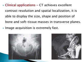
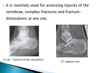
SlideShare a Scribd company logo
 Imaging modalities remains a keystone in
diagnosing orthopaedic conditions.
 Imaging started with the discovery of X-rays by W.
Rontgen in 1895.
 In general, imaging test have a high sensitivity –
few false negatives, but low specificity – high false
positive rate.
 Plain X-ray.
 Contrast Radiograph.
 Ultrasonography.
 Computed tomography.
 Magnetic Resonance Imaging.
 Scintigraphy.
 SPECT.
 PET
 High energy electromagnetic waves.
 Wavelength shorter than visible light.
 Structure –
◦ Positive anode
◦ Negative cathode
◦ Vacuum tube.
 Denser the tissue, greater X ray attenuation, more
bright.
 Commonly the first line investigation.
 Rule of 2 –
◦ 2 orthogonal views
◦ 2 joints.
◦ 2 sides
◦ 2 occasions
◦ 2 abnormalities
◦ 2 opinion.
 PACS - This is the system whereby all digitally
coded images are filed, stored.
 A convenient sequence for examination is: the patient –
the soft tissues – the bones – the joints.
 The patient –
◦ Identity,
◦ history and
◦ clinical findings.
 The soft tissues –
◦ Generalized change,
◦ Localized change.
 The bones –
◦ Shape
◦ Generalized change – density, sclerotic or lytic.
◦ Localized change - lesion’s size, site, shape, density.
 The joints - The radiographic ‘joint’ consists of the
articulating bones and the ‘space’ between them.
◦ The joint space.
◦ Shape.
◦ Erosions.
 Narrowing of the joint space +
subchondral sclerosis and cysts
+ osteophytes = osteoarthritis.
 Narrowing of the joint space +
osteoporosis + periarticular
erosions = inflammatory
arthritis
 Fracture/ Dislocation/ Subluxation.
 Soft tissue shadows.
 Evaluation of implants.
 Guiding image in case of close reduction.
 Identifying infection and tumours.
 Exposure of the patient to ionizing radiation.
 As a diagnostic tool, conventional radiography
provides poor soft-tissue contrast: for example, it
cannot distinguish between muscles, tendons,
ligaments and hyaline cartilage.
 The contrast media used in orthopaedics are
mostly iodine-based liquids which can be injected
into sinuses, joint cavities or the spinal theca.
 Ionic, water-soluble iodides permit much more
detailed imaging and, although also somewhat
irritant and neurotoxic, are rapidly absorbed and
excreted.
 Sinography –
 Arthrography – Intra-articular loose bodies will
produce filling defects in the opaque contrast
medium.
 Myelography -
 Introduction - The frequency used for therapeutic
ultrasound is usually 0.5 MHz (LIPUS) while those for
diagnostic ultrasound range between 2 MHz and 10
MHz.
 Higher the frequency used, the greater the spatial
resolution.
 Higher frequency probes (5 MHz–7.5 MHz) -
 Lower frequency probes(3–5 MHz).
 “A” (amplitude) type scanning –
 “B” (brightness) type scanning –
 Hypoechoic – Fat, Hyaline cartilage,
Synovium/capsule, Nerve.
 Hyperechoic – Tendon, Muscle, Fascia,
Ligament, Bone/periosteum.
 Advantages of ultrasonography –
1. Safety is the foremost advantage.
2. Real time imaging capability allowing dynamic
evaluation
3. Low cost
4. Easy accessibility
5. Portability
6. Non invasiveness
7. Multiplanar imaging.
 Diagnostic –
◦ Tendons –
 Tears
 Tendinosis, tendinitis and tenosynovitis
 Peritendinitis.
◦ Cysts –
 Therapeutic -
o Enthesopathy
o Bursitis
o Postoperative
o Interventional
o Aspiration of cysts, abscess and hematoma, etc.
o Injection of sclerosant into cysts.
 Plain tomography –
 Like plain tomography, CT produces sectional
images through selected tissue planes – but with
much greater resolution.
 The images are trans-axial.
 Region of interest is selected and a series of cross-
sectional images is produced and digitally
recorded.
 Clinical applications - CT achieves excellent
contrast resolution and spatial localization, it is
able to display the size, shape and position of
bone and soft-tissue masses in transverse planes.
 Image acquisition is extremely fast.
 It is routinely used for assessing injuries of the
vertebrae, complex fractures and fracture-
dislocations at any site.
X-ray - fracture of the calcaneum
CT sagittal view
 Limitations - it provides relatively poor soft-
tissue contrast when compared with MRI.
 A major disadvantage of this technique is the
relatively high radiation exposure to which the
patient is subjected.
 Introduction - Magnetic resonance imaging
produces cross-sectional images of any body part
in any plane. It yields superb soft-tissue contrast,
allowing different soft tissues to be clearly
distinguished, e.g. ligaments, tendons, muscle and
hyaline cartilage.
 MRI physics – Different speeds of tissue excitation
with radiofrequency pulses (repetition times, or
TR) and different intervals between recording
these signals (time to echo, or TE) will yield
anatomical pictures with varying ‘weighting’ and
characteristics.
 T1 weighted Image - high spatial resolution and
provide good anatomical- looking pictures.
 Short TR ( <1000) and TE (around 10).
 Fat Bright and Fluid dark.
 T2 weighted (T2W) images - give more information
about the physiological characteristics of the
tissue.
 Long TR (around 4000) and TE (around 90).
 Fat Dark and Fluid Bright.
 Proton density (PD) images - ‘balanced’ or
‘intermediate’.
 Yield excellent anatomical detail for orthopaedic
imaging.
 Medium TR and Medium TE.
 Fat bright (Dark if PDFS) , Fluid also bright and
Muscle medium.
 Fast spin-echo technique - This reduces length of
T2 sequence by two-thirds. But this produces
some blurring artifact.
 Gradient-echo technique – Fat Dark, Muscle Dark
and fluid Dark.
 Used selectively for cartilage imaging.
 Knee –
◦ Meniscal tears
◦ Degenerative conditions of meniscus
◦ Ligament tears
◦ Patellar or quadriceps tendon tears
◦ Tendinitis
◦ Cysts
◦ Synovial pathology
◦ Occult fractures
◦ Popliteal mass
◦ Physeal injuries in children.
 Shoulder –
◦ Rotator cuff tear
◦ Impingement syndromes
◦ Instability
◦ Osteonecrosis
◦ Pathological conditions of the tendon
 Spine –
◦ Evaluation of intervertebral disk disease
◦ Discitis
◦ Pyogenic and tuberculous infections
 Foot and ankle –
◦ Tendinopathy
◦ Articular disorders
◦ Osseous pathological conditions
◦ Avascular necrosis
◦ Fracture
◦ Osteochondral injuries
◦ Osteomyelitis
◦ Tendon injury
◦ Ligament injury.
 Hip –
◦ Avascular necrosis
◦ Transient osteoporosis of the hip
◦ Occult fractures
• Presence of intracerebral aneurysm clips
• Implanted cardiac pacemakers and automatic defibrillators
• Bio stimulators
• Implanted infusion devices and internal hearing aids
• Metallic orbital foreign bodies
• Metal external fixation devices
• Cochlear implants
• Ferromagnetic foreign bodies like shell shrapnel.
First-trimester pregnancy
Imaging In Orthopaedics.pptx

More Related Content

What's hot

Assessing bone loss in instability lf 2016
Assessing bone loss in instability   lf 2016Assessing bone loss in instability   lf 2016
Assessing bone loss in instability lf 2016Lennard Funk
 
Imaging of shoulder - Dr. Vishal Sankpal
Imaging of shoulder - Dr. Vishal SankpalImaging of shoulder - Dr. Vishal Sankpal
Imaging of shoulder - Dr. Vishal SankpalVishal Sankpal
 
MSK Imaging Guidelines
MSK Imaging GuidelinesMSK Imaging Guidelines
MSK Imaging Guidelineskkallas
 
Radial head replacement best evidence
Radial head replacement best evidenceRadial head replacement best evidence
Radial head replacement best evidenceorthoprinciples
 
Newer implants for geriatric hip fractures
Newer implants for geriatric hip fracturesNewer implants for geriatric hip fractures
Newer implants for geriatric hip fracturesArjun Viegas
 
Radiology in orthopaedics
Radiology in orthopaedicsRadiology in orthopaedics
Radiology in orthopaedicsYeswanth Mohan
 
Acromion - Os-acromiale Fractures and Insufficiency
Acromion - Os-acromiale Fractures and InsufficiencyAcromion - Os-acromiale Fractures and Insufficiency
Acromion - Os-acromiale Fractures and InsufficiencyWrightington Upper Limb Unit
 
Navigation Assisted Total Knee Replacement
Navigation Assisted Total Knee ReplacementNavigation Assisted Total Knee Replacement
Navigation Assisted Total Knee ReplacementMurtuza Rassiwala
 
how to read spine mri
 how to read spine mri how to read spine mri
how to read spine mriFK UNCEN
 
Bone Mineral Density
Bone Mineral DensityBone Mineral Density
Bone Mineral DensityZarooon
 
Ankle fractures final
Ankle fractures finalAnkle fractures final
Ankle fractures finalRavi Malhotra
 
Role of arthroscopic synovectomy of knee joint in ra
Role of arthroscopic synovectomy of knee joint in raRole of arthroscopic synovectomy of knee joint in ra
Role of arthroscopic synovectomy of knee joint in raDr. Prakhar chhawchharia
 
Bone and joint infection presentation
Bone and joint infection presentationBone and joint infection presentation
Bone and joint infection presentationAmit Rauniyar
 
Meniscus - Anatomy, function and injury
Meniscus - Anatomy, function and injuryMeniscus - Anatomy, function and injury
Meniscus - Anatomy, function and injuryAsish Rajak
 
Assessment Of Glenoid Bone Loss In Recurrent Shoulder Dislocation
Assessment Of Glenoid Bone LossIn Recurrent Shoulder Dislocation Assessment Of Glenoid Bone LossIn Recurrent Shoulder Dislocation
Assessment Of Glenoid Bone Loss In Recurrent Shoulder Dislocation Samir Dwidmuthe
 
Surgical Approaches to distal humerus fractures - DR.S.SENTHIL SAILESH, M.S...
 Surgical Approaches to distal  humerus fractures - DR.S.SENTHIL SAILESH, M.S... Surgical Approaches to distal  humerus fractures - DR.S.SENTHIL SAILESH, M.S...
Surgical Approaches to distal humerus fractures - DR.S.SENTHIL SAILESH, M.S...Senthil sailesh
 

What's hot (20)

Assessing bone loss in instability lf 2016
Assessing bone loss in instability   lf 2016Assessing bone loss in instability   lf 2016
Assessing bone loss in instability lf 2016
 
Imaging of shoulder - Dr. Vishal Sankpal
Imaging of shoulder - Dr. Vishal SankpalImaging of shoulder - Dr. Vishal Sankpal
Imaging of shoulder - Dr. Vishal Sankpal
 
MSK Imaging Guidelines
MSK Imaging GuidelinesMSK Imaging Guidelines
MSK Imaging Guidelines
 
MRI in orthopaedics
MRI in orthopaedicsMRI in orthopaedics
MRI in orthopaedics
 
Radial head replacement best evidence
Radial head replacement best evidenceRadial head replacement best evidence
Radial head replacement best evidence
 
Newer implants for geriatric hip fractures
Newer implants for geriatric hip fracturesNewer implants for geriatric hip fractures
Newer implants for geriatric hip fractures
 
Radiology in orthopaedics
Radiology in orthopaedicsRadiology in orthopaedics
Radiology in orthopaedics
 
Acromion - Os-acromiale Fractures and Insufficiency
Acromion - Os-acromiale Fractures and InsufficiencyAcromion - Os-acromiale Fractures and Insufficiency
Acromion - Os-acromiale Fractures and Insufficiency
 
Navigation Assisted Total Knee Replacement
Navigation Assisted Total Knee ReplacementNavigation Assisted Total Knee Replacement
Navigation Assisted Total Knee Replacement
 
Ankle arthrodesis
Ankle arthrodesisAnkle arthrodesis
Ankle arthrodesis
 
how to read spine mri
 how to read spine mri how to read spine mri
how to read spine mri
 
Bone Mineral Density
Bone Mineral DensityBone Mineral Density
Bone Mineral Density
 
Ankle fractures final
Ankle fractures finalAnkle fractures final
Ankle fractures final
 
Role of arthroscopic synovectomy of knee joint in ra
Role of arthroscopic synovectomy of knee joint in raRole of arthroscopic synovectomy of knee joint in ra
Role of arthroscopic synovectomy of knee joint in ra
 
Bone and joint infection presentation
Bone and joint infection presentationBone and joint infection presentation
Bone and joint infection presentation
 
Meniscus - Anatomy, function and injury
Meniscus - Anatomy, function and injuryMeniscus - Anatomy, function and injury
Meniscus - Anatomy, function and injury
 
Assessment Of Glenoid Bone Loss In Recurrent Shoulder Dislocation
Assessment Of Glenoid Bone LossIn Recurrent Shoulder Dislocation Assessment Of Glenoid Bone LossIn Recurrent Shoulder Dislocation
Assessment Of Glenoid Bone Loss In Recurrent Shoulder Dislocation
 
Surgical Approaches to distal humerus fractures - DR.S.SENTHIL SAILESH, M.S...
 Surgical Approaches to distal  humerus fractures - DR.S.SENTHIL SAILESH, M.S... Surgical Approaches to distal  humerus fractures - DR.S.SENTHIL SAILESH, M.S...
Surgical Approaches to distal humerus fractures - DR.S.SENTHIL SAILESH, M.S...
 
Non union scaphoid 1
Non union scaphoid 1Non union scaphoid 1
Non union scaphoid 1
 
Painful shoulder
Painful shoulderPainful shoulder
Painful shoulder
 

Similar to Imaging In Orthopaedics.pptx

1 diagnostic imaging
1 diagnostic imaging1 diagnostic imaging
1 diagnostic imagingSimba Syed
 
Imaging in orthopaedics
Imaging  in  orthopaedicsImaging  in  orthopaedics
Imaging in orthopaedicsBipulBorthakur
 
Foundations of Diagnostic Imaging for Physical Therapist
Foundations of Diagnostic Imaging for Physical TherapistFoundations of Diagnostic Imaging for Physical Therapist
Foundations of Diagnostic Imaging for Physical TherapistDana Tew
 
Neuroimaging Lecture
Neuroimaging LectureNeuroimaging Lecture
Neuroimaging Lecturetest
 
approach to radiology of spinal cord.pptx
approach to radiology of spinal cord.pptxapproach to radiology of spinal cord.pptx
approach to radiology of spinal cord.pptxshaitansingh8
 
Tendon elastography
Tendon elastography  Tendon elastography
Tendon elastography Tehreem Zahra
 
Imaging of eye and orbit
Imaging of eye and orbitImaging of eye and orbit
Imaging of eye and orbitShrikant Nagare
 
COMPUTED TOMOGRAPHY
COMPUTED TOMOGRAPHY COMPUTED TOMOGRAPHY
COMPUTED TOMOGRAPHY vamsi krishna
 
PATHOLOGICAL INVESTIGATIONS AND IMAGING TECHNIQUES IN NEUROMUSCULAR DISORDERS...
PATHOLOGICAL INVESTIGATIONS AND IMAGING TECHNIQUES IN NEUROMUSCULAR DISORDERS...PATHOLOGICAL INVESTIGATIONS AND IMAGING TECHNIQUES IN NEUROMUSCULAR DISORDERS...
PATHOLOGICAL INVESTIGATIONS AND IMAGING TECHNIQUES IN NEUROMUSCULAR DISORDERS...shuchij10
 
MRI-in Spine PPT.pptx
MRI-in Spine PPT.pptxMRI-in Spine PPT.pptx
MRI-in Spine PPT.pptxssusere26312
 
Bonescan 22-08-2014
Bonescan 22-08-2014Bonescan 22-08-2014
Bonescan 22-08-2014Amol Gulhane
 
3_2018_12_16!11_22_57_PM.pptx
3_2018_12_16!11_22_57_PM.pptx3_2018_12_16!11_22_57_PM.pptx
3_2018_12_16!11_22_57_PM.pptxKhaVuong2
 
MRI KNEE OF ORTHOPEDIC IMPORTANCE
MRI KNEE OF ORTHOPEDIC IMPORTANCEMRI KNEE OF ORTHOPEDIC IMPORTANCE
MRI KNEE OF ORTHOPEDIC IMPORTANCEBenthungo Tungoe
 
Computed tomography
Computed tomographyComputed tomography
Computed tomographysaanvi2011
 
diagnosticimaginginoromaxillofacialsurgery-141214161540-conversion-gate02.pptx
diagnosticimaginginoromaxillofacialsurgery-141214161540-conversion-gate02.pptxdiagnosticimaginginoromaxillofacialsurgery-141214161540-conversion-gate02.pptx
diagnosticimaginginoromaxillofacialsurgery-141214161540-conversion-gate02.pptxSamarElgendi
 
Spom Imaging
Spom ImagingSpom Imaging
Spom ImagingClaraha
 
Imaging of Soft tissue pathology
Imaging of Soft tissue pathologyImaging of Soft tissue pathology
Imaging of Soft tissue pathologyRakesh Ca
 

Similar to Imaging In Orthopaedics.pptx (20)

1 diagnostic imaging
1 diagnostic imaging1 diagnostic imaging
1 diagnostic imaging
 
Imaging in orthopaedics
Imaging  in  orthopaedicsImaging  in  orthopaedics
Imaging in orthopaedics
 
Foundations of Diagnostic Imaging for Physical Therapist
Foundations of Diagnostic Imaging for Physical TherapistFoundations of Diagnostic Imaging for Physical Therapist
Foundations of Diagnostic Imaging for Physical Therapist
 
Neuroimaging Lecture
Neuroimaging LectureNeuroimaging Lecture
Neuroimaging Lecture
 
approach to radiology of spinal cord.pptx
approach to radiology of spinal cord.pptxapproach to radiology of spinal cord.pptx
approach to radiology of spinal cord.pptx
 
Tendon elastography
Tendon elastography  Tendon elastography
Tendon elastography
 
Imaging of eye and orbit
Imaging of eye and orbitImaging of eye and orbit
Imaging of eye and orbit
 
COMPUTED TOMOGRAPHY
COMPUTED TOMOGRAPHY COMPUTED TOMOGRAPHY
COMPUTED TOMOGRAPHY
 
Imaging in sports injury
Imaging in sports injuryImaging in sports injury
Imaging in sports injury
 
PATHOLOGICAL INVESTIGATIONS AND IMAGING TECHNIQUES IN NEUROMUSCULAR DISORDERS...
PATHOLOGICAL INVESTIGATIONS AND IMAGING TECHNIQUES IN NEUROMUSCULAR DISORDERS...PATHOLOGICAL INVESTIGATIONS AND IMAGING TECHNIQUES IN NEUROMUSCULAR DISORDERS...
PATHOLOGICAL INVESTIGATIONS AND IMAGING TECHNIQUES IN NEUROMUSCULAR DISORDERS...
 
MRI-in Spine PPT.pptx
MRI-in Spine PPT.pptxMRI-in Spine PPT.pptx
MRI-in Spine PPT.pptx
 
Bonescan 22-08-2014
Bonescan 22-08-2014Bonescan 22-08-2014
Bonescan 22-08-2014
 
3_2018_12_16!11_22_57_PM.pptx
3_2018_12_16!11_22_57_PM.pptx3_2018_12_16!11_22_57_PM.pptx
3_2018_12_16!11_22_57_PM.pptx
 
MRI KNEE OF ORTHOPEDIC IMPORTANCE
MRI KNEE OF ORTHOPEDIC IMPORTANCEMRI KNEE OF ORTHOPEDIC IMPORTANCE
MRI KNEE OF ORTHOPEDIC IMPORTANCE
 
Computed tomography
Computed tomographyComputed tomography
Computed tomography
 
diagnosticimaginginoromaxillofacialsurgery-141214161540-conversion-gate02.pptx
diagnosticimaginginoromaxillofacialsurgery-141214161540-conversion-gate02.pptxdiagnosticimaginginoromaxillofacialsurgery-141214161540-conversion-gate02.pptx
diagnosticimaginginoromaxillofacialsurgery-141214161540-conversion-gate02.pptx
 
Spom Imaging
Spom ImagingSpom Imaging
Spom Imaging
 
Ultrasonography
UltrasonographyUltrasonography
Ultrasonography
 
CT and MRI
CT and MRICT and MRI
CT and MRI
 
Imaging of Soft tissue pathology
Imaging of Soft tissue pathologyImaging of Soft tissue pathology
Imaging of Soft tissue pathology
 

Recently uploaded

1130525--家醫計畫2.0糖尿病照護研討會-社團法人高雄市醫師公會.pdf
1130525--家醫計畫2.0糖尿病照護研討會-社團法人高雄市醫師公會.pdf1130525--家醫計畫2.0糖尿病照護研討會-社團法人高雄市醫師公會.pdf
1130525--家醫計畫2.0糖尿病照護研討會-社團法人高雄市醫師公會.pdfKs doctor
 
Alcohol_Dr. Jeenal Mistry MD Pharmacology.pdf
Alcohol_Dr. Jeenal Mistry MD Pharmacology.pdfAlcohol_Dr. Jeenal Mistry MD Pharmacology.pdf
Alcohol_Dr. Jeenal Mistry MD Pharmacology.pdfDr Jeenal Mistry
 
Book Trailer: PGMEE in a Nutshell (CEE MD/MS PG Entrance Examination)
Book Trailer: PGMEE in a Nutshell (CEE MD/MS PG Entrance Examination)Book Trailer: PGMEE in a Nutshell (CEE MD/MS PG Entrance Examination)
Book Trailer: PGMEE in a Nutshell (CEE MD/MS PG Entrance Examination)Dr. Aryan (Anish Dhakal)
 
Blue Printing in medical education by Dr.Mumtaz Ali.pptx
Blue Printing in medical education by Dr.Mumtaz Ali.pptxBlue Printing in medical education by Dr.Mumtaz Ali.pptx
Blue Printing in medical education by Dr.Mumtaz Ali.pptxNeurosurgeon Mumtaz Ali Narejo
 
Relationship between vascular system disfunction, neurofluid flow and Alzheim...
Relationship between vascular system disfunction, neurofluid flow and Alzheim...Relationship between vascular system disfunction, neurofluid flow and Alzheim...
Relationship between vascular system disfunction, neurofluid flow and Alzheim...Catherine Liao
 
5cl adbb 5cladba cheap and fine Telegram: +85297504341
5cl adbb 5cladba cheap and fine Telegram: +852975043415cl adbb 5cladba cheap and fine Telegram: +85297504341
5cl adbb 5cladba cheap and fine Telegram: +85297504341Sherrylee83
 
Aptopadesha Pramana / Pariksha: The Verbal Testimony
Aptopadesha Pramana / Pariksha: The Verbal TestimonyAptopadesha Pramana / Pariksha: The Verbal Testimony
Aptopadesha Pramana / Pariksha: The Verbal TestimonyDr KHALID B.M
 
Impact of cancers therapies on the loss in cardiac function, myocardial fffic...
Impact of cancers therapies on the loss in cardiac function, myocardial fffic...Impact of cancers therapies on the loss in cardiac function, myocardial fffic...
Impact of cancers therapies on the loss in cardiac function, myocardial fffic...Catherine Liao
 
Non-Invasive assessment of arterial stiffness in advanced heart failure patie...
Non-Invasive assessment of arterial stiffness in advanced heart failure patie...Non-Invasive assessment of arterial stiffness in advanced heart failure patie...
Non-Invasive assessment of arterial stiffness in advanced heart failure patie...Catherine Liao
 
PT MANAGEMENT OF URINARY INCONTINENCE.pptx
PT MANAGEMENT OF URINARY INCONTINENCE.pptxPT MANAGEMENT OF URINARY INCONTINENCE.pptx
PT MANAGEMENT OF URINARY INCONTINENCE.pptxdrtabassum4
 
Temporal, Infratemporal & Pterygopalatine BY Dr.RIG.pptx
Temporal, Infratemporal & Pterygopalatine BY Dr.RIG.pptxTemporal, Infratemporal & Pterygopalatine BY Dr.RIG.pptx
Temporal, Infratemporal & Pterygopalatine BY Dr.RIG.pptxDr. Rabia Inam Gandapore
 
Factors Affecting child behavior in Pediatric Dentistry
Factors Affecting child behavior in Pediatric DentistryFactors Affecting child behavior in Pediatric Dentistry
Factors Affecting child behavior in Pediatric DentistryDr Simran Deepak Vangani
 
"Central Hypertension"‚ in China: Towards the nation-wide use of SphygmoCor t...
"Central Hypertension"‚ in China: Towards the nation-wide use of SphygmoCor t..."Central Hypertension"‚ in China: Towards the nation-wide use of SphygmoCor t...
"Central Hypertension"‚ in China: Towards the nation-wide use of SphygmoCor t...Catherine Liao
 
Young at heart: Cardiovascular health stations to empower healthy lifestyle b...
Young at heart: Cardiovascular health stations to empower healthy lifestyle b...Young at heart: Cardiovascular health stations to empower healthy lifestyle b...
Young at heart: Cardiovascular health stations to empower healthy lifestyle b...Catherine Liao
 
Antiulcer drugs Advance Pharmacology .pptx
Antiulcer drugs Advance Pharmacology .pptxAntiulcer drugs Advance Pharmacology .pptx
Antiulcer drugs Advance Pharmacology .pptxRohit chaurpagar
 
Effects of vaping e-cigarettes on arterial health
Effects of vaping e-cigarettes on arterial healthEffects of vaping e-cigarettes on arterial health
Effects of vaping e-cigarettes on arterial healthCatherine Liao
 
THORACOTOMY . SURGICAL PERSPECTIVES VOL 1
THORACOTOMY . SURGICAL PERSPECTIVES VOL 1THORACOTOMY . SURGICAL PERSPECTIVES VOL 1
THORACOTOMY . SURGICAL PERSPECTIVES VOL 1DR SETH JOTHAM
 
linearity concept of significance, standard deviation, chi square test, stude...
linearity concept of significance, standard deviation, chi square test, stude...linearity concept of significance, standard deviation, chi square test, stude...
linearity concept of significance, standard deviation, chi square test, stude...KavyasriPuttamreddy
 
DECIPHERING COMMON ECG FINDINGS IN ED.pptx
DECIPHERING COMMON ECG FINDINGS IN ED.pptxDECIPHERING COMMON ECG FINDINGS IN ED.pptx
DECIPHERING COMMON ECG FINDINGS IN ED.pptxdrwaque
 

Recently uploaded (20)

1130525--家醫計畫2.0糖尿病照護研討會-社團法人高雄市醫師公會.pdf
1130525--家醫計畫2.0糖尿病照護研討會-社團法人高雄市醫師公會.pdf1130525--家醫計畫2.0糖尿病照護研討會-社團法人高雄市醫師公會.pdf
1130525--家醫計畫2.0糖尿病照護研討會-社團法人高雄市醫師公會.pdf
 
Alcohol_Dr. Jeenal Mistry MD Pharmacology.pdf
Alcohol_Dr. Jeenal Mistry MD Pharmacology.pdfAlcohol_Dr. Jeenal Mistry MD Pharmacology.pdf
Alcohol_Dr. Jeenal Mistry MD Pharmacology.pdf
 
Book Trailer: PGMEE in a Nutshell (CEE MD/MS PG Entrance Examination)
Book Trailer: PGMEE in a Nutshell (CEE MD/MS PG Entrance Examination)Book Trailer: PGMEE in a Nutshell (CEE MD/MS PG Entrance Examination)
Book Trailer: PGMEE in a Nutshell (CEE MD/MS PG Entrance Examination)
 
Blue Printing in medical education by Dr.Mumtaz Ali.pptx
Blue Printing in medical education by Dr.Mumtaz Ali.pptxBlue Printing in medical education by Dr.Mumtaz Ali.pptx
Blue Printing in medical education by Dr.Mumtaz Ali.pptx
 
Relationship between vascular system disfunction, neurofluid flow and Alzheim...
Relationship between vascular system disfunction, neurofluid flow and Alzheim...Relationship between vascular system disfunction, neurofluid flow and Alzheim...
Relationship between vascular system disfunction, neurofluid flow and Alzheim...
 
5cl adbb 5cladba cheap and fine Telegram: +85297504341
5cl adbb 5cladba cheap and fine Telegram: +852975043415cl adbb 5cladba cheap and fine Telegram: +85297504341
5cl adbb 5cladba cheap and fine Telegram: +85297504341
 
Aptopadesha Pramana / Pariksha: The Verbal Testimony
Aptopadesha Pramana / Pariksha: The Verbal TestimonyAptopadesha Pramana / Pariksha: The Verbal Testimony
Aptopadesha Pramana / Pariksha: The Verbal Testimony
 
Impact of cancers therapies on the loss in cardiac function, myocardial fffic...
Impact of cancers therapies on the loss in cardiac function, myocardial fffic...Impact of cancers therapies on the loss in cardiac function, myocardial fffic...
Impact of cancers therapies on the loss in cardiac function, myocardial fffic...
 
Non-Invasive assessment of arterial stiffness in advanced heart failure patie...
Non-Invasive assessment of arterial stiffness in advanced heart failure patie...Non-Invasive assessment of arterial stiffness in advanced heart failure patie...
Non-Invasive assessment of arterial stiffness in advanced heart failure patie...
 
PT MANAGEMENT OF URINARY INCONTINENCE.pptx
PT MANAGEMENT OF URINARY INCONTINENCE.pptxPT MANAGEMENT OF URINARY INCONTINENCE.pptx
PT MANAGEMENT OF URINARY INCONTINENCE.pptx
 
Temporal, Infratemporal & Pterygopalatine BY Dr.RIG.pptx
Temporal, Infratemporal & Pterygopalatine BY Dr.RIG.pptxTemporal, Infratemporal & Pterygopalatine BY Dr.RIG.pptx
Temporal, Infratemporal & Pterygopalatine BY Dr.RIG.pptx
 
Factors Affecting child behavior in Pediatric Dentistry
Factors Affecting child behavior in Pediatric DentistryFactors Affecting child behavior in Pediatric Dentistry
Factors Affecting child behavior in Pediatric Dentistry
 
"Central Hypertension"‚ in China: Towards the nation-wide use of SphygmoCor t...
"Central Hypertension"‚ in China: Towards the nation-wide use of SphygmoCor t..."Central Hypertension"‚ in China: Towards the nation-wide use of SphygmoCor t...
"Central Hypertension"‚ in China: Towards the nation-wide use of SphygmoCor t...
 
Young at heart: Cardiovascular health stations to empower healthy lifestyle b...
Young at heart: Cardiovascular health stations to empower healthy lifestyle b...Young at heart: Cardiovascular health stations to empower healthy lifestyle b...
Young at heart: Cardiovascular health stations to empower healthy lifestyle b...
 
Antiulcer drugs Advance Pharmacology .pptx
Antiulcer drugs Advance Pharmacology .pptxAntiulcer drugs Advance Pharmacology .pptx
Antiulcer drugs Advance Pharmacology .pptx
 
Contact dermaititis (irritant and allergic).pdf
Contact dermaititis (irritant and allergic).pdfContact dermaititis (irritant and allergic).pdf
Contact dermaititis (irritant and allergic).pdf
 
Effects of vaping e-cigarettes on arterial health
Effects of vaping e-cigarettes on arterial healthEffects of vaping e-cigarettes on arterial health
Effects of vaping e-cigarettes on arterial health
 
THORACOTOMY . SURGICAL PERSPECTIVES VOL 1
THORACOTOMY . SURGICAL PERSPECTIVES VOL 1THORACOTOMY . SURGICAL PERSPECTIVES VOL 1
THORACOTOMY . SURGICAL PERSPECTIVES VOL 1
 
linearity concept of significance, standard deviation, chi square test, stude...
linearity concept of significance, standard deviation, chi square test, stude...linearity concept of significance, standard deviation, chi square test, stude...
linearity concept of significance, standard deviation, chi square test, stude...
 
DECIPHERING COMMON ECG FINDINGS IN ED.pptx
DECIPHERING COMMON ECG FINDINGS IN ED.pptxDECIPHERING COMMON ECG FINDINGS IN ED.pptx
DECIPHERING COMMON ECG FINDINGS IN ED.pptx
 

Imaging In Orthopaedics.pptx

  • 1.
  • 2.  Imaging modalities remains a keystone in diagnosing orthopaedic conditions.  Imaging started with the discovery of X-rays by W. Rontgen in 1895.  In general, imaging test have a high sensitivity – few false negatives, but low specificity – high false positive rate.
  • 3.  Plain X-ray.  Contrast Radiograph.  Ultrasonography.  Computed tomography.  Magnetic Resonance Imaging.  Scintigraphy.  SPECT.  PET
  • 4.  High energy electromagnetic waves.  Wavelength shorter than visible light.  Structure – ◦ Positive anode ◦ Negative cathode ◦ Vacuum tube.  Denser the tissue, greater X ray attenuation, more bright.  Commonly the first line investigation.
  • 5.  Rule of 2 – ◦ 2 orthogonal views ◦ 2 joints. ◦ 2 sides ◦ 2 occasions ◦ 2 abnormalities ◦ 2 opinion.  PACS - This is the system whereby all digitally coded images are filed, stored.
  • 6.  A convenient sequence for examination is: the patient – the soft tissues – the bones – the joints.  The patient – ◦ Identity, ◦ history and ◦ clinical findings.  The soft tissues – ◦ Generalized change, ◦ Localized change.
  • 7.  The bones – ◦ Shape ◦ Generalized change – density, sclerotic or lytic. ◦ Localized change - lesion’s size, site, shape, density.  The joints - The radiographic ‘joint’ consists of the articulating bones and the ‘space’ between them. ◦ The joint space. ◦ Shape. ◦ Erosions.
  • 8.  Narrowing of the joint space + subchondral sclerosis and cysts + osteophytes = osteoarthritis.  Narrowing of the joint space + osteoporosis + periarticular erosions = inflammatory arthritis
  • 9.  Fracture/ Dislocation/ Subluxation.  Soft tissue shadows.  Evaluation of implants.  Guiding image in case of close reduction.  Identifying infection and tumours.
  • 10.  Exposure of the patient to ionizing radiation.  As a diagnostic tool, conventional radiography provides poor soft-tissue contrast: for example, it cannot distinguish between muscles, tendons, ligaments and hyaline cartilage.
  • 11.  The contrast media used in orthopaedics are mostly iodine-based liquids which can be injected into sinuses, joint cavities or the spinal theca.  Ionic, water-soluble iodides permit much more detailed imaging and, although also somewhat irritant and neurotoxic, are rapidly absorbed and excreted.
  • 12.  Sinography –  Arthrography – Intra-articular loose bodies will produce filling defects in the opaque contrast medium.  Myelography -
  • 13.  Introduction - The frequency used for therapeutic ultrasound is usually 0.5 MHz (LIPUS) while those for diagnostic ultrasound range between 2 MHz and 10 MHz.  Higher the frequency used, the greater the spatial resolution.  Higher frequency probes (5 MHz–7.5 MHz) -  Lower frequency probes(3–5 MHz).
  • 14.  “A” (amplitude) type scanning –  “B” (brightness) type scanning –  Hypoechoic – Fat, Hyaline cartilage, Synovium/capsule, Nerve.  Hyperechoic – Tendon, Muscle, Fascia, Ligament, Bone/periosteum.
  • 15.  Advantages of ultrasonography – 1. Safety is the foremost advantage. 2. Real time imaging capability allowing dynamic evaluation 3. Low cost 4. Easy accessibility 5. Portability 6. Non invasiveness 7. Multiplanar imaging.
  • 16.  Diagnostic – ◦ Tendons –  Tears  Tendinosis, tendinitis and tenosynovitis  Peritendinitis. ◦ Cysts –  Therapeutic - o Enthesopathy o Bursitis o Postoperative o Interventional o Aspiration of cysts, abscess and hematoma, etc. o Injection of sclerosant into cysts.
  • 17.  Plain tomography –  Like plain tomography, CT produces sectional images through selected tissue planes – but with much greater resolution.  The images are trans-axial.  Region of interest is selected and a series of cross- sectional images is produced and digitally recorded.
  • 18.  Clinical applications - CT achieves excellent contrast resolution and spatial localization, it is able to display the size, shape and position of bone and soft-tissue masses in transverse planes.  Image acquisition is extremely fast.
  • 19.  It is routinely used for assessing injuries of the vertebrae, complex fractures and fracture- dislocations at any site. X-ray - fracture of the calcaneum CT sagittal view
  • 20.  Limitations - it provides relatively poor soft- tissue contrast when compared with MRI.  A major disadvantage of this technique is the relatively high radiation exposure to which the patient is subjected.
  • 21.  Introduction - Magnetic resonance imaging produces cross-sectional images of any body part in any plane. It yields superb soft-tissue contrast, allowing different soft tissues to be clearly distinguished, e.g. ligaments, tendons, muscle and hyaline cartilage.
  • 22.  MRI physics – Different speeds of tissue excitation with radiofrequency pulses (repetition times, or TR) and different intervals between recording these signals (time to echo, or TE) will yield anatomical pictures with varying ‘weighting’ and characteristics.
  • 23.  T1 weighted Image - high spatial resolution and provide good anatomical- looking pictures.  Short TR ( <1000) and TE (around 10).  Fat Bright and Fluid dark.
  • 24.  T2 weighted (T2W) images - give more information about the physiological characteristics of the tissue.  Long TR (around 4000) and TE (around 90).  Fat Dark and Fluid Bright.
  • 25.  Proton density (PD) images - ‘balanced’ or ‘intermediate’.  Yield excellent anatomical detail for orthopaedic imaging.  Medium TR and Medium TE.  Fat bright (Dark if PDFS) , Fluid also bright and Muscle medium.
  • 26.  Fast spin-echo technique - This reduces length of T2 sequence by two-thirds. But this produces some blurring artifact.  Gradient-echo technique – Fat Dark, Muscle Dark and fluid Dark.  Used selectively for cartilage imaging.
  • 27.  Knee – ◦ Meniscal tears ◦ Degenerative conditions of meniscus ◦ Ligament tears ◦ Patellar or quadriceps tendon tears ◦ Tendinitis ◦ Cysts ◦ Synovial pathology ◦ Occult fractures ◦ Popliteal mass ◦ Physeal injuries in children.
  • 28.
  • 29.
  • 30.  Shoulder – ◦ Rotator cuff tear ◦ Impingement syndromes ◦ Instability ◦ Osteonecrosis ◦ Pathological conditions of the tendon  Spine – ◦ Evaluation of intervertebral disk disease ◦ Discitis ◦ Pyogenic and tuberculous infections
  • 31.
  • 32.
  • 33.  Foot and ankle – ◦ Tendinopathy ◦ Articular disorders ◦ Osseous pathological conditions ◦ Avascular necrosis ◦ Fracture ◦ Osteochondral injuries ◦ Osteomyelitis ◦ Tendon injury ◦ Ligament injury.  Hip – ◦ Avascular necrosis ◦ Transient osteoporosis of the hip ◦ Occult fractures
  • 34. • Presence of intracerebral aneurysm clips • Implanted cardiac pacemakers and automatic defibrillators • Bio stimulators • Implanted infusion devices and internal hearing aids • Metallic orbital foreign bodies • Metal external fixation devices • Cochlear implants • Ferromagnetic foreign bodies like shell shrapnel. First-trimester pregnancy