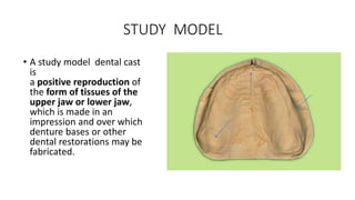
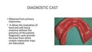
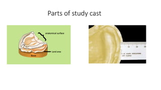
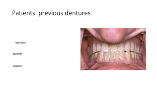
Diagnostic cast, Radiographs
Dr Ayesha Sadaf
ONE MINUTE PAPER
Q : A 60 year old male patient came for replacement of missing teeth.
On intraoral examination you found out patient is edentulous. His
maxillary residual ridge is low well rounded and mandible has knife edge
ridge. Maxillary and mandibular arches belong to which order of A
Atwoods classification?
a) Maxilla order 4 and mandible order 5
b)Maxilla order 5 and mandible order 4
c)Maxilla order 1 and mandible order 2
d) Maxilla order 6 and mandible order 7
Pre Extraction records
• Patient photographs
• Diagnostic casts
• Radiographs
• Pic of close relatives
• Previous dentures
Patients Photographs
DIAGNOSTIC CASTS
A diagnostic dental cast is a
cast/ model of a person’s
teeth that a dental
professional uses as a guide
in the application of
corrective or restorative
dentistry. Such diagnostic
casts are often referred to
as study models.
STUDY MODEL
• A study model dental cast
is
a positive reproduction of
the form of tissues of the
upper jaw or lower jaw,
which is made in an
impression and over which
denture bases or other
dental restorations may be
fabricated.
DIAGNOSTIC CAST
• Obtained from primary
impression.
• It allows the evaluation of
hard and soft tissue
anatomy without the
presence of the patient.
Diagnostic casts provide
the base from which
custom impression trays
are fabricated.
Parts of study cast
Significance of diagnostic casts
It allows the evaluation of hard and soft
tissue anatomy without the presence of
the patient
SIGNIFICANCE OF DIAGNOSTIC CAST
Measurement of arch
width and length
Patients previous dentures
• Gives information
about denture hygiene
• Maintenance at home
(home care)
Material of denture
Patients previous dentures
occlusion
aesthetics
Shade of teeth
Tooth
arrangement
Patients previous dentures
retention
stability
support
Diagnostic Radiographs
To Understand
residual ridge
morphology
Tooth roots and
pathologies
Pattern of Bone Resorption
MAXILLOMANDIBULAR
RELATIONS
IMPACTED TEETH
PATTERN OF BONE
RESORPTION
• Show the relative thickness of the submucosa covering the bone in
edentulous regions,
• the location of the mandibular canal, and
• the mental foramina in relation to the basal seat for dentures
More superficial
location of
mental
foramen, more
will be the bone
resorption
Gives an idea
about quality of
bone
More
radiopaque
bone, denser it
will appear on
radiograph
Cone beam computed tomography
3 dimensional View
Diagnostic casts , Radiographs and photographs helps in selecting
impression technique, planning pre-prosthetic surgery and selection of
teeth., ultimately formulating a treatment plan.
Patient specific care
How to read panaromic radiograph

Diagnostic cast.pptx